Scientific Literature Review
Tin and Tin Oxide as Used in Cosmetics
February 17, 2012
All interested persons are provided 60 days from the above date to comment on this Tentative Report and to identify
additional published data that should be included or provide unpublished data which can be made public and included.
Information may be submitted without identifying the source or the trade name of the cosmetic product containing the
ingredient. All unpublished data submitted to CIR will be discussed in open meetings, will be available at the CIR office for
review by any interested party and may be cited in a peer-reviewed scientific journal. Please submit data, comments, or
requests to the CIR Director, Dr. F. Alan Andersen.
The 2012 Cosmetic Ingredient Review Expert Panel members are: Chair, Wilma F. Bergfeld, M.D., F.A.C.P.; Donald V.
Belsito, M.D.; Curtis D. Klaassen, Ph.D.; Daniel C. Liebler, Ph.D.; Ronald A Hill, Ph.D. James G. Marks, Jr., M.D.; Ronald
C. Shank, Ph.D.; Thomas J. Slaga, Ph.D.; and Paul W. Snyder, D.V.M., Ph.D. The CIR Director is F. Alan Andersen, Ph.D.
This report was prepared by Wilbur Johnson, Jr., M.S., Manager/Lead Specialist.
© Cosmetic Ingredient Review
1101 17
TH
 STREET, NW, SUITE 412 " WASHINGTON, DC 20036‐4702 " PH 202.331.0651 " FAX 202.331.0088 " CIRINFO@CIR‐SAFETY.ORG 
Table of Contents
INTRODUCTION .......................................................................................................................................................... 1 
CHEMISTRY ................................................................................................................................................................. 1 
DEFINITION AND STRUCTURE .................................................................................................................................................. 1 
PHYSICAL AND CHEMICAL PROPERTIES ................................................................................................................................... 1 
METHOD OF MANUFACTURE .................................................................................................................................................... 1 
IMPURITIES .............................................................................................................................................................................. 1 
ii
USE ................................................................................................................................................................................ 2 
COSMETIC ................................................................................................................................................................................ 2 
NONCOSMETIC ......................................................................................................................................................................... 2 
TOXICOKINETICS ...................................................................................................................................................... 2 
ORAL STUDIES ......................................................................................................................................................................... 2 
INTRAVENOUS/INTRAPERITONEAL STUDY ............................................................................................................................... 3 
SUBCUTANEOUS STUDY ........................................................................................................................................................... 4 
PBPK MODEL .......................................................................................................................................................................... 4 
BIOAVAILABILITY .................................................................................................................................................................... 4 
TOXICOLOGY .............................................................................................................................................................. 4 
ACUTE TOXICITY ..................................................................................................................................................................... 4 
REPEATED DOSE TOXICITY ...................................................................................................................................................... 5 
CYTOTOXICITY ........................................................................................................................................................................ 5 
EFFECT ON ACETYLCHOLINESTERASE ..................................................................................................................................... 6 
RESPIRATORY IRRITATION ....................................................................................................................................................... 6 
OCULAR IRRITATION ............................................................................................................................................................... 6 
SKIN IRRITATION AND SENSITIZATION ..................................................................................................................................... 6 
ALLERGENICITY ....................................................................................................................................................................... 6 
OCCUPATIONAL STUDIES/CASE REPORTS ................................................................................................................................ 7 
REPRODUCTIVE AND DEVELOPMENTAL TOXICITY .......................................................................................... 8 
GENOTOXICITY .......................................................................................................................................................... 8 
CARCINOGENICITY ................................................................................................................................................... 8 
HEALTH EFFECTS ASSESSMENT ............................................................................................................................. 9 
DATA NEEDS .............................................................................................................................................................. 10 
SUMMARY .................................................................................................................................................................... 9 
1
INTRODUCTION
Published and unpublished data relevant to evaluating the safety of tin oxide and tin as used in cosmetics are
summarized in this literature review. Tin oxide functions as an abrasive, bulking agent, and opacifying agent and tin
functions as a surface modifier in cosmetic products.1
CHEMISTRY	
Definition and Structure
As given in the International Cosmetic Ingredient Dictionary and Handbook,1
tin oxide (CAS Nos. 1332-29-2 and
18282-10-5), the dioxide of tin, is an inorganic oxide that conforms to the following structure:
Other names for this chemical include: stannic oxide, white tin oxide, tin dioxide, stannic anhydride, and flowers of tin.1,2
Tin (CAS No. 7440-31-5) is a metallic element with the symbol Sn.
Physical and Chemical Properties
Tin is a silver-white metal that is malleable and somewhat ductile. It has a highly crystalline structure and exists in
two allotropic forms at normal pressures. Gray tin exists below 13.2 °C and has a cubic structure. At 13.2 °C, gray tin is
converted to white tin, which has a tetragonal structure.3,4
The white form is known as the common, stable form at room
temperature.5
In compounds, tin can exist in the +2 or +4 oxidation state.3,4
In compounds, tin in divalent and tetravalent
oxidation states are designated as stannous and stannic, respectively. The Stock Oxidation-Number system denotes the
oxidation state using Roman numerals in parentheses following the metal’s name: tin(II) and tin(IV).6
The cosmetic
ingredient, tin oxide is tin(IV) oxide.
Chemical and physical properties of Tin and Tin Oxide are found in Table 1.
Method of Manufacture
The earth's crust contains approximately 2 to 3 ppm tin, comprising 0.0006% of the earth's crust.2,7
The most
important tin-containing mineral is cassiterite, SnO2. Other tin minerals are stannite, teallite, cylindrite, and canfieldite.
After tin-containing ores are mined, they undergo further separation processing, resulting in concentrates containing 70–77%
tin by weight, almost pure cassiterite, and are ready for smelting.8
Elemental tin is obtained from cassiterite by reduction
with coal in a reverberatory furnace.5
Although tin oxide occurs naturally in mineral form, this is not the source of the
commercial product. It is manufactured directly from tin metal by thermal oxidation (from mined or recycled tin), either by
exposing molten tin to air in a furnace at elevated temperatures, or by blowing tin powder in a stream of air through a furnace
at approximately 700°C.
According to one source, the commercial production of tin oxides yielded the following grades: average particle
size of 0.3 µm (bulk density = 0.72 g/cm3
), average particle size of 0.4 µm (bulk density = 1.15 g/cm3
), and average particle
size of 0.5 µm (bulk density = 1.35 g/cm3
).9
Each grade is > 99.0% pure and has a specific gravity of 6.9.
Impurities
Commercially available metallic tin is approximately 99.8% pure.5
2
USE
Cosmetic
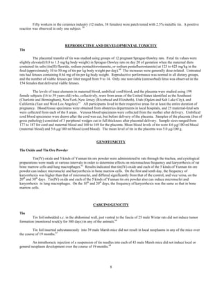
Tin oxide functions as an abrasive, bulking, and opacifying agent and tin functions as a surface modifier in cosmetic
products.1
According to information supplied to the Food and Drug Administration (FDA) by industry as part of the
Voluntary Cosmetic Registration Program (VCRP) in 2011, tin oxide was being used in cosmetic products, whereas,
elemental tin was not.10
These data are summarized in Table 2. Results from a survey of ingredient use concentrations
provided by the Personal Care Products Council (also included in Table 2) in 2011 indicate that tin oxide was being used at
concentrations up to 1% (rinse-off products) and up to 5 % (leave-on products).11
Cosmetic products containing tin oxide may be applied to the skin and hair, or, incidentally, may come in contact
with the eyes and mucous membranes. Products containing these ingredients may be applied as frequently as several times
per day and may come in contact with the skin or hair for variable periods following application. Daily or occasional use
may extend over many years.
Tin oxide is used in dusting powders and cosmetic sprays, other fragrance preparations, and body and hand sprays,
and could possibly be inhaled. In practice, 95% to 99% of the droplets/particles released from cosmetic sprays have
aerodynamic equivalent diameters >10 µm, with propellant sprays yielding a greater fraction of droplets/particles below 10
µm when compared with pump sprays.12,13
Therefore, most droplets/particles incidentally inhaled from cosmetic sprays
would be deposited in the nasopharyngeal region and would not be respirable (i.e., they would not enter the lungs) to any
appreciable amount.14,15
However, the potential for inhalation toxicity is not limited to respirable droplets/particles deposited
in the lungs. Inhaled droplets/particles deposited in the nasopharyngeal and thoracic regions of the respiratory tract may
cause toxic effects, depending on their chemical and other properties.
Noncosmetic	 	
Tin(IV) oxide is used in a variety of manufacturing applications, including polishing glass and other metals.2
Elemental tin is present mainly in solder alloys used in the electronics industry, and is also used as a protective coating for
other metals, especially those used for food containers.16
Foods usually contain tin at levels < 4 µg/g, but higher levels may
be found in processed foods because of tin-based preservatives and stabilizers and/or leaching from containers.17
The Food
and Agriculture Organization of the World Health Organization’s Joint Expert Committee on Food Additives has established
a provisional tolerable weekly intake of 14 mg Sn/kg body weight.18
The European Union has established maximum levels
for certain contaminants, inorganic tin included, to achieve a high level of public health protection, especially for sensitive
population groups such as children or individuals with allergies.19
Maximum levels of 200 mg/kg and 100 mg/kg were
established for inorganic tin in canned foods and canned beverages, respectively.
TOXICOKINETICS	
Oral Studies
Tin
After a single gavage dose of 20 mg/kg body weight of radiolabeled 113
Sn(II) or 113
Sn(IV) as the fluoride or citrate,
the tissue distribution of tin in female Charles River (CD) or Cox Charles River rats after 48 h as a percentage of the
administered tin(II) or tin(IV), respectively, was as follows: 1.0% and 0.24% (skeleton), 0.08% and 0.02% (liver), and
0.09% and 0.02% (kidneys).20
Female rats excreted 95% of the radiolabeled tin in the feces and less than 1% in the urine.
When oral tin doses of 20 mg/kg body weight were administered 6 days/week for 4 weeks, only the bone contained higher tin
concentrations after day 28 when compared to tin concentrations after day 1. The half-life of tin in the femur was estimated
to be 34 to 40 days. It was concluded that, of the soft tissues, only liver and kidneys were likely to accumulate significant
amounts of tin as a result of the oral ingestion of tin salts. 113
Sn was found in the brain of rats at 48 h post-administration of
113
Sn(II) or 113
Sn(IV) (as the citrate or fluoride) as a single oral dose (4 mg), as oral doses of 20 mg/kg body weight on 6
days/week for 4 weeks, or as a single i.v. dose (0.4 mg).
Tin(II) chloride was injected orally (intagastric [i.g.], using stomach tube) into mice, Sprague-Dawley rats, African
white-tailed rats, monkeys, and dogs.21
Less than 5% was absorbed from the gut, and bone was the chief site of tin
deposition.
3
The absorption of tin from the gastrointestinal tract and its distribution in the tissues was studied using groups of
male Wistar rats dosed orally(gastric intubation) with 113
Sn(II) chloride together with the following other food components:
sucrose, ascorbic acid, and potassium nitrate (given with the tin salt either separately or together), ethanol (given as 20%
solution), a solution of albumin, and an emulsion of sunflower oil and 1% Tween 20.22
In all groups, 90% to 99% of the
administered radioactivity was excreted in the feces within 48 h, at which time fecal excretion and retention in the alimentary
tract accounted for 98.7% to 99.9% of the dose. Only traces of 113
Sn were detected in organs and tissues examined,
irrespective of the other components administered with the tin salt.
In another study, rats reportedly absorbed 7.65% of a single oral dose of tin(IV) chloride.23
The recovery of 99% of
administered tin in the feces and the lack of detectable urinary tin in the 24 h following ingestion of tin (7 to 20 mg/kg body
weight) in orange juice by rats and cats indicated very low gastrointestinal absorption of tin.24
The concentrations of tin in the tibias (µg/g) of rats fed diets supplemented with tin(II) chloride (100 to 2,000 mg of
tin per kg of diet) were more than 5 times greater than the tin concentrations in the kidneys and nearly 20 times greater than
the concentrations in the liver.25
No other organs were analyzed. Tin accumulated in the tibia and kidneys in a dose-
dependent manner.
Male Wistar rats were given either 100 mg SnCl2 per liter (0.44 mM), 250 mg/l (1.11 mM) or 500 mg/l (2.22 mM)
in drinking water for 18 weeks.26
Control rats received tap water. The tin content of the right cerebral hemispheres ranged
from 5 to 10 pmol/g wet weight in control rats, and the following ranges were reported for the remaining groups: 7 to 19
pmol/g wet weight (0.44 mM in drinking water), 5 to 22 pmol/g (1.11 mM in drinking water), and 16 to 60 pmol/g (2.22 mM
in drinking water). At the highest dose (2.22 mM), tin accumulated in the cerebrum throughout the experiment. In the right
cerebral hemispheres, tin concentrations greater than the 1.11mM dose were only found after 15 and 18 weeks. Tin did not
increase in the right cerebral hemispheres after dosing with 0.44 mM. After one week at the highest dose (2.22 mM), blood
tin increased promptly, and there was no evidence of further accumulation. Blood tin at the 0.44 mM dose level did not
differ from controls. Effects on cerebral and muscle acetylcholinesterase activity reported in this study are included in the
Toxicology section later in the report text. Data from other animal studies suggest that inorganic tin does not readily cross
the blood-brain barrier.21,27,26
Studies with the radioactive tin isotope 113
Sn were performed using rats and rabbits to determine the kinetics of tin
and of its absorption and excretion following oral administration.28
The absorption of tin in the gastrointestinal tract was
small and was dependent upon the amount administered. An increased uptake of tin was not found in any organ; absorbed tin
was excreted via the kidneys. This information is from an English summary of a German publication. Additional details will
be included after this publication has been translated.
Eight healthy volunteers were fed mixed diets containing 0.11 mg tin (control diet) and 49.67 mg tin (test diet) daily
for 20days. 30
The tin content of the control diet was typical of that found in diets that contained fresh and frozen foods. The
tin content of the test diet was typical of the amount found in diets that containing several servings of certain canned foods.
When fed the test and control diets, 3% and 50% of their dietary tin intake, respectively, was absorbed .
When 9 healthy volunteers were given diets consisting of fresh foods (10 mg tin per day), or cold-stored canned
foods (26 mg of tin per day), or warm-stored canned foods (163 mg of tin per day) for 24 days, fecal excretion accounted for
the whole dose, and none was detected in the urine.31
Four human volunteers (2 males, 2 females) with tin blood levels of < 2 ng/ml (< 17 nmol/liter) each consumed 60
mg of tin in fruit juice from an unlacquered can, and blood samples were taken after 2 h, 5 h, and 24 h.32
The 2 females had
detectable tin blood levels (3 ng/ml) only in the 5-h blood samples. However, the 2 males had peak blood tin concentrations
of 4.7 ng/ml after 2 h and 3.9 ng/ml after 24 h.
Intravenous/Intraperitoneal Study
Intravenous (i.v.) dosing of 113
Sn(II) or 113
Sn(IV) (each as the citrate) in rats resulted in the excretion of significant
fractions of administered tin in the urine. 20
The presence of tin in the feces after i.v. dosing indicated that the biliary system
can contribute significantly to tin clearance.
4
Tin(II) chloride was injected by intraperitoneal (i.p.), and intravenous (i.v.) routes into mice, Sprague-Dawley rats,
and African white-tailed rats and i.v. into monkeys and dogs.21
Less than 5% was absorbed, and bone was the chief site of tin
deposition.
Subcutaneous Study
Ten female Wistar rats received repeated subcutaneous (s.c.) doses (2 mg Sn/kg) of tin(II) chloride every other day
(7 doses total).33
A second group of animals was exposed (same method) to tin(II) chloride labeled with 113
Sn. The control
group consisted of 6 rats. The animals were killed 24 h after the last dose. Approximately 60% of the metal was retained in
the body. Of this amount, approximately 95% accumulated in the skin and hair. The total dose of tin administered in the
study was approximately 3,400 µg/animal, of which an average of 2,035 µg was retained. In the remaining organs and
tissues, tin concentrations (expressed as µg Sn/g tissue) were lower by 1 to 2 orders of magnitude, which corresponded to
2.57 to 0.0001% of the retained dose. Most of the 113
Sn was retained in the kidneys (0.20 ± 0.35 µg Sn/kg) and muscles +
bones (0.59 ± 0.18 µg Sn/kg).
PBPK Model
According to The International Commission on Radiological Protection (ICRP) physiologically based
pharmacokinetic (PBPK) model,34
the fraction of ingested tin that is absorbed from the gastrointestinal tract (uptake to blood)
is assumed to be 0.02. Absorbed tin is assumed to enter the blood, from where 50% is immediately transferred to excreta
(specific routes not specified in the model), 35% is transferred to bone mineral, and 15% is uniformly distributed to all other
tissues. Tin in any tissue or organ is retained with elimination half-times of 4 (20% of tissue burden), 25 (20%), and 400
(60%) days.
The ICRP also provided classifications for clearance of inhaled tin compounds in the respiratory tract, for use in an
inhalation model.29,34
Sulfides, oxides, hydroxides, halides, and nitrates of tin, and stannic phosphate were assigned Type M,
and all other tin compounds were assigned to Type F. With Type F compounds, rapid absorption (100%) is assumed to occur
within 10 minutes of material deposition in the bronchi (BB), bronchiole (bb), and alveolar interstitial (AI) regions. Fifty
percent of Type F compounds deposited in the extrathoracic region transfer to the gastrointestinal tract (ET2). During
breathing through the nose, approximately 25% of the tin deposited in the extrathoracic region is absorbed rapidly, and
breathing through the mouth yields 50% absorption. With Type M compounds, approximately 70% of the tin deposited in AI
regions is eventually transferred to the blood, approximately 10% of the tin deposited in BB and bb is absorbed rapidly, and
5% of the tin is deposited in ET2. During breathing through the nose, approximately 2.5% of the tin deposit in the
extrathoracic region is absorbed rapidly and 5% is absorbed rapidly during breathing through the mouth.
Bioavailability
Overall, the oral, intravenous, subcutaneous and the modeling data for tin suggest that this element is not
bioavailable to any great extent. CIR is aware of additional studies on the bioavailability of tin and tin compounds in the
published literature.35,36,37,38
Relevant information from these studies will be added at the draft report stage.
TOXICOLOGY
Acute Toxicity	
Parenteral Studies
Tin
Reportedly, the results of studies in which animals (species not stated) were injected intravenously (i.v.) with tin
indicated a lethal dose of 100 mg/kg body weight.39
Using a laryngeal tube, tin dust (50 mg, in saline) was injected into the trachea of rats (number and strain not stated)
and then blown into the lungs.40
At 3 months post-injection, microscopic examination revealed many areas with dust cells
lining the alveoli. Tin particles were observed in phagocytes, in the pleural lymphatics, and in the mediastinal lymph glands.
In another experiment, tin dust was injected i.v. into the tail vein of mice (number and strain not stated). Particles of tin dust
5
were found in the lungs, spleen, and, particularly, in the liver. No cellular reactions were observed and all mice were very
healthy. In both mice and rats, no fibrous response in the lungs was observed up to a year post-evaluation.
Repeated Dose Toxicity
Oral Studies
Tin Oxide
Various tin compounds were fed to weanling Wistar rats for either 4 or 13 weeks. Groups of 10 males and 10
females were fed diets containing 0, 0.03, 0.10, 0.30, or 1.0% of the following: tin(IV) oxide, tin(II) chloride, tin(II)
orthophosphate, tin(II) sulfate, tin(II) sulfide, tin(II) oleate, tin(II) oxalate or tin(II) tartrate for 28 days.41
Additional groups
of 10 males and 10 females were fed either tin(II) chloride or tin(II) oxide in the diet at the levels previously mentioned for
90 days. Endpoints monitored included: mortality, growth, food consumption and utilization, hematology, urinalysis, serum
biochemistries, and gross and microscopic pathology. No compound-related adverse effects were observed among rats fed
tin(IV) oxide, tin(II) sulfide, or tin(II) oleate for 4 weeks. Anemia and reductions in growth, food consumption and food use
efficiency were observed, however, among rats fed either 0.3 or 1% of tin(II) chloride, tin(II) orthophosphate, tin(II) sulfate,
tin(II) oxalate, or tin(II) tartrate. Microscopic evidence of liver damage (homogeneous liver cell cytoplasm; slight but
definite oval cell type hyperplasia of bile ducts) was also observed in male and females fed 1.0% of the same compounds.
Similar hepatic changes, though of lesser intensity and frequency, were observed among rats fed 0.3% tin(II) chloride, tin(II)
oxalate or tin(II) orthophosphate. Females given tin(II) orthophosphate had a dose-related increase in relative liver weight at
≥ 0.1%.
No compound-related adverse effects were observed among rats fed tin(II) oxide for 13 weeks. In the 13-week
study with tin(II) chloride, rats fed 1.0% were killed after 8 weeks on test because of high mortality. Necropsy of these rats
revealed anemia, distinct liver changes (described above), severe pancreatic atrophy, enteritis, moderate testicular
degeneration, “ a spongy state of the white matter of the brain”, and acute broncho-pneumonia. It was speculated that some
of these changes were due to starvation. Poor appetite and reduced growth were also observed among rats fed 0.3% stannous
chloride, but these changes were observed only for the first 2 weeks. Thereafter, growth and food consumption among rats
fed 0.3% were similar to controls. Slight anemia (males only) and other changes (described above) were also observed
among rats fed 0.3%. No compound-related adverse effects were observed among rats fed 0.03 or 0.1% tin(II) chloride for
13 weeks. The authors concluded that 0.1% of tin compounds in the diet (22 to 33 mg of tin/kg/day; estimated by
investigators) was a “no-effect-level.” 41
Tin
The tolerability of tin migrated from canned food (tomato soup) packaging at concentrations < 0.5 mg/kg (tomato
soup without tin) and 201 mg Sn/kg and 267 mg Sn/kg in 250 ml tomato soup was evaluated using 24 normal volunteers.42
Tomato soup samples (250 ml) were heated to the temperature range of 65ºC to 70ºC and consumed within 15 minutes after
nominal dosing time (8:00 a.m.) in a 3-way crossover design with wash out periods of at least 48 h between the different dose
levels. There were 3 treatment periods, and the subjects were evaluated for adverse effects at 4 h post-dosing. No clinically
significant adverse effects were reported. Similarly, no toxic effects were observed when 9 volunteers were given canned
food with tin concentrations of 13, 33, and 204 mg/kg for three 24-day periods, respectively.31
However, toxicity (e.g.,
nausea, vomiting, and diarrhea) in humans due to the consumption of food highly contaminated with tin has been
reported.5,43,24,44,45,46
At the 64th
Joint FAO/WHO Expert Committee on Food Additives (JECFA) meeting in 2005, the Committee
reiterated its opinion, expressed at the 33rd
and 55th
Committee meetings, that the available data for humans indicated that
inorganic tin at concentrations > 150 mg/kg in canned beverages or > 250 mg/kg in canned foods may produce acute gastric
irritation in certain individuals.16
Therefore, ingestion of inorganic tin at concentrations equal to the proposed standard for
canned beverages (200 mg/kg) may lead to adverse reactions.
Cytotoxicity
Metallic tin (powdered form; particle size = < 125 µm) was not cytotoxic in an in vitro cell culture test involving
human fibroblasts incubated for 24 h.47
6
Effect on Acetylcholinesterase
Male Wistar rats were given either 100 mg SnCl2 per liter (0.44 mM), 250 mg/l (1.11 mM) or 500 mg/l (2.22 mM)
in drinking water for 18 weeks.26
At the 2 higher doses, tin exposure caused a dose-dependent increase in cerebral (left
cerebral hemispheres) and muscle acetylcholinesterase activity.
Respiratory Irritation
Reportedly, tin powder is moderately irritating to the airways.39
Ocular Irritation
Tin Oxide
The ocular irritation potential of an eyeshadow containing 0.3% tin oxide was evaluated using 34 female subjects
(18 to 65 years old), 3 of whom withdrew for reasons unrelated to conduct of the study.48
The participants were instructed to
use the test material at least once daily for 4 weeks. A comprehensive ocular examination was performed at the end of the 4-
week period. There were no adverse events, and all ophthalmologic examinations remained within normal limits. Study
results did not indicate a potential for ocular irritation or hypersensitivity.
Tin
Reportedly, tin powder is moderately irritating to the eyes.39
Skin Irritation and Sensitization
Tin Oxide
The skin irritation and sensitization potential of a powder eye shadow containing 0.3% tin oxide was evaluated in a
repeated insult patch test (RIPT) using 111 male and female subjects (18 to 75 years old), 98 of whom completed the study.49
Withdrawal from the study was not related to application of the test material. A 1" x 1" semi-occlusive patch containing 0.2
g of the test material was applied to the upper back (between the scapulae) of each subject 3 times per week for a total of 9,
24 h induction applications. After a 2-week non-treatment period, challenge patches were applied for 24 h to a new test site.
Reactions were scored at 24 h and 72 h post-application. No reactions were observed, and it was concluded that the test
material did not have skin irritation or allergic contact sensitization potential.
The skin irritation and sensitization potential of a lipstick containing 0.5% tin oxide was also evaluated in an RIPT
(same procedure) using 112 male and female subjects (16 to 79 years old), 103 of whom completed the study. Withdrawal
from the study was not related to application of the test material. No reactions were observed, and it was concluded that the
test material did not have the potential for causing dermal irritation or allergic contact sensitization.50
In another study, the
skin irritation and sensitization potential of a lipgloss product containing 0.35% tin oxide was evaluated in an RIPT (amount
per patch not stated) using 112 male and female subjects (18 to 70 years old), 108 of whom completed the study.51
The test
protocol was identical to that used in the preceding test, with the exception that challenge sites were evaluated at 24 h, 48 h,
and 72 h post-application. No reactions were observed, and it was concluded that the test material did not demonstrate a
clinically significant potential for eliciting dermal irritation or sensitization.
Tin
Seventy-three nickel-sensitive patients (age range not stated) were patch-tested with copper discs (12 mm diameter)
plated with a tin coating.52
The discs were applied directly to skin of the upper back and secured with Scanpor® tape for 48
h. Reactions were scored at 48 h and 72 h post-application. Positive reactions were observed in 6 subjects (age range: 7 to
74 years). Five subjects had a ++ allergic reaction and 1 subject had a +++ allergic reaction to the tin discs. An additional 4
subjects were classified as having doubtful reactions. Patch test results also indicated that it is unlikely that metallic tin is a
skin irritant. It was noted that if pure metallic tin were an irritant, one would have expected a higher number of doubtful
reactions.
Allergenicity
7
Intraperitoneal (i.p.) or intravenous (i.v.) injections of metallic tin powder (200 mg in saline) produced a striking
plasmacellular hyperplasia in the draining lymph nodes and spleen of Lewis rats.53,54
The lymph node response to metallic
tin varied from a very mild response to insoluble foreign particles to a marked granulomatous hyperplasia (August rats) and
intense plasmacellular hyperplasia (Lewis rats and F1 hybrids of Lewis rats).55
Occupational Studies/Case Reports	 	
Tin Oxide
Two-hundred fifteen workers (ages not stated) were exposed to tin oxide fumes at a plant, 95% of whom had at least
3 years of service.56
Of the 215 workers that received chest X-rays, 121 had changes identified as pneumoconiosis. None of
the X-ray films were suggestive of massive fibrosis or significant emphysema, and there was no evidence of massive fibrosis
or nodulation. There were no differences in the following between the 121 pneumoconiotic workers and 94 non-
pneumoconiotic workers: respiratory symptoms, vital capacity, chest expansion, loss time due to chest illness, and incidence
of tuberculosis.
A clinical study of 19 male employees (most < 30 years old) exposed to tin oxide dust and fumes at a plant was
performed.57
Impairment of pulmonary function was not observed in any of the subjects, and there were no reports of work
disability from any clinical cause. Physical examinations did not reveal any abnormal lung findings or significant findings in
general. All of the values for vital capacity, maximal breathing capacity, resting minute volume, and respiratory reserve were
within normal limits. The absence of alteration in these ventilatory tests indicae that there was no significant degree of
obstructive emphysema or of diffuse pulmonary fibrosis. Based on the methods used, it was noted that the only type of
pulmonary function alteration that could have escaped detection would have been impaired diffusion of the alveolo-capillary
block type, which is found in cases of asbestosis and berylliosis.
Based on chest roentgenograms, one subject was classified as completely normal, 8 were classified as stannosis
suspects, and 10 were classified as having tin oxide pneumoconiosis. It was noted that subjects with less than 3 years of
exposure may be classified as either normal or suspects, but do not present with pulmonary nodulation. After 3 years of
exposure to tin oxide, nodular stenosis was found in all cases, and advanced stages occurred with increasing frequency as the
years of exposure increased. Of the 10 employees with roentgenographic changes classified as stannosis, 6 had been exposed
to tin oxide fume. The most advanced changes were observed in 4 of these 6 employees. One of the 4 subjects probably had
been exposed exclusively to tin oxide dust and had first stage stannosis, and the remaining 3 subjects (exposure to dust and
fumes) had varying degrees of change. Six of the 10 employees with lung changes were asymptomatic and the following
signs were reported for the remaining 4: moderate anorexia (2 subjects), cough with serious expectoration (1 subject), and
scapular pain (1 subject). For the 10 cases of stannosis, the hemograms and sedimentation rates were within normal limits.
Traces of albumin in the urine were reported for 3 of the cases, and the blood Kahn reaction was normal in all cases. The
authors noted that the results of this study corroborate the conclusion that tin oxide fume, and not tin oxide dust, is more
likely to be the cause of stannosis.57
Stannosis is the form of pneumoconiosis (non-fibrotic form) that results from the inhalation of tin in the form of tin
oxide fumes or dust. Tin oxide accumulates in the pulmonary parenchyma. Lung radiography results for a man (age not
stated) who had worked in the smelter of a tin mine for 26 years revealed moderately profuse small nodules, some of which
were metallic in density.58
The patient was asymptomatic and clinically normal. Lung function tests were not performed.
Results 8 years later revealed an increase in the profusion of small opacities, particularly in the left mid-zone of the lung. The
patient remained asymptomatic. Another case report involved a 55-year-old male employee of a detinning plant for 15 years.
He was exposed to tin fumes as well as clouds of coal dust on the job, and lung function test results yielded a forced vital
capacity of 90% and a forced expiratory volume that was 96% of predicted values. Lung radiography results revealed very
profuse bilateral nodules (~ 3 mm in diameter). At lung biopsy, focal aggregations of macrophages containing dust particles
(black particles) were observed in some of the air spaces and in the perivascular and peribronchiolar connective tissue.
Electron probe analysis results indicated that tin was present in the dust.58
A 50-year-old female (non-smoker) with stannosis was exposed to tin fume for 33 years. There were also exposures
to biomass fuels and asbestos. A chest X-ray revealed common nolular lesions and thorax high resolution computed
tomography revealed widespread interlobular thickening and peribronchial thickening. Subpleural nodules with metallic
density were observed in the upper and middle lobe of the right lung. Bronchial lavage cytology was defined as class II, and
histiocytic cells and focal fibrosis were detected on transbronchial lung biopsy. The patient died 6 months later due to
respiratory failure.
Tin
8
Fifty workers in the ceramics industry (12 males, 38 females) were patch tested with 2.5% metallic tin. A positive
reaction was observed in only one subject. 59
REPRODUCTIVE AND DEVELOPMENTAL TOXICITY
Tin
The placental transfer of tin was studied using groups of 12 pregnant Sprague-Dawley rats. Fetal tin values were
slightly elevated (0.8 to 1.3 mg/kg body weight) in Sprague-Dawley rats on day 20 of gestation when the maternal diets
contained tin salts (tin(II) fluoride, sodium pentachlorostannite, or sodium pentafluorostannite) at 125 to 625 mg/kg in the
feed (approximately 10 to 50 mg of tin per kg body weight per day).60
The increases were generally dose-related. Untreated
rats had fetuses containing 0.64 mg of tin per kg body weight. Reproductive performance was normal in all dietary groups,
and the number of viable fetuses per litter ranged from 9 to 14. Only one nonviable (unresorbed) fetus was observed in the
154 females that delivered viable fetuses.
The levels of trace elements in maternal blood, umbilical cord blood, and the placenta were studied using 198
female subjects (16 to 39 years old) who, collectively, were from areas of the United States identified as the Southeast
(Charlotte and Birmingham), NewYork-New Jersey (Riverhead and Elizabeth), Utah (Ogden and Salt Lake City), and
California (East and West Los Angeles).61
All participants lived in their respective areas for at least the entire duration of
pregnancy. Blood/tissue specimens were obtained from obstetrics departments in local hospitals, and 25 maternal-fetal sets
were collected from each of the 8 areas. Venous blood specimens were collected from the mother after delivery. Umbilical
cord blood specimens were drawn after the cord was cut, but before delivery of the placenta. Samples of the placenta (free of
gross pathology) consisted of 3 peripheral wedges cut in full thickness after placental delivery. Sample sizes ranged from
177 to 187 for cord and maternal blood and 160 to 169 for the placenta. Mean blood levels of tin were 4.6 µg/100 ml blood
(maternal blood) and 5.6 µg/100 ml blood (cord blood). The mean level of tin in the placenta was 5.0 µg/100 g.
GENOTOXICITY
Tin Oxide and Tin Ore Powder
Tin(IV) oxide and 5 kinds of Yunnan tin ore powder were administered to rats through the trachea, and cytological
preparations were made at various intervals in order to determine effects on micronucleus frequency and karyorrhexis of rat
bone marrow cells and lung macrophages.62
Results indicated that tin(IV) oxide and each of the 5 kinds of Yunnan tin ore
powder can induce micronuclei and karyorrhexis in bone marrow cells. On the first and tenth day, the frequency of
karyorrhexis was higher than that of micronuclei, and differed significantly from that of the control, and vice versa, on the
20th
and 30th
days. Tin(IV) oxide and each of the 5 kinds of Yunnan tin ore powder also can induce micronuclei and
karyorrhexis in lung macrophages. On the 10th
and 20th
days, the frequency of karyorrhexis was the same as that in bone
marrow cells.
CARCINOGENICITY
Tin
Tin foil imbedded s.c. in the abdominal wall, just ventral to the fascia of 25 male Wistar rats did not induce tumor
formation (monitored weekly for 300 days) in any of the animals.66
Tin foil inserted subcutaneously into 39 male Marsh mice did not result in local neoplasms in any of the mice over
the course of 19 months.67
An intrathoracic injection of a suspension of tin needles into each of 43 male Marsh mice did not induce local or
general neoplasm development over the course of 19 months.68
9
HEALTH EFFECTS ASSESSMENT	 	
According to a health effects assessment on tin and tin compounds by the U. S. Environmental Protection Agency,
an oral reference dose (RfD) of 0.62 mg Sn/kg/day or (43.4 mg Sn/day) was recommended.66
The RfD is defined as an
estimate of an exposure level that would not be expected to cause adverse effects when exposure occurs for a significant
portion of the lifespan.
In a toxicological profile for tin and tin compounds, prepared by the Agency for Toxic Substances and Disease
Registry of the U.S. Department of Health and Human Services, a minimal risk level (MRL) of 0.3 mg/kg/day was derived
for intermediate-duration oral exposure (15 to 364 days) to inorganic tin.29
An MRL is defined as an estimate of daily human
exposure to a substance that is likely to be without an appreciable risk of adverse effects (noncarcinogenic) over a specified
duration of exposure.
In a Belgian case-control study (n = 272 men and women), a significantly increased risk (odds ratio 3.72, 95%
confidence interval 1.22 to 11.3) of chronic renal failure was found for occupational exposure to tin (Nuyts et al. 1995). 67
Exposures were reconstructed from self-reported occupational histories by 3 industrial hygienists independently.
The National Institute for Occupational Safety and Health (NIOSH) has established a recommended exposure limit
for tin and tin (IV) oxide of 2 mg/m3
of air as a time-weighted average for up to a 10-h workday during a 40-h work week.68
SUMMARY
The safety of tin oxide (dioxide of tin) and elemental tin in cosmetics is reviewed in this report. Elemental tin is
obtained from cassiterite by reduction with coal in a reverberatory furnace. Tin oxide is manufactured directly from tin metal
by thermal oxidation. According to information supplied to the Food and Drug Administration (FDA) by industry as part of
the Voluntary Cosmetic Registration Program (VCRP) in 2011, tin oxide, but not elemental tin, was being used in cosmetics.
Furthermore, results from a survey of ingredient use concentrations provided by the Personal Care Products Council in 2011
indicate that tin oxide was being used at concentrations up to 1% in rinse-off products and up to 5% in leave-on products.
The results of toxicokinetic studies involving rats, cats, monkeys, and dogs indicated that more than 90% of orally
or parenterally (i.v. and i.p.) administered tin (as tin salts) was excreted. The skeleton was the main site of deposition, but tin
was also deposited in the liver and kidneys. There was also very little absorption of tin from the GI tract of man. Following
s.c. dosing in rats, approximately 60% of the metal was retained in the body. Of this amount, approximately 95%
accumulated in the skin and hair. PBPK modeling (ingestion) indicated that tin in any tissue or organ is retained with
elimination half-times of 4 (20% of tissue burden), 25 (20% of tissue burden), and 400 (60% of tissue burden) days. An
inhalation model for tin compounds (oxides included) indicated that 70% of the tin was deposited in alveolar interstitial
regions and eventually transferred to the blood. Approximately 10% of the tin deposited in the bronchi and bronchioles was
absorbed rapidly.
Reportedly, i.v. dosing of animals yielded a lethal dose of 100 mg/kg body weight. No test substance-related
adverse effects were observed in rats fed tin(IV) oxide at dietary concentrations up to 1.0% for 4 or 13 weeks. Toxic effects
were not observed in 9 volunteers given canned food that yielded a maximum dose of 204 mg Sn/kg over a 24-h period.
Elemental tin was not cytotoxic to human fibroblasts in vitro.
Reportedly, tin powder is moderately irritating to the airways. An eyeshadow containing 0.3% tin oxide did not
cause ocular irritation in 31 subjects who participated in a 4-week product use study (daily applications). In repeated insult
patch tests, neither a lipstick (0.5% tin oxide, 103 subjects), lipgloss product (0.35% tin oxide, 108 subjects), nor a powder
eye shadow (0.3% tin oxide, 98 subjects) induced skin irritation or allergic contact sensitization. Of 73 nickel-sensitive
patients patch-tested with copper disks plated with a pure tin coating, 6 had allergic reactions and 4 had doubtful reactions.
Tin was not classified as a skin irritant. Intraperitoneal or i.v. injections of metallic tin powder (200 mg in saline) induced
plasmacellular hyperplasia in the spleen and draining lymph nodes.
In occupational settings, stannosis has been observed in workers exposed to tin oxide fumes. In a case-control
study (272 men and women), a significantly increased risk (odds ratio = 3.72) of chronic renal failure was found for
occupational exposure to tin. A positive reaction was observed in 1 of 50 workers in the ceramics industry patch-tested with
2.5% metallic tin.
10
In pregnant rats, dietary exposure to tin salts resulted in a dose-related elevation (0.8 to 1.3 mg/kg body weight) of
tin values in fetuses. Tin in maternal blood, umbilical cord blood, and the placenta was detected in a study involving 198
female subjects from various areas of the United States.
Tin(IV) oxide administered intratracheally induced micronuclei and karyorrhexis in rat bone marrow cells in vivo.
The s.c. implantation of tin foil into the abdominal wall of 25 rats did not induce malignant tumor formation during the 300-
day latent period necessary for tumor formation. Local neoplasms also did not develop during a 19-month observation period
after 39 mice were surgically implanted with pure tin foil. Similarly, over the same observation period, neither local nor
general neoplasm development was noted in any of 43 mice injected intrathoracically with a tin needle saline suspension over
a period of 19 months.
DATA NEEDS
Additional data that may be useful in addressing the safety of these ingredients as used in cosmetics include:
1. Dermal penetration of tin and tin oxide, preferably from actual or representative cosmetic formulations.
2. Information on the particle size of tin and tin oxide as used in cosmetics, particularly identification of any nano-
sized particles.
11
Table 1. Properties of Tin Oxide and Tin69
Chemical
Form Molecular
Weight logP Density Water Solubility Boiling Point Melting Point
Tin Oxide
Gray
tetragonal
crystals 150.71 NA* 6.85 g/cm3
Insoluble NA 1630⁰C
Tin Cubic crystals
(gray tin);
silvery
tetragonal
crystals (white
tin)
118.71 NA 7.287 g/cm3
(white
tin); 5.769 g/cm3
(gray tin)
Insoluble 2602⁰C (gray
and white tin)
231.93⁰C (white
tin); gray tin
transition to
white tin at
13.2⁰C
*NA = Not Available
Table 2. Frequency and Concentration of Use
According to Duration and Type of Exposure Provided in 201110,11
Tin Oxide
# of Uses Conc. (%)
Exposure Type
Eye Area 223 0.003 to 5
Incidental Ingestion 342 0.008 to 1
Incidental Inhalation-sprays 15 0.0005 to 0.08
Incidental Inhalation-powders 42 0.0005 to 1
Dermal Contact 472 0.000003 to 5
Deodorant (underarm) NR NR
Hair - Non-Coloring 9 0.0008 to 0.4
Hair-Coloring NR 0.04
Nail 45 0.002 to 1
Mucous Membrane 376 0.0005 to 1
Baby Products NR NR
Duration of Use
Leave-On 833 0.000003 to 5
Rinse off 38 0.0003 to 1
Diluted for (bath) use 1 NR
Totals***/Conc. Range 872 0.000003 to 5
NR = Not Reported; Totals = Rinse-off + Leave-on Product Uses
NOTE: Because each ingredient may be used in cosmetics with multiple exposure
types, the sum of all exposure type uses may not be equal to sum total uses.
12
References
1. Gottschalck TE and Breslawec HP. International Cosmetic Ingredient Dictionary and Handbook.
14 ed. Washington, DC: Personal Care Products Council, 2012.
2. O'Neil, M. J. The Merck Index. Whitehouse Station, NJ: Merck & Co., Inc., 2010.
3. Kirk-Othmer Encyclopedia of Chemical Technology. New York: John Wiley & Sons, 1997.
4. CRC Handbook of Chemistry and Physics. 81st ed. Boca Raton: CRC Press, 2000.
5. Schafer, S. G. and Femfert, U. Tin--a toxic heavy metal? A review of the literature. Regul
Toxicol Pharmacol. 1984;4(1):57-69.
6. Smith, P. A. S. Nomenclature. Kroschwitz, J. I. and Howe-Grant M. In: KirK-Othmer
Encyclopedia of Chemical Technology. Vol. 17. New York: John Wiley & Sons;
1996:238-259.
7. Bulten, E. J. and Meinema H. A. Tin. Merian, E. In: Metals and their compounds in the
environment. Weinheim: VCH; 1991:1243-1259.
8. Gaver, C. C. Jr. Tin and Tin Alloys. Kroschwitz, J. I. and Howe-Grant M. In: Kirk-Othmer
Encyclopedia of Chemical Technology. Vol. 24. New York: John Wiley & Sons;
1997:105-122.
9. Evans, C. J. Tin(IV) oxide: Many Uses. Chapter: 132. In: Tin and Its Uses. 1982:5-8.
10. Food and Drug Administration (FDA). Information supplied to FDA by industry as part of the
VCRP FDA database. 2011. Washington, D.C.: FDA.
11. Personal Care Products Council. Concentration of use by FDA product category. Tin oxide.
Unpublished data submitted by the Personal Care Products Council on 1-11-2012. 2011.
12. Rothe H. Special aspects of cosmetic spray evaluation. 2011.
13. Johnsen MA. The Influence of Particle Size. Spray Technology and Marketing. 2004;24-27.
14. Rothe H, Fautz R, Gerber E, Neumann L, Rettinger K, Schuh W, and Gronewold C. Special
aspects of cosmetic spray safety evaluations: Principles on inhalation risk assessment.
Toxicol Lett. 2011;205(2):97-104.
15. Bremmer HJ, Prud'homme de Lodder LCH, and van Engelen JGM. Cosmetics Fact Sheet: To
assess the risks for the consumer; Updated version for ConsExpo 4. 20200.
http://www.rivm.nl/bibliotheek/rapporten/320104001.pdf. Date Accessed 8-24-2011.
Report No. RIVM 320104001/2006. pp. 1-77.
16. World Health Organization. Concise International Chemical Assessment Document 65. Tin and
inorganic tin compounds. Geneva, World Health Organization. 2005. pp. 1-54.
13
17. Rader, J. I. Anti-nutritive effects of dietary tin. Adv Exp Med Biol. 1991;289:509-524.
18. Shimbo, S., Matsuda-Inoguchi, N., Watanabe, T., Sakurai, K., Date, C., Nishimura, A.,
Nakatsuka, H., Saito, H., Arisawa, K., and Ikeda, M. Dietary intake of tin in Japan, and
the effects on intake of canned food and beverage consumption. Food Addit Contam.
2007;24(5):535-545.
19. European Union. Maximum levels for certain contaminants. Inorganic tin.
http://europa.eu/legislation_summaries/food_safety/contamination_environmental_factor
s/121290_en.htm.
20. Hiles, R. A. Absorption, distribution and excretion of inorganic tin in rats. Toxicol Appl
Pharmacol. 1974;27(2):366-379.
21. Furchner, J. E. and Drake, G. A. Comparative metabolism of radionuclides in mammals--XI.
Retention of 113
Sn in the mouse, rat, monkey and dog. Health Phys. 1976;31(3):219-224.
22. Fritsch, P. DeSaint-Blanquat G. and Derache R. Effect of various dietary components on
absorption and tissue distribution of orally administered tin in rats. Food Cosmet.Toxicol.
1977;15:147-149.
23. Kojima, S., Saito, K., and Kiyozumi, M. [Studies on poisonous metals. IV. Absorption of stannic
chloride from rat alimentary tract and effect of various food components on its absorption
(author's transl)]. Yakugaku Zasshi. 1978;98(4):495-502.
24. Benoy, C. J., Hooper, P. A., and Schneider, R. The toxicity of tin in canned fruit juices and solid
foods. Food Cosmet Toxicol. 1971;9(5):645-656.
25. Johnson, M. A. and Greger J. L. Tin, copper, iron, and calcium metabolism of rats fed various
dietary levels of inorganic tin and zinc. Journal of Nutrition. 1985;115:615-624.
26. Savolainen, H. and Valkonen, S. Dose-dependent brain tin concentration in rats given stannous
chloride in drinking water. Toxicol Lett (Amst). 1986;30(1):35-40.
27. Hasset, J. M. Johnson D. Myers J. A. Al-Mudamgha A. Melcer M. E. Kutscher C. L. and
Sembrat M. M. The exposure of rats to inorganic tin: Behavioral and systemic effects of
different levels and modes of exposure. Trace Substances and Environmental Health.
1984;8:487-496.
28. Kutzner, J. and Brod K. H. Resorption and excretion of tin after oral administration of tin-133.
Nucl.Med. 1971;10:286-297.
29. Agency for Toxic Substances and Disease Registry (ASTDR). Toxicological Profile for Tin and
Tin Compounds. Atlanta, ASTDR. 2005.
30. Johnson, M. A. and Greger J. L. Effects of dietary tin on tin and calcium metabolism in adult
males. Am.J.Clin.Nutr. 1982;35:655-660.
31. Calloway, D. H. and McMullen, J. J. Fecal excretion of iron and tin by men fed stored canned
foods. Am J Clin Nutr. 1966;18(1):1-6.
14
32. Byrne, A. R. and Kosta L. On the vanadium and tin contents of diet and human blood. The
Science of the Total Environment. 1979;13:87-90.
33. Chmielnicka, J. Szymanska J. A. and Sniec J. Distributiion of tin in rats and disturbances in the
metabolism of zinc and copper due to repeated exposure to SnCl2. Arch.Toxicol.
1981;47:263-268.
34. International Commission on Radiological Protection (ICRP). Metabolic data for tin. In: Limits
for Intakes of Radionuclides by Workers (Publication30:Part 3). Ann ICRP.
1981;6(2/3):43-45.
35. Rudel, H. Case study: Bioavailability of tin and tin compounds. Ecotoxicology and
Environmental Safety. 2003;56(1):180-189.
36. Caussy, D., Gochfeld, M., Gurzau, E., Neagu, C., and Ruedel, H. Lessons from case studies of
metals: investigating exposure, bioavailability, and risk. Ecotoxicol.Environ Saf.
2003;56(1):45-51.
37. Duffield, J. R. Morris C. R. Morrish D. M. Vesey J. A. and Williams D. R. The speciation and
bioavailability of tin in biofluids. Cell Biology. 1990;37:147-167.
38. Solomons, N. W., Marchini, J. S., Duarte-Favaro, R. M., Vannuchi, H., and Dutra de Oliveira, J.
E. Studies on the bioavailability of zinc in humans: intestinal interaction of tin and zinc.
Am J Clin Nutr. 1983;37(4):566-571.
39. Karimuddin, T. Tin, Alloys and Compounds. In: Encyclopedia of Occupational Health and
Safety. Vol. 2. National Institute for Occupational Safety and Health; 1983:2177-2179.
40. Robertson, J. A. Pneumoconiosis due to tin oxide. Chapter: 14. In: Industrial Pulmonary
Diseases Symposium. London: J. and A. Churchill Ltd.; 1960:168-184.
41. DeGroot, A. P. Feron V. J. and Til H. P. Short-term toxicity studies on some salts and oxides of
tin in rats. Food Cosmet.Toxicol. 1972;11(1):19-30.
42. Boogaard, P. J., Boisset, M., Blunden, S., Davies, S., Ong, T. J., and Taverne, J. P. Comparative
assessment of gastrointestinal irritant potency in man of tin(II) chloride and tin migrated
from packaging. Food Chem Toxicol. 2003;41(12):1663-1670.
43. Barker, W. H., Jr. and Runte, V. Tomato juice-associated gastroenteritis, Washington and
Oregon, 1969. Am J Epidemiol. 1972;96(3):219-226.
44. Warburton, S., Udler, W., Ewert, R. M., and Haynes, W. S. Outbreak of foodborne illness
attributed to tin. Public Health Rep. 1962;77:798-800.
45. Cheftel, H. and Truffert, L. [Trace elements and their toxicity in human alimentation]. Ann Nutr
Aliment. 1972;26(3):B521-B526.
46. Omori, Y. Takanaka A. Tanaka S. Ikeda Y. and Furuya T. Experimental studies on toxicity of tin
in canned orange juice. J.Food Hyg.Soc. 1973;14:69-74.
15
47. Kaga, M., Seale, N. S., Hanawa, T., Ferracane, J. L., Waite, D. E., and Okabe, T. Cytotoxicity of
amalgams, alloys, and their elements and phases. Dent.Mater. 1991;7(1):68-72.
48. Consumer Product Testing Co. Ophthalmological in-use safety evaluation of an eye shadow
containing 0.3% tin oxide. Experiment Reference Number: C11-2693.01. Unpublished
data submitted by the Personal Care Products Council on 1-9-2012. 2011. pp.1-6.
49. Consumer Product Testing Co. Repeated insult patch test of an eye shadow containing 0.3% tin
oxide. Experiment Reference Number: C11-1390.03. Unpublished data submitted by the
Personal Care Products Council on 1-9-2012. 2011. pp.1-13.
50. Consumer Product Testing Co. Repeated insult patch test of a lipstick containing 0.5% tin oxide.
Experiment Reference Number: C11-0704.03. Unpublished data submitted by the
Personal Care Products Council on 1-9-2012. 2011. pp.1-13.
51. Clinical Research laboratories, Inc. Repeated insult patch test of a lipgloss containing 0.35% tin
oxide. CRL Study Number: CRL34408-4. Unpublished data submitted by the Personal
Care Products Council on 1-9-2012. 2011. pp.1-13.
52. Menne, T., Andersen, K. E., Kaaber, K., Osmundsen, P. E., Andersen, J. R., Yding, F., and
Valeur, G. Tin an overlooked contact sensitizer. Contact Dermatitis. 1987;16(1):9-10.
53. Levine, S. and Sowinski, R. Plasmacellular lymphadenopathy produced in rats by tin. Exp Mol
Pathol. 1982;36(1):86-98.
54. Levine, S., Sowinski, R., and Koulish, S. Plasmacellular and granulomatous splenomegaly
produced in rats by tin. Exp Mol Pathol.. 1983;39(3):364-376.
55. Levine, S. and Saltzman, A. Metallic tin-induced lymphadenopathy in rat strains and hybrids.
Biol Trace Elem.Res. 1996;52(3):303-308.
56. Robertson, A. J. and Whitaker P. H. Radiological changes in pneumoconiosis due to tin oxide.
Journal of the Faculty of Radiologists. 1955;6(4):224-233.
57. Schuler, P., Cruz, E., Guijon, C., Maturana, V., and Valenzuela, A. Stannosis: benign
pneumoconiosis owing to inhalation of tin dust and fume. II. Clinical study. Ind Med
Surg. 1958;27(9):432-435.
58. Sluis-Cremer, G. K., Thomas, R. G., Goldstein, B., and Solomon, A. Stannosis. A report of 2
cases. S Afr Med J. 1989;75(3):124-126.
59. Gaddoni, G. Baldassari L. Francesconi E. and Motolese A. Contact dermatitis among decorators
and enamellers in hand-made ceramic decorations. Contact Dermatitis. 1993;28:127-
128.
60. Theuer, R. C. Mahoney A. W. and Sarett H. P. Placental transfer of fluoride and tin in rats given
various fluoride and tin salts. Journal of Nutrition. 1971;101:525-532.
61. Creason, J. P. Svendsgaard D. Bumgarner J. Pinkerton C. and Hinners T. Maternal-fetal tissue
levels of 16 trace elements in 8 selected continental United States communities. Trace
Subst.Environ.Health. 1976;10:53-62.
16
62. Liu, A. He W. Lin S. Xiong X. Zhao G. Sun L. Ji X. Liu C. and Xuan X. Micronuclei of rat bone
marrow cells and lung macrophages induced by five kinds of Yunnan tin ore powder and
four kinds of metallic compound. Zool.Res. 1991;12(3):309-314.
63. Oppenheimer, B. S. Oppenheimer E. T. Danishefsky I. and Stout A. P. Carcinogenic effect or
metals in rodents. Cancer Research. 1956;16:439-441.
64. Bischoff, F. and Bryson G. Inert foreign-body implants in the mouse CNS. Research
Communications in Psychology, Psychiatry and Behavior. 1976;1:187-190.
65. Bischoff, F. and Bryson, G. Toxicologic studies of tin needles at the intrathoracic site of mice.
Res Commun Chem Pathol Pharmacol. 1976;15(2):331-340.
66. Environmental Protection Agency. Health effects assessment for tin and compounds. Springfield,
National Technical Information Service. 1987. Report No. PB88178926.
67. Nuyts, G. D., Van, Vlem E., Thys, J., De, Leersnijder D., D'Haese, P. C., Elseviers, M. M., and
De Broe, M. E. New occupational risk factors for chronic renal failure. Lancet.
1995;346(8966):7-11.
68. National Institute for Occupational Safety and Health (NIOSH). NIOSH Pocket Guide to
Chemical Hazards. http://www.cdc.gov/niosh/npg/.
69. CRC Handbook of Chemistry and Physics. 90th ed. Boca Raton: CRC Press, 2010.

Tin and Tin Oxide as Used in Cosmetics

  • 1.
    Scientific Literature Review Tinand Tin Oxide as Used in Cosmetics February 17, 2012 All interested persons are provided 60 days from the above date to comment on this Tentative Report and to identify additional published data that should be included or provide unpublished data which can be made public and included. Information may be submitted without identifying the source or the trade name of the cosmetic product containing the ingredient. All unpublished data submitted to CIR will be discussed in open meetings, will be available at the CIR office for review by any interested party and may be cited in a peer-reviewed scientific journal. Please submit data, comments, or requests to the CIR Director, Dr. F. Alan Andersen. The 2012 Cosmetic Ingredient Review Expert Panel members are: Chair, Wilma F. Bergfeld, M.D., F.A.C.P.; Donald V. Belsito, M.D.; Curtis D. Klaassen, Ph.D.; Daniel C. Liebler, Ph.D.; Ronald A Hill, Ph.D. James G. Marks, Jr., M.D.; Ronald C. Shank, Ph.D.; Thomas J. Slaga, Ph.D.; and Paul W. Snyder, D.V.M., Ph.D. The CIR Director is F. Alan Andersen, Ph.D. This report was prepared by Wilbur Johnson, Jr., M.S., Manager/Lead Specialist. © Cosmetic Ingredient Review 1101 17 TH  STREET, NW, SUITE 412 " WASHINGTON, DC 20036‐4702 " PH 202.331.0651 " FAX 202.331.0088 " CIRINFO@CIR‐SAFETY.ORG  Table of Contents INTRODUCTION .......................................................................................................................................................... 1  CHEMISTRY ................................................................................................................................................................. 1  DEFINITION AND STRUCTURE .................................................................................................................................................. 1  PHYSICAL AND CHEMICAL PROPERTIES ................................................................................................................................... 1  METHOD OF MANUFACTURE .................................................................................................................................................... 1  IMPURITIES .............................................................................................................................................................................. 1 
  • 2.
    ii USE ................................................................................................................................................................................ 2  COSMETIC ................................................................................................................................................................................ 2  NONCOSMETIC ......................................................................................................................................................................... 2  TOXICOKINETICS ...................................................................................................................................................... 2  ORAL STUDIES ......................................................................................................................................................................... 2  INTRAVENOUS/INTRAPERITONEAL STUDY ............................................................................................................................... 3  SUBCUTANEOUSSTUDY ........................................................................................................................................................... 4  PBPK MODEL .......................................................................................................................................................................... 4  BIOAVAILABILITY .................................................................................................................................................................... 4  TOXICOLOGY .............................................................................................................................................................. 4  ACUTE TOXICITY ..................................................................................................................................................................... 4  REPEATED DOSE TOXICITY ...................................................................................................................................................... 5  CYTOTOXICITY ........................................................................................................................................................................ 5  EFFECT ON ACETYLCHOLINESTERASE ..................................................................................................................................... 6  RESPIRATORY IRRITATION ....................................................................................................................................................... 6  OCULAR IRRITATION ............................................................................................................................................................... 6  SKIN IRRITATION AND SENSITIZATION ..................................................................................................................................... 6  ALLERGENICITY ....................................................................................................................................................................... 6  OCCUPATIONAL STUDIES/CASE REPORTS ................................................................................................................................ 7  REPRODUCTIVE AND DEVELOPMENTAL TOXICITY .......................................................................................... 8  GENOTOXICITY .......................................................................................................................................................... 8  CARCINOGENICITY ................................................................................................................................................... 8  HEALTH EFFECTS ASSESSMENT ............................................................................................................................. 9  DATA NEEDS .............................................................................................................................................................. 10  SUMMARY .................................................................................................................................................................... 9 
  • 3.
    1 INTRODUCTION Published and unpublisheddata relevant to evaluating the safety of tin oxide and tin as used in cosmetics are summarized in this literature review. Tin oxide functions as an abrasive, bulking agent, and opacifying agent and tin functions as a surface modifier in cosmetic products.1 CHEMISTRY Definition and Structure As given in the International Cosmetic Ingredient Dictionary and Handbook,1 tin oxide (CAS Nos. 1332-29-2 and 18282-10-5), the dioxide of tin, is an inorganic oxide that conforms to the following structure: Other names for this chemical include: stannic oxide, white tin oxide, tin dioxide, stannic anhydride, and flowers of tin.1,2 Tin (CAS No. 7440-31-5) is a metallic element with the symbol Sn. Physical and Chemical Properties Tin is a silver-white metal that is malleable and somewhat ductile. It has a highly crystalline structure and exists in two allotropic forms at normal pressures. Gray tin exists below 13.2 °C and has a cubic structure. At 13.2 °C, gray tin is converted to white tin, which has a tetragonal structure.3,4 The white form is known as the common, stable form at room temperature.5 In compounds, tin can exist in the +2 or +4 oxidation state.3,4 In compounds, tin in divalent and tetravalent oxidation states are designated as stannous and stannic, respectively. The Stock Oxidation-Number system denotes the oxidation state using Roman numerals in parentheses following the metal’s name: tin(II) and tin(IV).6 The cosmetic ingredient, tin oxide is tin(IV) oxide. Chemical and physical properties of Tin and Tin Oxide are found in Table 1. Method of Manufacture The earth's crust contains approximately 2 to 3 ppm tin, comprising 0.0006% of the earth's crust.2,7 The most important tin-containing mineral is cassiterite, SnO2. Other tin minerals are stannite, teallite, cylindrite, and canfieldite. After tin-containing ores are mined, they undergo further separation processing, resulting in concentrates containing 70–77% tin by weight, almost pure cassiterite, and are ready for smelting.8 Elemental tin is obtained from cassiterite by reduction with coal in a reverberatory furnace.5 Although tin oxide occurs naturally in mineral form, this is not the source of the commercial product. It is manufactured directly from tin metal by thermal oxidation (from mined or recycled tin), either by exposing molten tin to air in a furnace at elevated temperatures, or by blowing tin powder in a stream of air through a furnace at approximately 700°C. According to one source, the commercial production of tin oxides yielded the following grades: average particle size of 0.3 µm (bulk density = 0.72 g/cm3 ), average particle size of 0.4 µm (bulk density = 1.15 g/cm3 ), and average particle size of 0.5 µm (bulk density = 1.35 g/cm3 ).9 Each grade is > 99.0% pure and has a specific gravity of 6.9. Impurities Commercially available metallic tin is approximately 99.8% pure.5
  • 4.
    2 USE Cosmetic Tin oxide functionsas an abrasive, bulking, and opacifying agent and tin functions as a surface modifier in cosmetic products.1 According to information supplied to the Food and Drug Administration (FDA) by industry as part of the Voluntary Cosmetic Registration Program (VCRP) in 2011, tin oxide was being used in cosmetic products, whereas, elemental tin was not.10 These data are summarized in Table 2. Results from a survey of ingredient use concentrations provided by the Personal Care Products Council (also included in Table 2) in 2011 indicate that tin oxide was being used at concentrations up to 1% (rinse-off products) and up to 5 % (leave-on products).11 Cosmetic products containing tin oxide may be applied to the skin and hair, or, incidentally, may come in contact with the eyes and mucous membranes. Products containing these ingredients may be applied as frequently as several times per day and may come in contact with the skin or hair for variable periods following application. Daily or occasional use may extend over many years. Tin oxide is used in dusting powders and cosmetic sprays, other fragrance preparations, and body and hand sprays, and could possibly be inhaled. In practice, 95% to 99% of the droplets/particles released from cosmetic sprays have aerodynamic equivalent diameters >10 µm, with propellant sprays yielding a greater fraction of droplets/particles below 10 µm when compared with pump sprays.12,13 Therefore, most droplets/particles incidentally inhaled from cosmetic sprays would be deposited in the nasopharyngeal region and would not be respirable (i.e., they would not enter the lungs) to any appreciable amount.14,15 However, the potential for inhalation toxicity is not limited to respirable droplets/particles deposited in the lungs. Inhaled droplets/particles deposited in the nasopharyngeal and thoracic regions of the respiratory tract may cause toxic effects, depending on their chemical and other properties. Noncosmetic Tin(IV) oxide is used in a variety of manufacturing applications, including polishing glass and other metals.2 Elemental tin is present mainly in solder alloys used in the electronics industry, and is also used as a protective coating for other metals, especially those used for food containers.16 Foods usually contain tin at levels < 4 µg/g, but higher levels may be found in processed foods because of tin-based preservatives and stabilizers and/or leaching from containers.17 The Food and Agriculture Organization of the World Health Organization’s Joint Expert Committee on Food Additives has established a provisional tolerable weekly intake of 14 mg Sn/kg body weight.18 The European Union has established maximum levels for certain contaminants, inorganic tin included, to achieve a high level of public health protection, especially for sensitive population groups such as children or individuals with allergies.19 Maximum levels of 200 mg/kg and 100 mg/kg were established for inorganic tin in canned foods and canned beverages, respectively. TOXICOKINETICS Oral Studies Tin After a single gavage dose of 20 mg/kg body weight of radiolabeled 113 Sn(II) or 113 Sn(IV) as the fluoride or citrate, the tissue distribution of tin in female Charles River (CD) or Cox Charles River rats after 48 h as a percentage of the administered tin(II) or tin(IV), respectively, was as follows: 1.0% and 0.24% (skeleton), 0.08% and 0.02% (liver), and 0.09% and 0.02% (kidneys).20 Female rats excreted 95% of the radiolabeled tin in the feces and less than 1% in the urine. When oral tin doses of 20 mg/kg body weight were administered 6 days/week for 4 weeks, only the bone contained higher tin concentrations after day 28 when compared to tin concentrations after day 1. The half-life of tin in the femur was estimated to be 34 to 40 days. It was concluded that, of the soft tissues, only liver and kidneys were likely to accumulate significant amounts of tin as a result of the oral ingestion of tin salts. 113 Sn was found in the brain of rats at 48 h post-administration of 113 Sn(II) or 113 Sn(IV) (as the citrate or fluoride) as a single oral dose (4 mg), as oral doses of 20 mg/kg body weight on 6 days/week for 4 weeks, or as a single i.v. dose (0.4 mg). Tin(II) chloride was injected orally (intagastric [i.g.], using stomach tube) into mice, Sprague-Dawley rats, African white-tailed rats, monkeys, and dogs.21 Less than 5% was absorbed from the gut, and bone was the chief site of tin deposition.
  • 5.
    3 The absorption oftin from the gastrointestinal tract and its distribution in the tissues was studied using groups of male Wistar rats dosed orally(gastric intubation) with 113 Sn(II) chloride together with the following other food components: sucrose, ascorbic acid, and potassium nitrate (given with the tin salt either separately or together), ethanol (given as 20% solution), a solution of albumin, and an emulsion of sunflower oil and 1% Tween 20.22 In all groups, 90% to 99% of the administered radioactivity was excreted in the feces within 48 h, at which time fecal excretion and retention in the alimentary tract accounted for 98.7% to 99.9% of the dose. Only traces of 113 Sn were detected in organs and tissues examined, irrespective of the other components administered with the tin salt. In another study, rats reportedly absorbed 7.65% of a single oral dose of tin(IV) chloride.23 The recovery of 99% of administered tin in the feces and the lack of detectable urinary tin in the 24 h following ingestion of tin (7 to 20 mg/kg body weight) in orange juice by rats and cats indicated very low gastrointestinal absorption of tin.24 The concentrations of tin in the tibias (µg/g) of rats fed diets supplemented with tin(II) chloride (100 to 2,000 mg of tin per kg of diet) were more than 5 times greater than the tin concentrations in the kidneys and nearly 20 times greater than the concentrations in the liver.25 No other organs were analyzed. Tin accumulated in the tibia and kidneys in a dose- dependent manner. Male Wistar rats were given either 100 mg SnCl2 per liter (0.44 mM), 250 mg/l (1.11 mM) or 500 mg/l (2.22 mM) in drinking water for 18 weeks.26 Control rats received tap water. The tin content of the right cerebral hemispheres ranged from 5 to 10 pmol/g wet weight in control rats, and the following ranges were reported for the remaining groups: 7 to 19 pmol/g wet weight (0.44 mM in drinking water), 5 to 22 pmol/g (1.11 mM in drinking water), and 16 to 60 pmol/g (2.22 mM in drinking water). At the highest dose (2.22 mM), tin accumulated in the cerebrum throughout the experiment. In the right cerebral hemispheres, tin concentrations greater than the 1.11mM dose were only found after 15 and 18 weeks. Tin did not increase in the right cerebral hemispheres after dosing with 0.44 mM. After one week at the highest dose (2.22 mM), blood tin increased promptly, and there was no evidence of further accumulation. Blood tin at the 0.44 mM dose level did not differ from controls. Effects on cerebral and muscle acetylcholinesterase activity reported in this study are included in the Toxicology section later in the report text. Data from other animal studies suggest that inorganic tin does not readily cross the blood-brain barrier.21,27,26 Studies with the radioactive tin isotope 113 Sn were performed using rats and rabbits to determine the kinetics of tin and of its absorption and excretion following oral administration.28 The absorption of tin in the gastrointestinal tract was small and was dependent upon the amount administered. An increased uptake of tin was not found in any organ; absorbed tin was excreted via the kidneys. This information is from an English summary of a German publication. Additional details will be included after this publication has been translated. Eight healthy volunteers were fed mixed diets containing 0.11 mg tin (control diet) and 49.67 mg tin (test diet) daily for 20days. 30 The tin content of the control diet was typical of that found in diets that contained fresh and frozen foods. The tin content of the test diet was typical of the amount found in diets that containing several servings of certain canned foods. When fed the test and control diets, 3% and 50% of their dietary tin intake, respectively, was absorbed . When 9 healthy volunteers were given diets consisting of fresh foods (10 mg tin per day), or cold-stored canned foods (26 mg of tin per day), or warm-stored canned foods (163 mg of tin per day) for 24 days, fecal excretion accounted for the whole dose, and none was detected in the urine.31 Four human volunteers (2 males, 2 females) with tin blood levels of < 2 ng/ml (< 17 nmol/liter) each consumed 60 mg of tin in fruit juice from an unlacquered can, and blood samples were taken after 2 h, 5 h, and 24 h.32 The 2 females had detectable tin blood levels (3 ng/ml) only in the 5-h blood samples. However, the 2 males had peak blood tin concentrations of 4.7 ng/ml after 2 h and 3.9 ng/ml after 24 h. Intravenous/Intraperitoneal Study Intravenous (i.v.) dosing of 113 Sn(II) or 113 Sn(IV) (each as the citrate) in rats resulted in the excretion of significant fractions of administered tin in the urine. 20 The presence of tin in the feces after i.v. dosing indicated that the biliary system can contribute significantly to tin clearance.
  • 6.
    4 Tin(II) chloride wasinjected by intraperitoneal (i.p.), and intravenous (i.v.) routes into mice, Sprague-Dawley rats, and African white-tailed rats and i.v. into monkeys and dogs.21 Less than 5% was absorbed, and bone was the chief site of tin deposition. Subcutaneous Study Ten female Wistar rats received repeated subcutaneous (s.c.) doses (2 mg Sn/kg) of tin(II) chloride every other day (7 doses total).33 A second group of animals was exposed (same method) to tin(II) chloride labeled with 113 Sn. The control group consisted of 6 rats. The animals were killed 24 h after the last dose. Approximately 60% of the metal was retained in the body. Of this amount, approximately 95% accumulated in the skin and hair. The total dose of tin administered in the study was approximately 3,400 µg/animal, of which an average of 2,035 µg was retained. In the remaining organs and tissues, tin concentrations (expressed as µg Sn/g tissue) were lower by 1 to 2 orders of magnitude, which corresponded to 2.57 to 0.0001% of the retained dose. Most of the 113 Sn was retained in the kidneys (0.20 ± 0.35 µg Sn/kg) and muscles + bones (0.59 ± 0.18 µg Sn/kg). PBPK Model According to The International Commission on Radiological Protection (ICRP) physiologically based pharmacokinetic (PBPK) model,34 the fraction of ingested tin that is absorbed from the gastrointestinal tract (uptake to blood) is assumed to be 0.02. Absorbed tin is assumed to enter the blood, from where 50% is immediately transferred to excreta (specific routes not specified in the model), 35% is transferred to bone mineral, and 15% is uniformly distributed to all other tissues. Tin in any tissue or organ is retained with elimination half-times of 4 (20% of tissue burden), 25 (20%), and 400 (60%) days. The ICRP also provided classifications for clearance of inhaled tin compounds in the respiratory tract, for use in an inhalation model.29,34 Sulfides, oxides, hydroxides, halides, and nitrates of tin, and stannic phosphate were assigned Type M, and all other tin compounds were assigned to Type F. With Type F compounds, rapid absorption (100%) is assumed to occur within 10 minutes of material deposition in the bronchi (BB), bronchiole (bb), and alveolar interstitial (AI) regions. Fifty percent of Type F compounds deposited in the extrathoracic region transfer to the gastrointestinal tract (ET2). During breathing through the nose, approximately 25% of the tin deposited in the extrathoracic region is absorbed rapidly, and breathing through the mouth yields 50% absorption. With Type M compounds, approximately 70% of the tin deposited in AI regions is eventually transferred to the blood, approximately 10% of the tin deposited in BB and bb is absorbed rapidly, and 5% of the tin is deposited in ET2. During breathing through the nose, approximately 2.5% of the tin deposit in the extrathoracic region is absorbed rapidly and 5% is absorbed rapidly during breathing through the mouth. Bioavailability Overall, the oral, intravenous, subcutaneous and the modeling data for tin suggest that this element is not bioavailable to any great extent. CIR is aware of additional studies on the bioavailability of tin and tin compounds in the published literature.35,36,37,38 Relevant information from these studies will be added at the draft report stage. TOXICOLOGY Acute Toxicity Parenteral Studies Tin Reportedly, the results of studies in which animals (species not stated) were injected intravenously (i.v.) with tin indicated a lethal dose of 100 mg/kg body weight.39 Using a laryngeal tube, tin dust (50 mg, in saline) was injected into the trachea of rats (number and strain not stated) and then blown into the lungs.40 At 3 months post-injection, microscopic examination revealed many areas with dust cells lining the alveoli. Tin particles were observed in phagocytes, in the pleural lymphatics, and in the mediastinal lymph glands. In another experiment, tin dust was injected i.v. into the tail vein of mice (number and strain not stated). Particles of tin dust
  • 7.
    5 were found inthe lungs, spleen, and, particularly, in the liver. No cellular reactions were observed and all mice were very healthy. In both mice and rats, no fibrous response in the lungs was observed up to a year post-evaluation. Repeated Dose Toxicity Oral Studies Tin Oxide Various tin compounds were fed to weanling Wistar rats for either 4 or 13 weeks. Groups of 10 males and 10 females were fed diets containing 0, 0.03, 0.10, 0.30, or 1.0% of the following: tin(IV) oxide, tin(II) chloride, tin(II) orthophosphate, tin(II) sulfate, tin(II) sulfide, tin(II) oleate, tin(II) oxalate or tin(II) tartrate for 28 days.41 Additional groups of 10 males and 10 females were fed either tin(II) chloride or tin(II) oxide in the diet at the levels previously mentioned for 90 days. Endpoints monitored included: mortality, growth, food consumption and utilization, hematology, urinalysis, serum biochemistries, and gross and microscopic pathology. No compound-related adverse effects were observed among rats fed tin(IV) oxide, tin(II) sulfide, or tin(II) oleate for 4 weeks. Anemia and reductions in growth, food consumption and food use efficiency were observed, however, among rats fed either 0.3 or 1% of tin(II) chloride, tin(II) orthophosphate, tin(II) sulfate, tin(II) oxalate, or tin(II) tartrate. Microscopic evidence of liver damage (homogeneous liver cell cytoplasm; slight but definite oval cell type hyperplasia of bile ducts) was also observed in male and females fed 1.0% of the same compounds. Similar hepatic changes, though of lesser intensity and frequency, were observed among rats fed 0.3% tin(II) chloride, tin(II) oxalate or tin(II) orthophosphate. Females given tin(II) orthophosphate had a dose-related increase in relative liver weight at ≥ 0.1%. No compound-related adverse effects were observed among rats fed tin(II) oxide for 13 weeks. In the 13-week study with tin(II) chloride, rats fed 1.0% were killed after 8 weeks on test because of high mortality. Necropsy of these rats revealed anemia, distinct liver changes (described above), severe pancreatic atrophy, enteritis, moderate testicular degeneration, “ a spongy state of the white matter of the brain”, and acute broncho-pneumonia. It was speculated that some of these changes were due to starvation. Poor appetite and reduced growth were also observed among rats fed 0.3% stannous chloride, but these changes were observed only for the first 2 weeks. Thereafter, growth and food consumption among rats fed 0.3% were similar to controls. Slight anemia (males only) and other changes (described above) were also observed among rats fed 0.3%. No compound-related adverse effects were observed among rats fed 0.03 or 0.1% tin(II) chloride for 13 weeks. The authors concluded that 0.1% of tin compounds in the diet (22 to 33 mg of tin/kg/day; estimated by investigators) was a “no-effect-level.” 41 Tin The tolerability of tin migrated from canned food (tomato soup) packaging at concentrations < 0.5 mg/kg (tomato soup without tin) and 201 mg Sn/kg and 267 mg Sn/kg in 250 ml tomato soup was evaluated using 24 normal volunteers.42 Tomato soup samples (250 ml) were heated to the temperature range of 65ºC to 70ºC and consumed within 15 minutes after nominal dosing time (8:00 a.m.) in a 3-way crossover design with wash out periods of at least 48 h between the different dose levels. There were 3 treatment periods, and the subjects were evaluated for adverse effects at 4 h post-dosing. No clinically significant adverse effects were reported. Similarly, no toxic effects were observed when 9 volunteers were given canned food with tin concentrations of 13, 33, and 204 mg/kg for three 24-day periods, respectively.31 However, toxicity (e.g., nausea, vomiting, and diarrhea) in humans due to the consumption of food highly contaminated with tin has been reported.5,43,24,44,45,46 At the 64th Joint FAO/WHO Expert Committee on Food Additives (JECFA) meeting in 2005, the Committee reiterated its opinion, expressed at the 33rd and 55th Committee meetings, that the available data for humans indicated that inorganic tin at concentrations > 150 mg/kg in canned beverages or > 250 mg/kg in canned foods may produce acute gastric irritation in certain individuals.16 Therefore, ingestion of inorganic tin at concentrations equal to the proposed standard for canned beverages (200 mg/kg) may lead to adverse reactions. Cytotoxicity Metallic tin (powdered form; particle size = < 125 µm) was not cytotoxic in an in vitro cell culture test involving human fibroblasts incubated for 24 h.47
  • 8.
    6 Effect on Acetylcholinesterase MaleWistar rats were given either 100 mg SnCl2 per liter (0.44 mM), 250 mg/l (1.11 mM) or 500 mg/l (2.22 mM) in drinking water for 18 weeks.26 At the 2 higher doses, tin exposure caused a dose-dependent increase in cerebral (left cerebral hemispheres) and muscle acetylcholinesterase activity. Respiratory Irritation Reportedly, tin powder is moderately irritating to the airways.39 Ocular Irritation Tin Oxide The ocular irritation potential of an eyeshadow containing 0.3% tin oxide was evaluated using 34 female subjects (18 to 65 years old), 3 of whom withdrew for reasons unrelated to conduct of the study.48 The participants were instructed to use the test material at least once daily for 4 weeks. A comprehensive ocular examination was performed at the end of the 4- week period. There were no adverse events, and all ophthalmologic examinations remained within normal limits. Study results did not indicate a potential for ocular irritation or hypersensitivity. Tin Reportedly, tin powder is moderately irritating to the eyes.39 Skin Irritation and Sensitization Tin Oxide The skin irritation and sensitization potential of a powder eye shadow containing 0.3% tin oxide was evaluated in a repeated insult patch test (RIPT) using 111 male and female subjects (18 to 75 years old), 98 of whom completed the study.49 Withdrawal from the study was not related to application of the test material. A 1" x 1" semi-occlusive patch containing 0.2 g of the test material was applied to the upper back (between the scapulae) of each subject 3 times per week for a total of 9, 24 h induction applications. After a 2-week non-treatment period, challenge patches were applied for 24 h to a new test site. Reactions were scored at 24 h and 72 h post-application. No reactions were observed, and it was concluded that the test material did not have skin irritation or allergic contact sensitization potential. The skin irritation and sensitization potential of a lipstick containing 0.5% tin oxide was also evaluated in an RIPT (same procedure) using 112 male and female subjects (16 to 79 years old), 103 of whom completed the study. Withdrawal from the study was not related to application of the test material. No reactions were observed, and it was concluded that the test material did not have the potential for causing dermal irritation or allergic contact sensitization.50 In another study, the skin irritation and sensitization potential of a lipgloss product containing 0.35% tin oxide was evaluated in an RIPT (amount per patch not stated) using 112 male and female subjects (18 to 70 years old), 108 of whom completed the study.51 The test protocol was identical to that used in the preceding test, with the exception that challenge sites were evaluated at 24 h, 48 h, and 72 h post-application. No reactions were observed, and it was concluded that the test material did not demonstrate a clinically significant potential for eliciting dermal irritation or sensitization. Tin Seventy-three nickel-sensitive patients (age range not stated) were patch-tested with copper discs (12 mm diameter) plated with a tin coating.52 The discs were applied directly to skin of the upper back and secured with Scanpor® tape for 48 h. Reactions were scored at 48 h and 72 h post-application. Positive reactions were observed in 6 subjects (age range: 7 to 74 years). Five subjects had a ++ allergic reaction and 1 subject had a +++ allergic reaction to the tin discs. An additional 4 subjects were classified as having doubtful reactions. Patch test results also indicated that it is unlikely that metallic tin is a skin irritant. It was noted that if pure metallic tin were an irritant, one would have expected a higher number of doubtful reactions. Allergenicity
  • 9.
    7 Intraperitoneal (i.p.) orintravenous (i.v.) injections of metallic tin powder (200 mg in saline) produced a striking plasmacellular hyperplasia in the draining lymph nodes and spleen of Lewis rats.53,54 The lymph node response to metallic tin varied from a very mild response to insoluble foreign particles to a marked granulomatous hyperplasia (August rats) and intense plasmacellular hyperplasia (Lewis rats and F1 hybrids of Lewis rats).55 Occupational Studies/Case Reports Tin Oxide Two-hundred fifteen workers (ages not stated) were exposed to tin oxide fumes at a plant, 95% of whom had at least 3 years of service.56 Of the 215 workers that received chest X-rays, 121 had changes identified as pneumoconiosis. None of the X-ray films were suggestive of massive fibrosis or significant emphysema, and there was no evidence of massive fibrosis or nodulation. There were no differences in the following between the 121 pneumoconiotic workers and 94 non- pneumoconiotic workers: respiratory symptoms, vital capacity, chest expansion, loss time due to chest illness, and incidence of tuberculosis. A clinical study of 19 male employees (most < 30 years old) exposed to tin oxide dust and fumes at a plant was performed.57 Impairment of pulmonary function was not observed in any of the subjects, and there were no reports of work disability from any clinical cause. Physical examinations did not reveal any abnormal lung findings or significant findings in general. All of the values for vital capacity, maximal breathing capacity, resting minute volume, and respiratory reserve were within normal limits. The absence of alteration in these ventilatory tests indicae that there was no significant degree of obstructive emphysema or of diffuse pulmonary fibrosis. Based on the methods used, it was noted that the only type of pulmonary function alteration that could have escaped detection would have been impaired diffusion of the alveolo-capillary block type, which is found in cases of asbestosis and berylliosis. Based on chest roentgenograms, one subject was classified as completely normal, 8 were classified as stannosis suspects, and 10 were classified as having tin oxide pneumoconiosis. It was noted that subjects with less than 3 years of exposure may be classified as either normal or suspects, but do not present with pulmonary nodulation. After 3 years of exposure to tin oxide, nodular stenosis was found in all cases, and advanced stages occurred with increasing frequency as the years of exposure increased. Of the 10 employees with roentgenographic changes classified as stannosis, 6 had been exposed to tin oxide fume. The most advanced changes were observed in 4 of these 6 employees. One of the 4 subjects probably had been exposed exclusively to tin oxide dust and had first stage stannosis, and the remaining 3 subjects (exposure to dust and fumes) had varying degrees of change. Six of the 10 employees with lung changes were asymptomatic and the following signs were reported for the remaining 4: moderate anorexia (2 subjects), cough with serious expectoration (1 subject), and scapular pain (1 subject). For the 10 cases of stannosis, the hemograms and sedimentation rates were within normal limits. Traces of albumin in the urine were reported for 3 of the cases, and the blood Kahn reaction was normal in all cases. The authors noted that the results of this study corroborate the conclusion that tin oxide fume, and not tin oxide dust, is more likely to be the cause of stannosis.57 Stannosis is the form of pneumoconiosis (non-fibrotic form) that results from the inhalation of tin in the form of tin oxide fumes or dust. Tin oxide accumulates in the pulmonary parenchyma. Lung radiography results for a man (age not stated) who had worked in the smelter of a tin mine for 26 years revealed moderately profuse small nodules, some of which were metallic in density.58 The patient was asymptomatic and clinically normal. Lung function tests were not performed. Results 8 years later revealed an increase in the profusion of small opacities, particularly in the left mid-zone of the lung. The patient remained asymptomatic. Another case report involved a 55-year-old male employee of a detinning plant for 15 years. He was exposed to tin fumes as well as clouds of coal dust on the job, and lung function test results yielded a forced vital capacity of 90% and a forced expiratory volume that was 96% of predicted values. Lung radiography results revealed very profuse bilateral nodules (~ 3 mm in diameter). At lung biopsy, focal aggregations of macrophages containing dust particles (black particles) were observed in some of the air spaces and in the perivascular and peribronchiolar connective tissue. Electron probe analysis results indicated that tin was present in the dust.58 A 50-year-old female (non-smoker) with stannosis was exposed to tin fume for 33 years. There were also exposures to biomass fuels and asbestos. A chest X-ray revealed common nolular lesions and thorax high resolution computed tomography revealed widespread interlobular thickening and peribronchial thickening. Subpleural nodules with metallic density were observed in the upper and middle lobe of the right lung. Bronchial lavage cytology was defined as class II, and histiocytic cells and focal fibrosis were detected on transbronchial lung biopsy. The patient died 6 months later due to respiratory failure. Tin
  • 10.
    8 Fifty workers inthe ceramics industry (12 males, 38 females) were patch tested with 2.5% metallic tin. A positive reaction was observed in only one subject. 59 REPRODUCTIVE AND DEVELOPMENTAL TOXICITY Tin The placental transfer of tin was studied using groups of 12 pregnant Sprague-Dawley rats. Fetal tin values were slightly elevated (0.8 to 1.3 mg/kg body weight) in Sprague-Dawley rats on day 20 of gestation when the maternal diets contained tin salts (tin(II) fluoride, sodium pentachlorostannite, or sodium pentafluorostannite) at 125 to 625 mg/kg in the feed (approximately 10 to 50 mg of tin per kg body weight per day).60 The increases were generally dose-related. Untreated rats had fetuses containing 0.64 mg of tin per kg body weight. Reproductive performance was normal in all dietary groups, and the number of viable fetuses per litter ranged from 9 to 14. Only one nonviable (unresorbed) fetus was observed in the 154 females that delivered viable fetuses. The levels of trace elements in maternal blood, umbilical cord blood, and the placenta were studied using 198 female subjects (16 to 39 years old) who, collectively, were from areas of the United States identified as the Southeast (Charlotte and Birmingham), NewYork-New Jersey (Riverhead and Elizabeth), Utah (Ogden and Salt Lake City), and California (East and West Los Angeles).61 All participants lived in their respective areas for at least the entire duration of pregnancy. Blood/tissue specimens were obtained from obstetrics departments in local hospitals, and 25 maternal-fetal sets were collected from each of the 8 areas. Venous blood specimens were collected from the mother after delivery. Umbilical cord blood specimens were drawn after the cord was cut, but before delivery of the placenta. Samples of the placenta (free of gross pathology) consisted of 3 peripheral wedges cut in full thickness after placental delivery. Sample sizes ranged from 177 to 187 for cord and maternal blood and 160 to 169 for the placenta. Mean blood levels of tin were 4.6 µg/100 ml blood (maternal blood) and 5.6 µg/100 ml blood (cord blood). The mean level of tin in the placenta was 5.0 µg/100 g. GENOTOXICITY Tin Oxide and Tin Ore Powder Tin(IV) oxide and 5 kinds of Yunnan tin ore powder were administered to rats through the trachea, and cytological preparations were made at various intervals in order to determine effects on micronucleus frequency and karyorrhexis of rat bone marrow cells and lung macrophages.62 Results indicated that tin(IV) oxide and each of the 5 kinds of Yunnan tin ore powder can induce micronuclei and karyorrhexis in bone marrow cells. On the first and tenth day, the frequency of karyorrhexis was higher than that of micronuclei, and differed significantly from that of the control, and vice versa, on the 20th and 30th days. Tin(IV) oxide and each of the 5 kinds of Yunnan tin ore powder also can induce micronuclei and karyorrhexis in lung macrophages. On the 10th and 20th days, the frequency of karyorrhexis was the same as that in bone marrow cells. CARCINOGENICITY Tin Tin foil imbedded s.c. in the abdominal wall, just ventral to the fascia of 25 male Wistar rats did not induce tumor formation (monitored weekly for 300 days) in any of the animals.66 Tin foil inserted subcutaneously into 39 male Marsh mice did not result in local neoplasms in any of the mice over the course of 19 months.67 An intrathoracic injection of a suspension of tin needles into each of 43 male Marsh mice did not induce local or general neoplasm development over the course of 19 months.68
  • 11.
    9 HEALTH EFFECTS ASSESSMENT According to a health effects assessment on tin and tin compounds by the U. S. Environmental Protection Agency, an oral reference dose (RfD) of 0.62 mg Sn/kg/day or (43.4 mg Sn/day) was recommended.66 The RfD is defined as an estimate of an exposure level that would not be expected to cause adverse effects when exposure occurs for a significant portion of the lifespan. In a toxicological profile for tin and tin compounds, prepared by the Agency for Toxic Substances and Disease Registry of the U.S. Department of Health and Human Services, a minimal risk level (MRL) of 0.3 mg/kg/day was derived for intermediate-duration oral exposure (15 to 364 days) to inorganic tin.29 An MRL is defined as an estimate of daily human exposure to a substance that is likely to be without an appreciable risk of adverse effects (noncarcinogenic) over a specified duration of exposure. In a Belgian case-control study (n = 272 men and women), a significantly increased risk (odds ratio 3.72, 95% confidence interval 1.22 to 11.3) of chronic renal failure was found for occupational exposure to tin (Nuyts et al. 1995). 67 Exposures were reconstructed from self-reported occupational histories by 3 industrial hygienists independently. The National Institute for Occupational Safety and Health (NIOSH) has established a recommended exposure limit for tin and tin (IV) oxide of 2 mg/m3 of air as a time-weighted average for up to a 10-h workday during a 40-h work week.68 SUMMARY The safety of tin oxide (dioxide of tin) and elemental tin in cosmetics is reviewed in this report. Elemental tin is obtained from cassiterite by reduction with coal in a reverberatory furnace. Tin oxide is manufactured directly from tin metal by thermal oxidation. According to information supplied to the Food and Drug Administration (FDA) by industry as part of the Voluntary Cosmetic Registration Program (VCRP) in 2011, tin oxide, but not elemental tin, was being used in cosmetics. Furthermore, results from a survey of ingredient use concentrations provided by the Personal Care Products Council in 2011 indicate that tin oxide was being used at concentrations up to 1% in rinse-off products and up to 5% in leave-on products. The results of toxicokinetic studies involving rats, cats, monkeys, and dogs indicated that more than 90% of orally or parenterally (i.v. and i.p.) administered tin (as tin salts) was excreted. The skeleton was the main site of deposition, but tin was also deposited in the liver and kidneys. There was also very little absorption of tin from the GI tract of man. Following s.c. dosing in rats, approximately 60% of the metal was retained in the body. Of this amount, approximately 95% accumulated in the skin and hair. PBPK modeling (ingestion) indicated that tin in any tissue or organ is retained with elimination half-times of 4 (20% of tissue burden), 25 (20% of tissue burden), and 400 (60% of tissue burden) days. An inhalation model for tin compounds (oxides included) indicated that 70% of the tin was deposited in alveolar interstitial regions and eventually transferred to the blood. Approximately 10% of the tin deposited in the bronchi and bronchioles was absorbed rapidly. Reportedly, i.v. dosing of animals yielded a lethal dose of 100 mg/kg body weight. No test substance-related adverse effects were observed in rats fed tin(IV) oxide at dietary concentrations up to 1.0% for 4 or 13 weeks. Toxic effects were not observed in 9 volunteers given canned food that yielded a maximum dose of 204 mg Sn/kg over a 24-h period. Elemental tin was not cytotoxic to human fibroblasts in vitro. Reportedly, tin powder is moderately irritating to the airways. An eyeshadow containing 0.3% tin oxide did not cause ocular irritation in 31 subjects who participated in a 4-week product use study (daily applications). In repeated insult patch tests, neither a lipstick (0.5% tin oxide, 103 subjects), lipgloss product (0.35% tin oxide, 108 subjects), nor a powder eye shadow (0.3% tin oxide, 98 subjects) induced skin irritation or allergic contact sensitization. Of 73 nickel-sensitive patients patch-tested with copper disks plated with a pure tin coating, 6 had allergic reactions and 4 had doubtful reactions. Tin was not classified as a skin irritant. Intraperitoneal or i.v. injections of metallic tin powder (200 mg in saline) induced plasmacellular hyperplasia in the spleen and draining lymph nodes. In occupational settings, stannosis has been observed in workers exposed to tin oxide fumes. In a case-control study (272 men and women), a significantly increased risk (odds ratio = 3.72) of chronic renal failure was found for occupational exposure to tin. A positive reaction was observed in 1 of 50 workers in the ceramics industry patch-tested with 2.5% metallic tin.
  • 12.
    10 In pregnant rats,dietary exposure to tin salts resulted in a dose-related elevation (0.8 to 1.3 mg/kg body weight) of tin values in fetuses. Tin in maternal blood, umbilical cord blood, and the placenta was detected in a study involving 198 female subjects from various areas of the United States. Tin(IV) oxide administered intratracheally induced micronuclei and karyorrhexis in rat bone marrow cells in vivo. The s.c. implantation of tin foil into the abdominal wall of 25 rats did not induce malignant tumor formation during the 300- day latent period necessary for tumor formation. Local neoplasms also did not develop during a 19-month observation period after 39 mice were surgically implanted with pure tin foil. Similarly, over the same observation period, neither local nor general neoplasm development was noted in any of 43 mice injected intrathoracically with a tin needle saline suspension over a period of 19 months. DATA NEEDS Additional data that may be useful in addressing the safety of these ingredients as used in cosmetics include: 1. Dermal penetration of tin and tin oxide, preferably from actual or representative cosmetic formulations. 2. Information on the particle size of tin and tin oxide as used in cosmetics, particularly identification of any nano- sized particles.
  • 13.
    11 Table 1. Propertiesof Tin Oxide and Tin69 Chemical Form Molecular Weight logP Density Water Solubility Boiling Point Melting Point Tin Oxide Gray tetragonal crystals 150.71 NA* 6.85 g/cm3 Insoluble NA 1630⁰C Tin Cubic crystals (gray tin); silvery tetragonal crystals (white tin) 118.71 NA 7.287 g/cm3 (white tin); 5.769 g/cm3 (gray tin) Insoluble 2602⁰C (gray and white tin) 231.93⁰C (white tin); gray tin transition to white tin at 13.2⁰C *NA = Not Available Table 2. Frequency and Concentration of Use According to Duration and Type of Exposure Provided in 201110,11 Tin Oxide # of Uses Conc. (%) Exposure Type Eye Area 223 0.003 to 5 Incidental Ingestion 342 0.008 to 1 Incidental Inhalation-sprays 15 0.0005 to 0.08 Incidental Inhalation-powders 42 0.0005 to 1 Dermal Contact 472 0.000003 to 5 Deodorant (underarm) NR NR Hair - Non-Coloring 9 0.0008 to 0.4 Hair-Coloring NR 0.04 Nail 45 0.002 to 1 Mucous Membrane 376 0.0005 to 1 Baby Products NR NR Duration of Use Leave-On 833 0.000003 to 5 Rinse off 38 0.0003 to 1 Diluted for (bath) use 1 NR Totals***/Conc. Range 872 0.000003 to 5 NR = Not Reported; Totals = Rinse-off + Leave-on Product Uses NOTE: Because each ingredient may be used in cosmetics with multiple exposure types, the sum of all exposure type uses may not be equal to sum total uses.
  • 14.
    12 References 1. Gottschalck TEand Breslawec HP. International Cosmetic Ingredient Dictionary and Handbook. 14 ed. Washington, DC: Personal Care Products Council, 2012. 2. O'Neil, M. J. The Merck Index. Whitehouse Station, NJ: Merck & Co., Inc., 2010. 3. Kirk-Othmer Encyclopedia of Chemical Technology. New York: John Wiley & Sons, 1997. 4. CRC Handbook of Chemistry and Physics. 81st ed. Boca Raton: CRC Press, 2000. 5. Schafer, S. G. and Femfert, U. Tin--a toxic heavy metal? A review of the literature. Regul Toxicol Pharmacol. 1984;4(1):57-69. 6. Smith, P. A. S. Nomenclature. Kroschwitz, J. I. and Howe-Grant M. In: KirK-Othmer Encyclopedia of Chemical Technology. Vol. 17. New York: John Wiley & Sons; 1996:238-259. 7. Bulten, E. J. and Meinema H. A. Tin. Merian, E. In: Metals and their compounds in the environment. Weinheim: VCH; 1991:1243-1259. 8. Gaver, C. C. Jr. Tin and Tin Alloys. Kroschwitz, J. I. and Howe-Grant M. In: Kirk-Othmer Encyclopedia of Chemical Technology. Vol. 24. New York: John Wiley & Sons; 1997:105-122. 9. Evans, C. J. Tin(IV) oxide: Many Uses. Chapter: 132. In: Tin and Its Uses. 1982:5-8. 10. Food and Drug Administration (FDA). Information supplied to FDA by industry as part of the VCRP FDA database. 2011. Washington, D.C.: FDA. 11. Personal Care Products Council. Concentration of use by FDA product category. Tin oxide. Unpublished data submitted by the Personal Care Products Council on 1-11-2012. 2011. 12. Rothe H. Special aspects of cosmetic spray evaluation. 2011. 13. Johnsen MA. The Influence of Particle Size. Spray Technology and Marketing. 2004;24-27. 14. Rothe H, Fautz R, Gerber E, Neumann L, Rettinger K, Schuh W, and Gronewold C. Special aspects of cosmetic spray safety evaluations: Principles on inhalation risk assessment. Toxicol Lett. 2011;205(2):97-104. 15. Bremmer HJ, Prud'homme de Lodder LCH, and van Engelen JGM. Cosmetics Fact Sheet: To assess the risks for the consumer; Updated version for ConsExpo 4. 20200. http://www.rivm.nl/bibliotheek/rapporten/320104001.pdf. Date Accessed 8-24-2011. Report No. RIVM 320104001/2006. pp. 1-77. 16. World Health Organization. Concise International Chemical Assessment Document 65. Tin and inorganic tin compounds. Geneva, World Health Organization. 2005. pp. 1-54.
  • 15.
    13 17. Rader, J.I. Anti-nutritive effects of dietary tin. Adv Exp Med Biol. 1991;289:509-524. 18. Shimbo, S., Matsuda-Inoguchi, N., Watanabe, T., Sakurai, K., Date, C., Nishimura, A., Nakatsuka, H., Saito, H., Arisawa, K., and Ikeda, M. Dietary intake of tin in Japan, and the effects on intake of canned food and beverage consumption. Food Addit Contam. 2007;24(5):535-545. 19. European Union. Maximum levels for certain contaminants. Inorganic tin. http://europa.eu/legislation_summaries/food_safety/contamination_environmental_factor s/121290_en.htm. 20. Hiles, R. A. Absorption, distribution and excretion of inorganic tin in rats. Toxicol Appl Pharmacol. 1974;27(2):366-379. 21. Furchner, J. E. and Drake, G. A. Comparative metabolism of radionuclides in mammals--XI. Retention of 113 Sn in the mouse, rat, monkey and dog. Health Phys. 1976;31(3):219-224. 22. Fritsch, P. DeSaint-Blanquat G. and Derache R. Effect of various dietary components on absorption and tissue distribution of orally administered tin in rats. Food Cosmet.Toxicol. 1977;15:147-149. 23. Kojima, S., Saito, K., and Kiyozumi, M. [Studies on poisonous metals. IV. Absorption of stannic chloride from rat alimentary tract and effect of various food components on its absorption (author's transl)]. Yakugaku Zasshi. 1978;98(4):495-502. 24. Benoy, C. J., Hooper, P. A., and Schneider, R. The toxicity of tin in canned fruit juices and solid foods. Food Cosmet Toxicol. 1971;9(5):645-656. 25. Johnson, M. A. and Greger J. L. Tin, copper, iron, and calcium metabolism of rats fed various dietary levels of inorganic tin and zinc. Journal of Nutrition. 1985;115:615-624. 26. Savolainen, H. and Valkonen, S. Dose-dependent brain tin concentration in rats given stannous chloride in drinking water. Toxicol Lett (Amst). 1986;30(1):35-40. 27. Hasset, J. M. Johnson D. Myers J. A. Al-Mudamgha A. Melcer M. E. Kutscher C. L. and Sembrat M. M. The exposure of rats to inorganic tin: Behavioral and systemic effects of different levels and modes of exposure. Trace Substances and Environmental Health. 1984;8:487-496. 28. Kutzner, J. and Brod K. H. Resorption and excretion of tin after oral administration of tin-133. Nucl.Med. 1971;10:286-297. 29. Agency for Toxic Substances and Disease Registry (ASTDR). Toxicological Profile for Tin and Tin Compounds. Atlanta, ASTDR. 2005. 30. Johnson, M. A. and Greger J. L. Effects of dietary tin on tin and calcium metabolism in adult males. Am.J.Clin.Nutr. 1982;35:655-660. 31. Calloway, D. H. and McMullen, J. J. Fecal excretion of iron and tin by men fed stored canned foods. Am J Clin Nutr. 1966;18(1):1-6.
  • 16.
    14 32. Byrne, A.R. and Kosta L. On the vanadium and tin contents of diet and human blood. The Science of the Total Environment. 1979;13:87-90. 33. Chmielnicka, J. Szymanska J. A. and Sniec J. Distributiion of tin in rats and disturbances in the metabolism of zinc and copper due to repeated exposure to SnCl2. Arch.Toxicol. 1981;47:263-268. 34. International Commission on Radiological Protection (ICRP). Metabolic data for tin. In: Limits for Intakes of Radionuclides by Workers (Publication30:Part 3). Ann ICRP. 1981;6(2/3):43-45. 35. Rudel, H. Case study: Bioavailability of tin and tin compounds. Ecotoxicology and Environmental Safety. 2003;56(1):180-189. 36. Caussy, D., Gochfeld, M., Gurzau, E., Neagu, C., and Ruedel, H. Lessons from case studies of metals: investigating exposure, bioavailability, and risk. Ecotoxicol.Environ Saf. 2003;56(1):45-51. 37. Duffield, J. R. Morris C. R. Morrish D. M. Vesey J. A. and Williams D. R. The speciation and bioavailability of tin in biofluids. Cell Biology. 1990;37:147-167. 38. Solomons, N. W., Marchini, J. S., Duarte-Favaro, R. M., Vannuchi, H., and Dutra de Oliveira, J. E. Studies on the bioavailability of zinc in humans: intestinal interaction of tin and zinc. Am J Clin Nutr. 1983;37(4):566-571. 39. Karimuddin, T. Tin, Alloys and Compounds. In: Encyclopedia of Occupational Health and Safety. Vol. 2. National Institute for Occupational Safety and Health; 1983:2177-2179. 40. Robertson, J. A. Pneumoconiosis due to tin oxide. Chapter: 14. In: Industrial Pulmonary Diseases Symposium. London: J. and A. Churchill Ltd.; 1960:168-184. 41. DeGroot, A. P. Feron V. J. and Til H. P. Short-term toxicity studies on some salts and oxides of tin in rats. Food Cosmet.Toxicol. 1972;11(1):19-30. 42. Boogaard, P. J., Boisset, M., Blunden, S., Davies, S., Ong, T. J., and Taverne, J. P. Comparative assessment of gastrointestinal irritant potency in man of tin(II) chloride and tin migrated from packaging. Food Chem Toxicol. 2003;41(12):1663-1670. 43. Barker, W. H., Jr. and Runte, V. Tomato juice-associated gastroenteritis, Washington and Oregon, 1969. Am J Epidemiol. 1972;96(3):219-226. 44. Warburton, S., Udler, W., Ewert, R. M., and Haynes, W. S. Outbreak of foodborne illness attributed to tin. Public Health Rep. 1962;77:798-800. 45. Cheftel, H. and Truffert, L. [Trace elements and their toxicity in human alimentation]. Ann Nutr Aliment. 1972;26(3):B521-B526. 46. Omori, Y. Takanaka A. Tanaka S. Ikeda Y. and Furuya T. Experimental studies on toxicity of tin in canned orange juice. J.Food Hyg.Soc. 1973;14:69-74.
  • 17.
    15 47. Kaga, M.,Seale, N. S., Hanawa, T., Ferracane, J. L., Waite, D. E., and Okabe, T. Cytotoxicity of amalgams, alloys, and their elements and phases. Dent.Mater. 1991;7(1):68-72. 48. Consumer Product Testing Co. Ophthalmological in-use safety evaluation of an eye shadow containing 0.3% tin oxide. Experiment Reference Number: C11-2693.01. Unpublished data submitted by the Personal Care Products Council on 1-9-2012. 2011. pp.1-6. 49. Consumer Product Testing Co. Repeated insult patch test of an eye shadow containing 0.3% tin oxide. Experiment Reference Number: C11-1390.03. Unpublished data submitted by the Personal Care Products Council on 1-9-2012. 2011. pp.1-13. 50. Consumer Product Testing Co. Repeated insult patch test of a lipstick containing 0.5% tin oxide. Experiment Reference Number: C11-0704.03. Unpublished data submitted by the Personal Care Products Council on 1-9-2012. 2011. pp.1-13. 51. Clinical Research laboratories, Inc. Repeated insult patch test of a lipgloss containing 0.35% tin oxide. CRL Study Number: CRL34408-4. Unpublished data submitted by the Personal Care Products Council on 1-9-2012. 2011. pp.1-13. 52. Menne, T., Andersen, K. E., Kaaber, K., Osmundsen, P. E., Andersen, J. R., Yding, F., and Valeur, G. Tin an overlooked contact sensitizer. Contact Dermatitis. 1987;16(1):9-10. 53. Levine, S. and Sowinski, R. Plasmacellular lymphadenopathy produced in rats by tin. Exp Mol Pathol. 1982;36(1):86-98. 54. Levine, S., Sowinski, R., and Koulish, S. Plasmacellular and granulomatous splenomegaly produced in rats by tin. Exp Mol Pathol.. 1983;39(3):364-376. 55. Levine, S. and Saltzman, A. Metallic tin-induced lymphadenopathy in rat strains and hybrids. Biol Trace Elem.Res. 1996;52(3):303-308. 56. Robertson, A. J. and Whitaker P. H. Radiological changes in pneumoconiosis due to tin oxide. Journal of the Faculty of Radiologists. 1955;6(4):224-233. 57. Schuler, P., Cruz, E., Guijon, C., Maturana, V., and Valenzuela, A. Stannosis: benign pneumoconiosis owing to inhalation of tin dust and fume. II. Clinical study. Ind Med Surg. 1958;27(9):432-435. 58. Sluis-Cremer, G. K., Thomas, R. G., Goldstein, B., and Solomon, A. Stannosis. A report of 2 cases. S Afr Med J. 1989;75(3):124-126. 59. Gaddoni, G. Baldassari L. Francesconi E. and Motolese A. Contact dermatitis among decorators and enamellers in hand-made ceramic decorations. Contact Dermatitis. 1993;28:127- 128. 60. Theuer, R. C. Mahoney A. W. and Sarett H. P. Placental transfer of fluoride and tin in rats given various fluoride and tin salts. Journal of Nutrition. 1971;101:525-532. 61. Creason, J. P. Svendsgaard D. Bumgarner J. Pinkerton C. and Hinners T. Maternal-fetal tissue levels of 16 trace elements in 8 selected continental United States communities. Trace Subst.Environ.Health. 1976;10:53-62.
  • 18.
    16 62. Liu, A.He W. Lin S. Xiong X. Zhao G. Sun L. Ji X. Liu C. and Xuan X. Micronuclei of rat bone marrow cells and lung macrophages induced by five kinds of Yunnan tin ore powder and four kinds of metallic compound. Zool.Res. 1991;12(3):309-314. 63. Oppenheimer, B. S. Oppenheimer E. T. Danishefsky I. and Stout A. P. Carcinogenic effect or metals in rodents. Cancer Research. 1956;16:439-441. 64. Bischoff, F. and Bryson G. Inert foreign-body implants in the mouse CNS. Research Communications in Psychology, Psychiatry and Behavior. 1976;1:187-190. 65. Bischoff, F. and Bryson, G. Toxicologic studies of tin needles at the intrathoracic site of mice. Res Commun Chem Pathol Pharmacol. 1976;15(2):331-340. 66. Environmental Protection Agency. Health effects assessment for tin and compounds. Springfield, National Technical Information Service. 1987. Report No. PB88178926. 67. Nuyts, G. D., Van, Vlem E., Thys, J., De, Leersnijder D., D'Haese, P. C., Elseviers, M. M., and De Broe, M. E. New occupational risk factors for chronic renal failure. Lancet. 1995;346(8966):7-11. 68. National Institute for Occupational Safety and Health (NIOSH). NIOSH Pocket Guide to Chemical Hazards. http://www.cdc.gov/niosh/npg/. 69. CRC Handbook of Chemistry and Physics. 90th ed. Boca Raton: CRC Press, 2010.