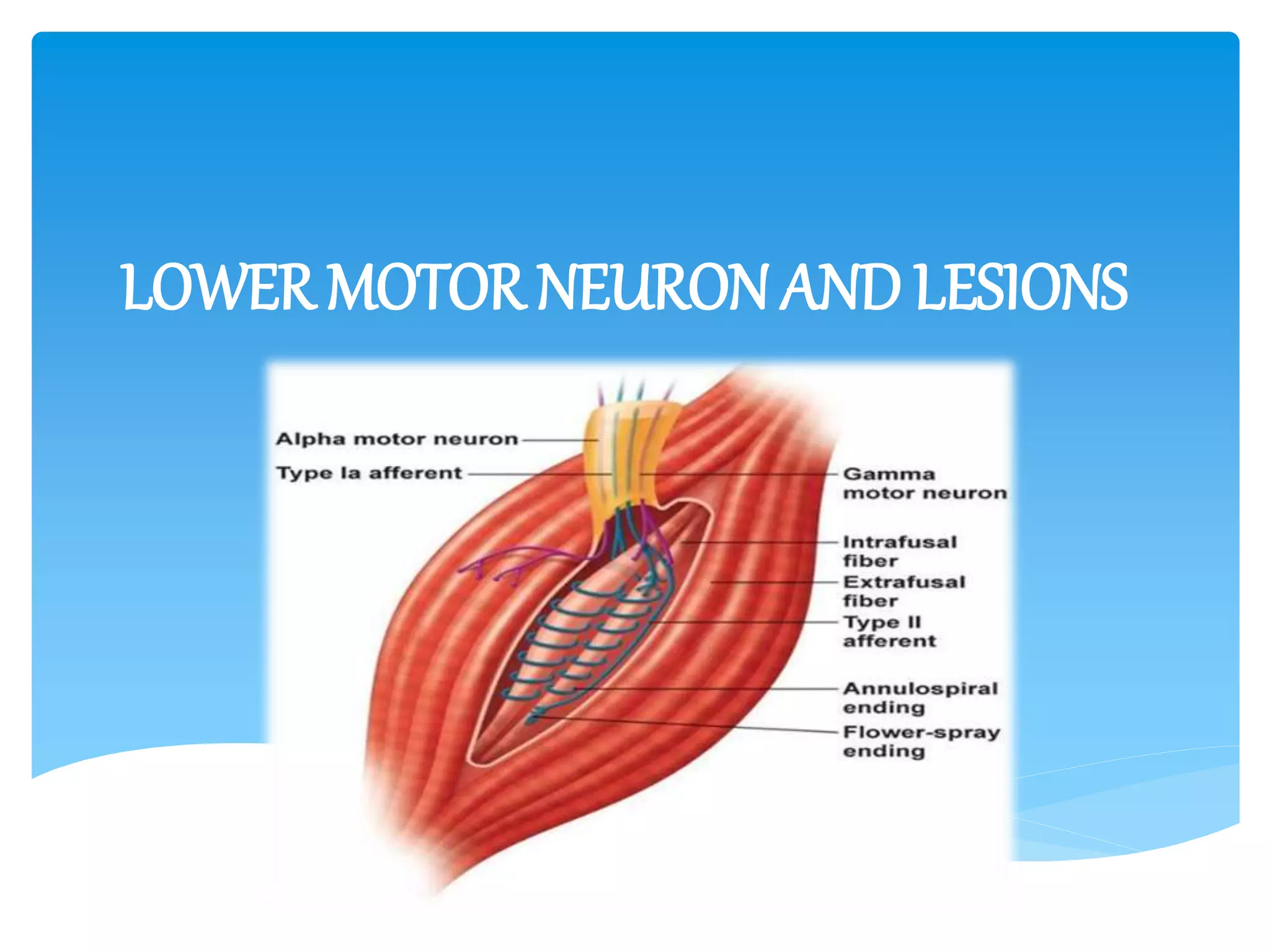
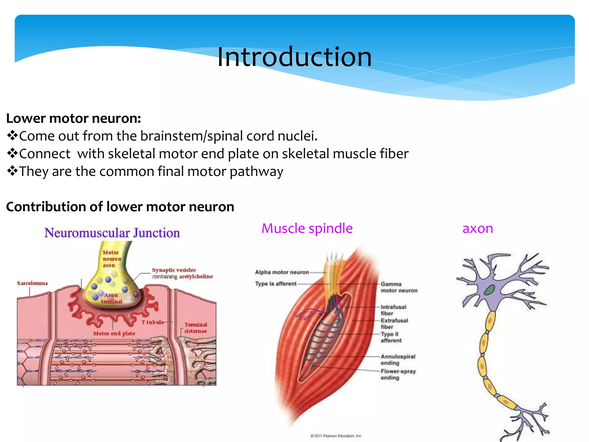
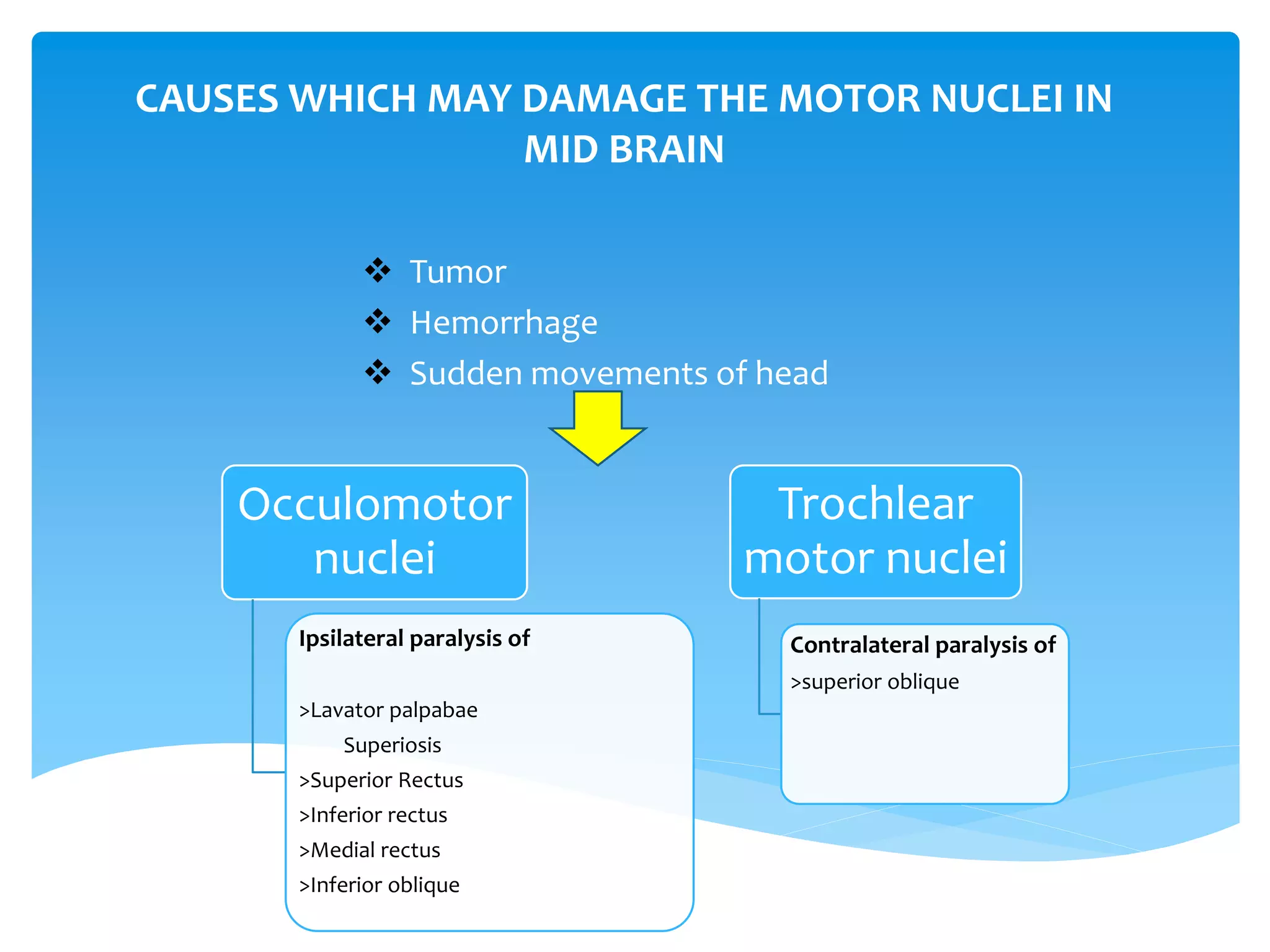
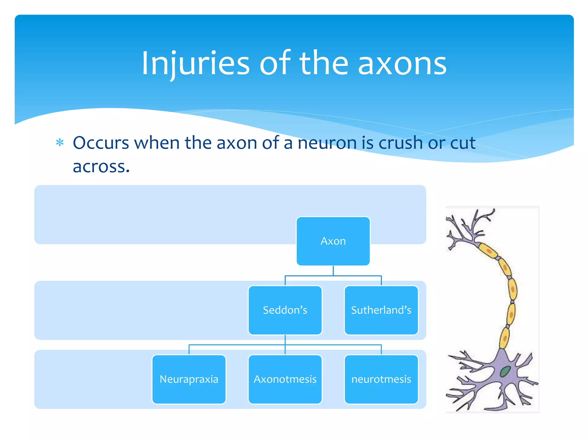
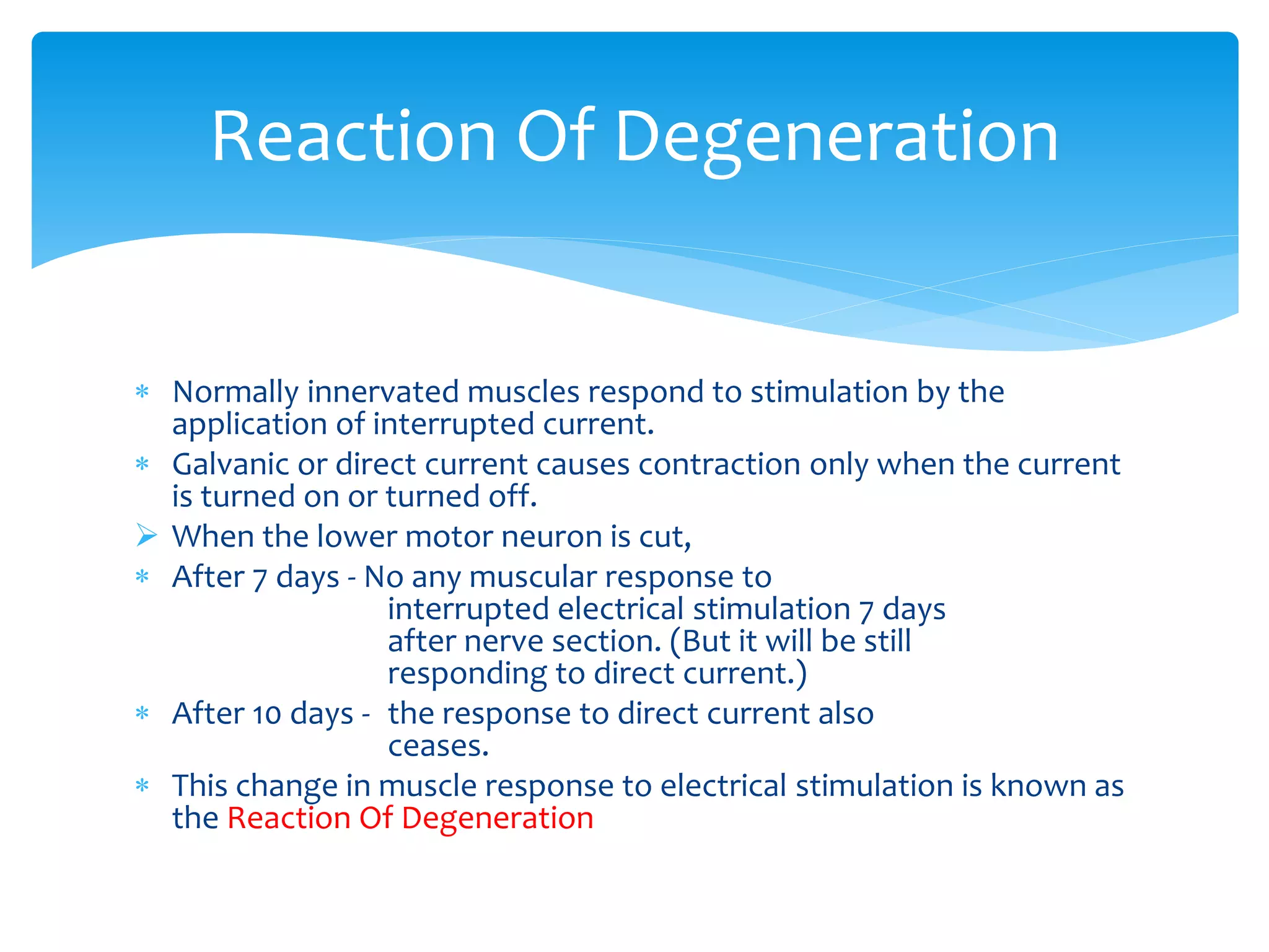
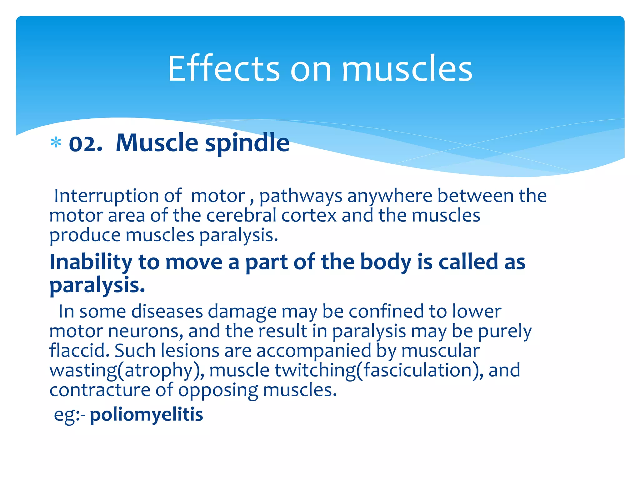
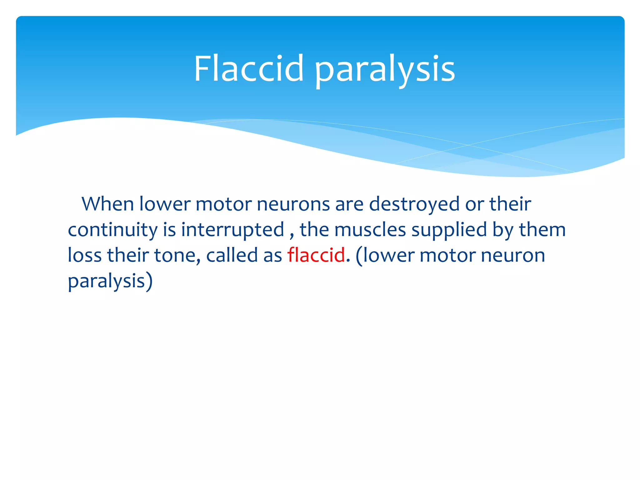
LOWER MOTOR NEURONAND LESIONS
Introduction
Lower motor neuron:
Come out from the brainstem/spinal cord nuclei.
Connect with skeletal motor end plate on skeletal muscle fiber
They are the common final motor pathway
Contribution of lower motor neuron
Muscle spindle axon
Lower motor neuron synapses with upper motor neuron,
Such as
Descending
tracts
Cortical
Pyram
idal
cortic
ospin
al
Extra
pyram
idal
Cortico
bulbur
Cortico
pontain
Cortico
mesencep
halic
Sub
cortical
Vesti
bulos
pinal
balance of
extensor
muscles
Ru
bro
spi
nal
Flex
or
Reticu
lo
spinal
Pons-
exten
sor
musc
les Med
ulla-
flexo
r
mus
cles
Mid
brain
Oliv
o
spin
al
Tec
tos
pina
l
Vis
ual
refl
ex
Aud
itor
y
refle
x
LOWER MOTOR NEURONS COMMON PATHWAY
Muscle spindle is sensory organ
Extra fusal fibers are contractile unit
α motor neuron innervates extra fusal fibers
ϒ motor neurons innervates muscle spindle
Ia fibers conduct information from muscle spindle
Information -> The proprioception
Ia fibers synapse with α MN within spinal cord , the degree
of contraction increase( α MN stimulates extra fusal fibers)
α MN -> NMJ -> Ca +2 influx -> AP-> actin-myosin function-> muscle contraction
Muscle spindle / intra fusal fibers-> stretch receptors
Deep tendon reflex
Strech on tendon-> EFF ->I α -> spinal cord-> α MN -> NMJ-> muscle contraction
When over stetch of tendon ->
Golgi tendon organ-> I b fibers -> SC -> innhibitory inter neuron ->
 Lesions may occur in these sites of lower motor
neurons
Lower motor neuron lesions
Lesions
Nuclei
Cranial
Nerves
Spinal
Nerves
Axon
Seddo
n’s
Sutherl
and’s
Muscle
Muscle
spindle
Motor
End Plate
Cranial motor nuclei lesions belong to lower motor
neuron lesions………
MID BRAIN
Occulomotor
nuclei
Trochlear nuclei
PONS
Trigeminal motor
nuclei
Abducent motor
nuclei
Facial motor nuclei
MEDULLA
OBLONGATA
Motor nuclei of
Glossopharyngeal
Motor nuclei of
Vagus
Hypoglossal nuclei
CAUSES WHICH MAY DAMAGE THE MOTOR NUCLEI IN
MID BRAIN
 Tumor
 Hemorrhage
 Sudden movements of head
Occulomotor
nuclei
Ipsilateral paralysis of
>Lavator palpabae
Superiosis
>Superior Rectus
>Inferior rectus
>Medial rectus
>Inferior oblique
Trochlear
motor nuclei
Contralateral paralysis of
>superior oblique
Damage of both Occulomotor and Trochlear nuclei will
result in impairment of occular movements
CAUSES WHICH MAY DAMAGE THE MOTOR NUCLEI OF
PONS
 Tumor
 Pontine hemorrhage
Facial motor
nuclei
Weakness of
facial muscles
in same side
Abducent
nuclei
Weakness of
lateral rectus
one or both
sides
Paralysis of
conjugate
occular
deviation
Trigeminal
motor nuclei
Weakness of
jaw muscles
CAUSES WHICH MAY DAMAGE THE MOTOR NUCLEI IN
MEDULLA OBLONGATA
 Raised pressure in posterior cranial fossa
> Glossopharyngeal motor nuclei
> Motor nuclei of vagus
> Hypoglossal nuclei
 Lateral medullary syndrome
> Abducent nuclei
> Nucleus ambiguus
 Medial medullary syndrome
> Hypoglossal nuclei – Ipsilateral paralysis of tongue
 Occurs when the axon of a neuron is crush or cut
across.
Injuries of the axons
Axon
Seddon’s
Neurapraxia Axonotmesis neurotmesis
Sutherland’s
 Neurapraxia
- Due to blockage of nerve conduction.
 Axonotmesis
- Disruption of nerve cell axon.
- Endoneurium is intact.
- With Wallerian degeneration.
 Neurotmesis
- Most serious nerve injury.
- Both the nerve and the nerve sheath are disrupted.
Seddon’s Classification
Degree of
injury
Myelin Axon Endo-
neurium
Peri-
neurium
Epi-
neurium
I +/-
II No Yes Yes No No
III No Yes Yes Yes No
IV No Yes Yes Yes Yes
V Yes Yes Yes Yes Yes
Sutherlands classification
 Normally innervated muscles respond to stimulation by the
application of interrupted current.
 Galvanic or direct current causes contraction only when the current
is turned on or turned off.
 When the lower motor neuron is cut,
 After 7 days - No any muscular response to
interrupted electrical stimulation 7 days
after nerve section. (But it will be still
responding to direct current.)
 After 10 days - the response to direct current also
ceases.
 This change in muscle response to electrical stimulation is known as
the Reaction Of Degeneration
Reaction Of Degeneration
Degeneration
Neural
Degeneration
Wallerian
degeneration
Axonal/Retrograde
degeneration
Transneural
degeneration
Reaction Of Degeneration
 Because of interruption of the efferent part of reflex
pathways, tendon reflexes are abolished.
Hyporeflexia
Loss Of Reflexes
 01 Motor end plate
Myasthenia gravis
The immune system inappropriately produce antibodies that
bind to and block some Ach receptors thereby decreasing the
number of functional of skeletal muscles. More receptors are
lost. Thus muscles become increasing weaker, fatigue more
easily, and many eventually cease to function.
Effects on muscles
 02. Muscle spindle
Interruption of motor , pathways anywhere between the
motor area of the cerebral cortex and the muscles
produce muscles paralysis.
Inability to move a part of the body is called as
paralysis.
In some diseases damage may be confined to lower
motor neurons, and the result in paralysis may be purely
flaccid. Such lesions are accompanied by muscular
wasting(atrophy), muscle twitching(fasciculation), and
contracture of opposing muscles.
eg:- poliomyelitis
Effects on muscles
When lower motor neurons are destroyed or their
continuity is interrupted , the muscles supplied by them
loss their tone, called as flaccid. (lower motor neuron
paralysis)
Flaccid paralysis
 Destruction of nerve supply. (motor
nerve)
 Abnormal excitability
 Sensitive to circulating Acetylcholine
 Irregular contraction of individual fiber
(fibrillation )
Muscular Atrophy
Jerky visible contraction of group of
muscle fibers.
Pathological discharge of spinal motor
neuron.
Muscular Fasciculation
This is a shortening of the paralyzed
muscles.
It occurs more often in the antagonist
muscles whose action is no longer
opposed by the paralyzed muscles.
Muscular Contracture
Lower motor neurons lesions
Causes-
Trauma
Infection
Vascular disorders
Degenerative disease
Neoplasm
fDestroy the cell bodies in
anterior gray matter or it’s axon
in anterior root of spinal nerve
28
Principal Features of UMNL & LMNL
UMNL:
(1) No muscle wasting, except
from disuse ( disuse atrophy)
(3) Spasticity ( hypertonia ) ,
called
“ clasp-knife spasticity ”
(4) Clonus present
(5) Brisk ( exaggerated ) tendon
jerks
(6) Extensor plantar reflex ,
Babinski sign ( dorsiflexion of
the big toe and fanning out of
the other toes )
(7) Absent abdominal reflexes
(8) No fasciculations
(9) No fibrillation potential in
EMG
{
LMNL:
(1) Marked muscle wasting
(atrophy )
(3) Flacidity (Hypotonia ) , hence
given the name “ flaccid
paralysis ”
(4) No clonus
(5) Diminished or absent tendon
reflexes
(6) Absent plantar reflex
(normally it is flexor ) .
(7) Absent abdominal reflexes
(8) Fasciculations may occur .
(9) Fibrillation potentials present
.
THANK YOU
Presented by 3rd group

vdocument.in_lower-motor-neuron-lesions.pptx

  • 1.
  • 2.
    Introduction Lower motor neuron: Comeout from the brainstem/spinal cord nuclei. Connect with skeletal motor end plate on skeletal muscle fiber They are the common final motor pathway Contribution of lower motor neuron Muscle spindle axon
  • 3.
    Lower motor neuronsynapses with upper motor neuron, Such as Descending tracts Cortical Pyram idal cortic ospin al Extra pyram idal Cortico bulbur Cortico pontain Cortico mesencep halic Sub cortical Vesti bulos pinal balance of extensor muscles Ru bro spi nal Flex or Reticu lo spinal Pons- exten sor musc les Med ulla- flexo r mus cles Mid brain Oliv o spin al Tec tos pina l Vis ual refl ex Aud itor y refle x
  • 4.
    LOWER MOTOR NEURONSCOMMON PATHWAY Muscle spindle is sensory organ Extra fusal fibers are contractile unit α motor neuron innervates extra fusal fibers ϒ motor neurons innervates muscle spindle Ia fibers conduct information from muscle spindle Information -> The proprioception Ia fibers synapse with α MN within spinal cord , the degree of contraction increase( α MN stimulates extra fusal fibers)
  • 5.
    α MN ->NMJ -> Ca +2 influx -> AP-> actin-myosin function-> muscle contraction Muscle spindle / intra fusal fibers-> stretch receptors Deep tendon reflex Strech on tendon-> EFF ->I α -> spinal cord-> α MN -> NMJ-> muscle contraction When over stetch of tendon -> Golgi tendon organ-> I b fibers -> SC -> innhibitory inter neuron ->
  • 6.
     Lesions mayoccur in these sites of lower motor neurons Lower motor neuron lesions Lesions Nuclei Cranial Nerves Spinal Nerves Axon Seddo n’s Sutherl and’s Muscle Muscle spindle Motor End Plate
  • 7.
    Cranial motor nucleilesions belong to lower motor neuron lesions……… MID BRAIN Occulomotor nuclei Trochlear nuclei PONS Trigeminal motor nuclei Abducent motor nuclei Facial motor nuclei MEDULLA OBLONGATA Motor nuclei of Glossopharyngeal Motor nuclei of Vagus Hypoglossal nuclei
  • 8.
    CAUSES WHICH MAYDAMAGE THE MOTOR NUCLEI IN MID BRAIN  Tumor  Hemorrhage  Sudden movements of head Occulomotor nuclei Ipsilateral paralysis of >Lavator palpabae Superiosis >Superior Rectus >Inferior rectus >Medial rectus >Inferior oblique Trochlear motor nuclei Contralateral paralysis of >superior oblique
  • 9.
    Damage of bothOcculomotor and Trochlear nuclei will result in impairment of occular movements
  • 10.
    CAUSES WHICH MAYDAMAGE THE MOTOR NUCLEI OF PONS  Tumor  Pontine hemorrhage Facial motor nuclei Weakness of facial muscles in same side Abducent nuclei Weakness of lateral rectus one or both sides Paralysis of conjugate occular deviation Trigeminal motor nuclei Weakness of jaw muscles
  • 11.
    CAUSES WHICH MAYDAMAGE THE MOTOR NUCLEI IN MEDULLA OBLONGATA  Raised pressure in posterior cranial fossa > Glossopharyngeal motor nuclei > Motor nuclei of vagus > Hypoglossal nuclei  Lateral medullary syndrome > Abducent nuclei > Nucleus ambiguus  Medial medullary syndrome > Hypoglossal nuclei – Ipsilateral paralysis of tongue
  • 14.
     Occurs whenthe axon of a neuron is crush or cut across. Injuries of the axons Axon Seddon’s Neurapraxia Axonotmesis neurotmesis Sutherland’s
  • 15.
     Neurapraxia - Dueto blockage of nerve conduction.  Axonotmesis - Disruption of nerve cell axon. - Endoneurium is intact. - With Wallerian degeneration.  Neurotmesis - Most serious nerve injury. - Both the nerve and the nerve sheath are disrupted. Seddon’s Classification
  • 16.
    Degree of injury Myelin AxonEndo- neurium Peri- neurium Epi- neurium I +/- II No Yes Yes No No III No Yes Yes Yes No IV No Yes Yes Yes Yes V Yes Yes Yes Yes Yes Sutherlands classification
  • 17.
     Normally innervatedmuscles respond to stimulation by the application of interrupted current.  Galvanic or direct current causes contraction only when the current is turned on or turned off.  When the lower motor neuron is cut,  After 7 days - No any muscular response to interrupted electrical stimulation 7 days after nerve section. (But it will be still responding to direct current.)  After 10 days - the response to direct current also ceases.  This change in muscle response to electrical stimulation is known as the Reaction Of Degeneration Reaction Of Degeneration
  • 19.
  • 20.
     Because ofinterruption of the efferent part of reflex pathways, tendon reflexes are abolished. Hyporeflexia Loss Of Reflexes
  • 21.
     01 Motorend plate Myasthenia gravis The immune system inappropriately produce antibodies that bind to and block some Ach receptors thereby decreasing the number of functional of skeletal muscles. More receptors are lost. Thus muscles become increasing weaker, fatigue more easily, and many eventually cease to function. Effects on muscles
  • 22.
     02. Musclespindle Interruption of motor , pathways anywhere between the motor area of the cerebral cortex and the muscles produce muscles paralysis. Inability to move a part of the body is called as paralysis. In some diseases damage may be confined to lower motor neurons, and the result in paralysis may be purely flaccid. Such lesions are accompanied by muscular wasting(atrophy), muscle twitching(fasciculation), and contracture of opposing muscles. eg:- poliomyelitis Effects on muscles
  • 23.
    When lower motorneurons are destroyed or their continuity is interrupted , the muscles supplied by them loss their tone, called as flaccid. (lower motor neuron paralysis) Flaccid paralysis
  • 24.
     Destruction ofnerve supply. (motor nerve)  Abnormal excitability  Sensitive to circulating Acetylcholine  Irregular contraction of individual fiber (fibrillation ) Muscular Atrophy
  • 25.
    Jerky visible contractionof group of muscle fibers. Pathological discharge of spinal motor neuron. Muscular Fasciculation
  • 26.
    This is ashortening of the paralyzed muscles. It occurs more often in the antagonist muscles whose action is no longer opposed by the paralyzed muscles. Muscular Contracture
  • 27.
    Lower motor neuronslesions Causes- Trauma Infection Vascular disorders Degenerative disease Neoplasm fDestroy the cell bodies in anterior gray matter or it’s axon in anterior root of spinal nerve
  • 28.
    28 Principal Features ofUMNL & LMNL UMNL: (1) No muscle wasting, except from disuse ( disuse atrophy) (3) Spasticity ( hypertonia ) , called “ clasp-knife spasticity ” (4) Clonus present (5) Brisk ( exaggerated ) tendon jerks (6) Extensor plantar reflex , Babinski sign ( dorsiflexion of the big toe and fanning out of the other toes ) (7) Absent abdominal reflexes (8) No fasciculations (9) No fibrillation potential in EMG { LMNL: (1) Marked muscle wasting (atrophy ) (3) Flacidity (Hypotonia ) , hence given the name “ flaccid paralysis ” (4) No clonus (5) Diminished or absent tendon reflexes (6) Absent plantar reflex (normally it is flexor ) . (7) Absent abdominal reflexes (8) Fasciculations may occur . (9) Fibrillation potentials present .
  • 29.