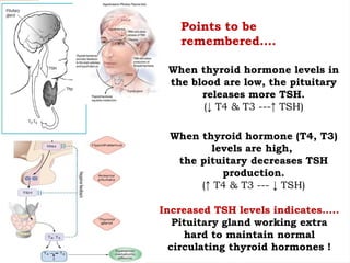
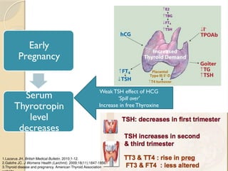
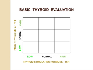
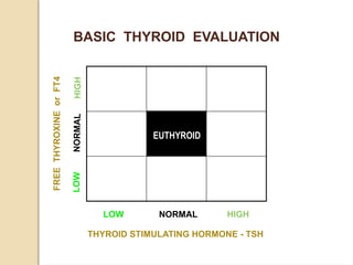
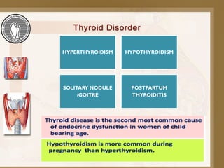
THYROID DISORDERS &
PREGNANCY
Thyroid Gland
One of the largest endocrine gland
International Journal of Health Sciences & Research.2013;3(5):29
Located front of the neck, below the larynx
2 inch long, Butterfly shaped gland
It has two lobes (Right & Left)
Average weight 25-30g in adults (slightly more in women)
The thyroid makes two thyroid hormones
• Thyroxine (T4)
• Triiodothyronine (T3)
Thyroid Gland
Functions
MOST OF FUNCTION DUE
TO T3
Growth & development
Increasing rate of
metabolism
Increase metabolic rate in
CVS → blood flow
Regulating cerebral
conducion in cns
Sleep
Lipid metabolism
One of the largest endocrine gland
The thyroid makes two thyroid
hormones
• Thyroxine (T4)
• Triiodothyronine (T3)
When thyroid hormone levels in
the blood are low, the pituitary
releases more TSH.
(↓ T4 & T3 ---↑ TSH)
When thyroid hormone (T4, T3)
levels are high,
the pituitary decreases TSH
production.
(↑ T4 & T3 --- ↓ TSH)
Points to be
remembered….
Increased TSH levels indicates…..
Pituitary gland working extra
hard to maintain normal
circulating thyroid hormones !
Early
Pregnancy
Serum
Thyrotropin
level
decreases
Weak TSH effect of HCG
‘Spill over’
Increase in free Thyroxine
1.Lazarus JH. British Medical Bulletin. 2010;1-12.
2.Galofre JC. J Womens Health (Larchmt). 2009;18(11):1847-1856.
3.Thyroid disease and pregnancy. American Thyroid Association
The Nine Square Game
To evaluate our Thyroid patient
As per the AACE and ITS Guidelines
LOW NORMAL HIGH
THYROID STIMULATING HORMONE - TSH
LOW
NOR
MAL
HIGH
FREE
THYROXINE
or
FT4
BASIC THYROID EVALUATION
LOW
NOR
MAL
HIGH
FREE
THYROXINE
or
FT4
EUTHYROID
LOW NORMAL HIGH
THYROID STIMULATING HORMONE - TSH
BASIC THYROID EVALUATION
LOW
NOR
MAL
HIGH
FREE
THYROXINE
or
FT4
EUTHYROID
SUB-CLINICAL
HYPERTHYROID
NON THYROID
ILLNESS - NTI
NTI or Pt.
on HYROID
HORMONES
SUB-CLINICAL
HYPOTHYROID
SECONDARY
HYPERTHYROID
SECONDARY
HYPOTHYROID
PRIMARY
HYPERTHYROID
PRIMARY
HYPOTHYROID
LOW NORMAL HIGH
THYROID STIMULATING HORMONE - TSH
BASIC THYROID EVALUATION
THYROID HORMONES
TEST REFERENCE RANGE
TSH Normal Range 0.3 - 4.0 mU/L
Free T4 Normal Range 0.7-2.1 ng/dL
TSH upper limit has been revised to 2.5 mU/L
HYPERTHYROIDISM HYPOTHYROIDISM
SOLITARY NODULE
/GOITRE
POSTPARTUM
THYROIDITIS
THYROID PHYSIOLOGY IN
NORMAL PREGNANCY
In women with normal thyroid function there is an
increase in thyroxine (T4) and triiodothyronine
(T3) production, which results in inhibition of
thyroid-stimulating hormone (TSH) in the first
trimester of pregnancy, due to a high human
chorionic gonadotropin (hCG) level that
stimulates the TSH receptor.
Why higher thyroxine requirements in
pregnancy?
 A large plasma volume and thus an
altered distribution of thyroid hormone
 increased thyroid hormone metabolism,
 increased renal clearance of iodide,
 higher levels of hepatic production of
thyroxine-binding globulin (TBG) in the
hyperestrogenic state
Although the fetal thyroid in humans is
histologically developed and can synthesize
thyroid hormone by the 10th to 12th week
of gestation, thyroid maturation takes longer,
and it is until midgestation that substantial
amounts of thyroid hormone are produced
by the fetal thyroid gland.
Maternal hypothyroidism during the first
trimester can thus have deleterious effects
on fetal neurodevelopment, which is
largely dependent on maternal thyroxine.
Epidemiologic and prospective evidence
suggests that early-gestation maternal
hypothyroidism can result in mental and
motor delay in children when assessed at 1
and 2 years of age, and in some cases may
even lead to profound mental retardation
and cretinism
The National Health and Nutrition
Examination Survey (NHANES 1999–
2002) indicates that 3.1% of women of
reproductive age in the United States may
have hypothyroidism.
The prevalence of overt hypothyroidism
in pregnancy is estimated to be between
0.3% and 1.5% in different studies
The most common cause of maternal
hypothyroidism worldwide is iodine deficiency,
but in developed countries autoimmune thyroid
disease or Hashimoto’s thyroiditis is more
prevalent.
The WHO recommends that pregnant and
lactating women have an iodine intake of 250
mg per day, which is 100 mg above that
recommended for nonpregnant adults, to
compensate for increased thyroxine
requirements, renal iodine losses, and fetal
iodine requirements in pregnancy
Hypothyroidism signs and symptoms may include:
 Fatigue
 Increased sensitivity to cold
 Constipation
 Dry skin
 Weight gain
 Puffy face
 Hoarseness
 Muscle weakness
• Elevated blood cholesterol level
• Muscle aches, tenderness and
stiffness
• Pain, stiffness or swelling in your
joints
• Heavier than normal or irregular
menstrual periods
• Thinning hair
• Slowed heart rate
• Depression
• Impaired memory
• Enlarged thyroid gland (goiter)
Some studies
 Thyroid autoimmunity is an important
marker for subsequent development of
hypothyroidism
 The measurement of antibody levels may
be helpful in determining the frequency of
monitoring thyroid function during the
remainder of gestation.
when euthyroid pregnant women with
TPO-Ab positivity were treated with
levothyroxine (low dose 0.5–1.0 mg/kg/d) to
normalize serum TSH, the rate of
miscarriage was similar to antibody negative
women (3.5% vs 2.4%), whereas untreated
TPO-Ab–positive pregnant women had a
significantly higher TSH and a higher
miscarriage rate of 13.8%.
 A TSH level above 6 mU/mL was associated
with a fourfold increase in fetal death in one
population-based study of more than 9000
pregnant women.
 A large cohort of TPO-Ab–negative
pregnant women showed that those with a
TSH of 2.5 mU/mL or less in the first
trimester had a lower rate of spontaneous
pregnancy loss (3.6%) than women with a
TSH between 2.5 and 5 mU/mL (6.1%).
 A cohort of more than 17,000 women
found that pregnancy in those with
subclinical hypothyroidism was 3 times
more likely to be complicated by placental
abruption and had twice the risk of
preterm delivery.
 The rate of miscarriage was 17% in
women with positive thyroid antibodies
compared with 8.4% in autoantibody-
negative women.
This observation suggests that the
upper limit of normal forTSH in the
first trimester of pregnancy should be
2.5 mU/mL.
Screening for thyroid disease in
pregnancy
 The Endocrine Society recommend targeted case-
finding in pregnant women with:
 a personal history of thyroid disease,
 a positive family history,
 type 1 diabetes mellitus or other autoimmune
disorders
 infertility
 history of miscarriage or preterm delivery
 clinical signs or symptoms of thyroid disease that
would classify them as high risk.
 up to one-third of hypothyroid women (TSH >4.2
mU/mL) will be missed with case-finding alone
in women with a prior history of
hypothyroidism the preconception and first-
trimester TSH level should not exceed 2.5
mU/mL. In the second and third trimesters, a
TSH of less than 3.0 mU/mL is desirable.
A pregnant woman receiving an established,
stable dose of levothyroxine will likely need a
30% to 50% dose augmentation by the
fourth through sixth week of gestation
ATA recommendation
The goal of LT4 treatment is to normalize
maternal serum TSH values within the
trimester-specific pregnancy reference range
(first trimester, 0.1–2.5 mIU/L; second
trimester, 0.2–3.0 mIU/L; third trimester,
0.3–3.0 mIU/L).
4 hours interval between Ca and Fe intake
and levothyroxin
Euthyroid women with TPO-Abs are
believed to be at high risk for developing
hypothyroidism later in pregnancy, and
should be monitored for TSH elevation and
receive prenatal counseling; however,
treatment with thyroid hormone is not yet
recommended in this population
Monitoring of thyroid function tests in
pregnant women with hypothyroidism
should occur frequently, as often as every 2
to 4 weeks throughout midgestation and 4
to 8 weeks in the second half of pregnancy,
as this time interval has been shown to
identify 90% of abnormal TSH values that
resulted in Levothyroxine dose adjustment.
HYPERTHYROIDISM
The differential diagnosis of thyrotoxicosis
in pregnancy is similar to that of
nonpregnant women: autoimmune thyroid
disease(eg, Graves’ disease, hashitoxicosis),
toxic adenoma or goiter, transient thyroiditis
(eg, subacute thyroiditis, silent thyroiditis),
iodine-induced hyperthyroidism and, rarely, a
pituitary adenoma secreting TSH or thyroid
hormone resistance
Symptoms of hyperthyroidism
 Rapid heartbeat (palpitations).
 Feeling shaky and/or nervous.
 Weight loss.
 Increased appetite.
 Diarrhea and/or more frequent bowel movements.
 Thin, warm and moist skin.
 Intolerance to heat and excessive sweating.
 Difficulty sleeping, such as insomnia.
 Enlarged thyroid gland (goiter).
 Hair loss and change in hair texture (brittle).
 Menstrual changes.
 Muscle weakness.
Graves’ disease can also cause eye disease symptoms,
including:
 Gritty, irritated eyes.
 Swelling of the tissues around your eyes
(puffy eyes).
 Bulging eyes.
 Light sensitivity.
 Pressure or pain in your eyes.
 Blurred or double vision.
 Additional consideration of hCG-
mediated hyperthyroidism including
gestational transient thyrotoxicosis (GTT)
and its association with hyperemesis
gravidarum becomes very important.
 Postpartum thyroiditis is also unique to
the pregnant cohort within several months
of delivery.
The most common cause of hyperthyroidism
in pregnancy, as in all women of reproductive
age, is autoimmune Graves’ disease with
circulating TSH receptor antibodies (TRAbs)
stimulating the thyroid gland andTSH
receptor. Graves’ disease may be challenging
to diagnose in the hypermetabolic state of
pregnancy because of similar symptoms in
both conditions.
Features suggestive of Graves’ hyperthyroidism in
normal pregnancy include:
a diffuse goiter, evidence of ophthalmopathy, TRAb
or thyroid-stimulating immunoglobulin (TSI) positivity,
and a family history of autoimmune thyroid disease.
The extent of elevation of thyroid function tests
typically is higher in Graves’ disease than in
hyperemesis gravidarum.
The immune-tolerant state of pregnancy is associated
over time with a decrease inTRAb titers, thus the
hyperthyroidism secondary to Graves’ disease may
subside in severity as gestation progresses
GestationalTransientThyrotoxicosis
GTT differs from Graves’ disease in that
it is a self-limited, nonautoimmune form
of hyperthyroidism with negativeTRAbs.
it is related to the elevation of hCG,
which can then cross-react with the TSH
receptor and induce thyroidal
iodothyronine secretion
GestationalTransientThyrotoxicosis
Women have a TSH in the low to normal
range for pregnancy or is frequently
undetectable, elevated free T4 and free T3
levels, and prolonged abnormally high hCG in
the first and second trimesters, occasionally
higher than 100,000 U/L.These patients
present with symptoms of hyperthyroidism in
about 50% of cases, and the emesis or
symptoms from thyrotoxicosis may be severe
enough to require hospitalization.
Hyperemesis gravidarum
characterized by excessive nausea and
vomiting, which causes a greater than 5%
weight loss, dehydration, and ketonuria in
early pregnancy, is a milder form of GTT
in that women have subclinical or very
mild overt hyperthyroidism in one-third
to two-thirds of cases.
 pregnant women with uncontrolled
hyperthyroidism:
 Preeclampsia,
 congestive heart failure,
 placental abruption and
 cesarean delivery,
 thyrotoxic periodic paralysis.
 Fetal risks of maternal thyrotoxicosis
include:
 spontaneous abortion,
 premature labor and
 low birth weight,
 stillbirth,
 congenital abnormalities
TREATMENT
During pregnancy, medical treatment is
preferred despite class D classification of
thionamides by the (FDA), because
radioactive iodine cannot be used and
surgery is typically reserved for severe
thyrotoxicosis that has failed medical
therapy. PTU had been the drug of choice
during pregnancy to avoid MMI-associated
scalp defects of aplasia cutis or
choanal/esophageal atresia.
Updated guidelines from the ATA and
American Association of Clinical
Endocrinologists are expected in 2011 to
delineate a limited therapeutic window for
PTU during the first trimester of pregnancy,
in the treatment of thyroid storm, and in
patients with minor reactions to MMI who
refuse radioactive iodine therapy or surgery;
otherwise MMI should be used in every
patient who chooses antithyroid drug
therapy for Graves’ disease
The goal of therapy is to use the lowest
dose of medication to maintain maternal
free T4 in the upper nonpregnant
reference range, as this minimizes the risk
of hypothyroidism to the fetus.
radioactive iodine administration for
diagnostic scans and treatment is
absolutely contraindicated in pregnant
patients and in women considering
becoming pregnant in the near future,
secondary to adverse effects on the fetus
and the fetal thyroid gland.
Thyrotoxicosis and primary
hyperthyroidism in the neonate is usually
transient, and remits with clearance of
maternal TRAbs in the first 3 to 6 months of
life. postpartum short-term levothyroxine
administration to the hypothyroid neonate is
recommended, and the hyperthyroidism
should only be treated with antithyroid
agents if the neonate has clinically severe
thyrotoxicosis.
When the mother is euthyroid after
treatment of Graves’ disease, is at risk of
recurrence, especially postpartum, and her
thyroid function should be monitored
during pregnancy and after delivery.
If the mother has had thyroid ablation or
surgery she may still be TRAb positive,
and antibodies may cause fetal
hyperthyroidism via passage through the
placenta.
TRAb titers should be measured before
pregnancy or at the end of the second
trimester in all pregnant women with current
or a history of Graves’ disease to assess the
risk of neonatal hyperthyroidism in the fetus.
If titers are high, fetal ultrasonography is
necessary to look for growth restriction,
hydrops, goiter, and cardiac failure in addition
to clinical monitoring of the fetus for
hyperthyroidism
 Subtotal thyroidectomy can usually be
performed in the second trimester of
pregnancy, in situations whereby:
 Hyperthyroidism is uncontrolled with ATDs
 Treatment requires persistently high doses
of antithyroid medication (generally
thought to be more than approximately
300 mg of PTU daily)
 If the mother has a serious adverse effect
to medical therapy.
Women who receive I-131 therapy for
Graves’ disease should wait at least 6
months before becoming pregnant, although
this is an empiric recommendation.These
patients should become hypothyroid within
2 to 3 months, and it may subsequently take
1 to 2 months to restore euthyroidism with
exogenous levothyroxine.
Lactation andTreatment of
Thyroid Dysfunction
Breastfeeding is considered safe in
mothers with hypothyroidism and
hyperthyroidism taking thyroid medications.
Although MMI and PTU both appear in
human milk, it is at very small
concentrations The decision whether to
breastfeed while taking antithyroid agents is
an individual decision that should be
discussed by the patient and her physician.
PostpartumThyroid Dysfunction
an autoimmune destructive thyroiditis (PPT)
with a period of thyrotoxicosis,
hypothyroidism, or both, followed by
recovery in the first year after delivery. It is
frequent and occurs in about 8% of women
worldwide, and is strongly associated with
TPO-Ab positivity. PPTD occurs most
frequently at about the fourth month after
delivery, although it can take place any time
between 1 and 12 months postpartum.
Up to 50% of pregnant women with TPO-
Abs will develop PPTD, and of these about
20% may develop permanent hypothyroidism
over the next several years.
Risk factors for subsequent and permanent
hypothyroidism include a postpartum peak
TSH greater than 20 mU/mL, a high titer of
TPO-Abs, hypoechogenicity on thyroid
ultrasonography, and a hypothyroid phase of
PPTD.
The hypothyroid phase of PPTD should be
considered for treatment with levothyroxine
at replacement doses, especially when TSH is
10 mU/mL or greater.
stop the medication after approximately 6
to 12 months of treatment to determine if
the hypothyroidism is permanent and then, if
normal, perform periodic monitoring.
The Endocrine Society recommends
screening for PPTD by measuring TSH at
3 and 6 months postpartum in all women
with TPO-Abs and type 1 diabetes
mellitus, as well as annually in those with a
history of PPTD in prior pregnancy.
Mothers who experience depression in
the postpartum period should also be
screened for hypothyroidism, and treated
accordingly.
Various thyroid diseases on pregnancy.ppt

Various thyroid diseases on pregnancy.ppt

  • 1.
  • 3.
    Thyroid Gland One ofthe largest endocrine gland International Journal of Health Sciences & Research.2013;3(5):29 Located front of the neck, below the larynx 2 inch long, Butterfly shaped gland It has two lobes (Right & Left) Average weight 25-30g in adults (slightly more in women) The thyroid makes two thyroid hormones • Thyroxine (T4) • Triiodothyronine (T3)
  • 4.
    Thyroid Gland Functions MOST OFFUNCTION DUE TO T3 Growth & development Increasing rate of metabolism Increase metabolic rate in CVS → blood flow Regulating cerebral conducion in cns Sleep Lipid metabolism One of the largest endocrine gland The thyroid makes two thyroid hormones • Thyroxine (T4) • Triiodothyronine (T3)
  • 5.
    When thyroid hormonelevels in the blood are low, the pituitary releases more TSH. (↓ T4 & T3 ---↑ TSH) When thyroid hormone (T4, T3) levels are high, the pituitary decreases TSH production. (↑ T4 & T3 --- ↓ TSH) Points to be remembered…. Increased TSH levels indicates….. Pituitary gland working extra hard to maintain normal circulating thyroid hormones !
  • 6.
    Early Pregnancy Serum Thyrotropin level decreases Weak TSH effectof HCG ‘Spill over’ Increase in free Thyroxine 1.Lazarus JH. British Medical Bulletin. 2010;1-12. 2.Galofre JC. J Womens Health (Larchmt). 2009;18(11):1847-1856. 3.Thyroid disease and pregnancy. American Thyroid Association
  • 7.
    The Nine SquareGame To evaluate our Thyroid patient As per the AACE and ITS Guidelines
  • 8.
    LOW NORMAL HIGH THYROIDSTIMULATING HORMONE - TSH LOW NOR MAL HIGH FREE THYROXINE or FT4 BASIC THYROID EVALUATION
  • 9.
    LOW NOR MAL HIGH FREE THYROXINE or FT4 EUTHYROID LOW NORMAL HIGH THYROIDSTIMULATING HORMONE - TSH BASIC THYROID EVALUATION
  • 10.
    LOW NOR MAL HIGH FREE THYROXINE or FT4 EUTHYROID SUB-CLINICAL HYPERTHYROID NON THYROID ILLNESS -NTI NTI or Pt. on HYROID HORMONES SUB-CLINICAL HYPOTHYROID SECONDARY HYPERTHYROID SECONDARY HYPOTHYROID PRIMARY HYPERTHYROID PRIMARY HYPOTHYROID LOW NORMAL HIGH THYROID STIMULATING HORMONE - TSH BASIC THYROID EVALUATION
  • 11.
    THYROID HORMONES TEST REFERENCERANGE TSH Normal Range 0.3 - 4.0 mU/L Free T4 Normal Range 0.7-2.1 ng/dL TSH upper limit has been revised to 2.5 mU/L
  • 12.
  • 13.
    THYROID PHYSIOLOGY IN NORMALPREGNANCY In women with normal thyroid function there is an increase in thyroxine (T4) and triiodothyronine (T3) production, which results in inhibition of thyroid-stimulating hormone (TSH) in the first trimester of pregnancy, due to a high human chorionic gonadotropin (hCG) level that stimulates the TSH receptor.
  • 14.
    Why higher thyroxinerequirements in pregnancy?  A large plasma volume and thus an altered distribution of thyroid hormone  increased thyroid hormone metabolism,  increased renal clearance of iodide,  higher levels of hepatic production of thyroxine-binding globulin (TBG) in the hyperestrogenic state
  • 16.
    Although the fetalthyroid in humans is histologically developed and can synthesize thyroid hormone by the 10th to 12th week of gestation, thyroid maturation takes longer, and it is until midgestation that substantial amounts of thyroid hormone are produced by the fetal thyroid gland. Maternal hypothyroidism during the first trimester can thus have deleterious effects on fetal neurodevelopment, which is largely dependent on maternal thyroxine.
  • 17.
    Epidemiologic and prospectiveevidence suggests that early-gestation maternal hypothyroidism can result in mental and motor delay in children when assessed at 1 and 2 years of age, and in some cases may even lead to profound mental retardation and cretinism
  • 18.
    The National Healthand Nutrition Examination Survey (NHANES 1999– 2002) indicates that 3.1% of women of reproductive age in the United States may have hypothyroidism. The prevalence of overt hypothyroidism in pregnancy is estimated to be between 0.3% and 1.5% in different studies
  • 19.
    The most commoncause of maternal hypothyroidism worldwide is iodine deficiency, but in developed countries autoimmune thyroid disease or Hashimoto’s thyroiditis is more prevalent. The WHO recommends that pregnant and lactating women have an iodine intake of 250 mg per day, which is 100 mg above that recommended for nonpregnant adults, to compensate for increased thyroxine requirements, renal iodine losses, and fetal iodine requirements in pregnancy
  • 20.
    Hypothyroidism signs andsymptoms may include:  Fatigue  Increased sensitivity to cold  Constipation  Dry skin  Weight gain  Puffy face  Hoarseness  Muscle weakness • Elevated blood cholesterol level • Muscle aches, tenderness and stiffness • Pain, stiffness or swelling in your joints • Heavier than normal or irregular menstrual periods • Thinning hair • Slowed heart rate • Depression • Impaired memory • Enlarged thyroid gland (goiter)
  • 21.
    Some studies  Thyroidautoimmunity is an important marker for subsequent development of hypothyroidism  The measurement of antibody levels may be helpful in determining the frequency of monitoring thyroid function during the remainder of gestation.
  • 22.
    when euthyroid pregnantwomen with TPO-Ab positivity were treated with levothyroxine (low dose 0.5–1.0 mg/kg/d) to normalize serum TSH, the rate of miscarriage was similar to antibody negative women (3.5% vs 2.4%), whereas untreated TPO-Ab–positive pregnant women had a significantly higher TSH and a higher miscarriage rate of 13.8%.
  • 23.
     A TSHlevel above 6 mU/mL was associated with a fourfold increase in fetal death in one population-based study of more than 9000 pregnant women.  A large cohort of TPO-Ab–negative pregnant women showed that those with a TSH of 2.5 mU/mL or less in the first trimester had a lower rate of spontaneous pregnancy loss (3.6%) than women with a TSH between 2.5 and 5 mU/mL (6.1%).
  • 24.
     A cohortof more than 17,000 women found that pregnancy in those with subclinical hypothyroidism was 3 times more likely to be complicated by placental abruption and had twice the risk of preterm delivery.  The rate of miscarriage was 17% in women with positive thyroid antibodies compared with 8.4% in autoantibody- negative women.
  • 25.
    This observation suggeststhat the upper limit of normal forTSH in the first trimester of pregnancy should be 2.5 mU/mL.
  • 26.
    Screening for thyroiddisease in pregnancy  The Endocrine Society recommend targeted case- finding in pregnant women with:  a personal history of thyroid disease,  a positive family history,  type 1 diabetes mellitus or other autoimmune disorders  infertility  history of miscarriage or preterm delivery  clinical signs or symptoms of thyroid disease that would classify them as high risk.  up to one-third of hypothyroid women (TSH >4.2 mU/mL) will be missed with case-finding alone
  • 27.
    in women witha prior history of hypothyroidism the preconception and first- trimester TSH level should not exceed 2.5 mU/mL. In the second and third trimesters, a TSH of less than 3.0 mU/mL is desirable. A pregnant woman receiving an established, stable dose of levothyroxine will likely need a 30% to 50% dose augmentation by the fourth through sixth week of gestation
  • 28.
    ATA recommendation The goalof LT4 treatment is to normalize maternal serum TSH values within the trimester-specific pregnancy reference range (first trimester, 0.1–2.5 mIU/L; second trimester, 0.2–3.0 mIU/L; third trimester, 0.3–3.0 mIU/L). 4 hours interval between Ca and Fe intake and levothyroxin
  • 29.
    Euthyroid women withTPO-Abs are believed to be at high risk for developing hypothyroidism later in pregnancy, and should be monitored for TSH elevation and receive prenatal counseling; however, treatment with thyroid hormone is not yet recommended in this population
  • 30.
    Monitoring of thyroidfunction tests in pregnant women with hypothyroidism should occur frequently, as often as every 2 to 4 weeks throughout midgestation and 4 to 8 weeks in the second half of pregnancy, as this time interval has been shown to identify 90% of abnormal TSH values that resulted in Levothyroxine dose adjustment.
  • 31.
    HYPERTHYROIDISM The differential diagnosisof thyrotoxicosis in pregnancy is similar to that of nonpregnant women: autoimmune thyroid disease(eg, Graves’ disease, hashitoxicosis), toxic adenoma or goiter, transient thyroiditis (eg, subacute thyroiditis, silent thyroiditis), iodine-induced hyperthyroidism and, rarely, a pituitary adenoma secreting TSH or thyroid hormone resistance
  • 32.
    Symptoms of hyperthyroidism Rapid heartbeat (palpitations).  Feeling shaky and/or nervous.  Weight loss.  Increased appetite.  Diarrhea and/or more frequent bowel movements.  Thin, warm and moist skin.  Intolerance to heat and excessive sweating.  Difficulty sleeping, such as insomnia.  Enlarged thyroid gland (goiter).  Hair loss and change in hair texture (brittle).  Menstrual changes.  Muscle weakness.
  • 33.
    Graves’ disease canalso cause eye disease symptoms, including:  Gritty, irritated eyes.  Swelling of the tissues around your eyes (puffy eyes).  Bulging eyes.  Light sensitivity.  Pressure or pain in your eyes.  Blurred or double vision.
  • 34.
     Additional considerationof hCG- mediated hyperthyroidism including gestational transient thyrotoxicosis (GTT) and its association with hyperemesis gravidarum becomes very important.  Postpartum thyroiditis is also unique to the pregnant cohort within several months of delivery.
  • 35.
    The most commoncause of hyperthyroidism in pregnancy, as in all women of reproductive age, is autoimmune Graves’ disease with circulating TSH receptor antibodies (TRAbs) stimulating the thyroid gland andTSH receptor. Graves’ disease may be challenging to diagnose in the hypermetabolic state of pregnancy because of similar symptoms in both conditions.
  • 36.
    Features suggestive ofGraves’ hyperthyroidism in normal pregnancy include: a diffuse goiter, evidence of ophthalmopathy, TRAb or thyroid-stimulating immunoglobulin (TSI) positivity, and a family history of autoimmune thyroid disease. The extent of elevation of thyroid function tests typically is higher in Graves’ disease than in hyperemesis gravidarum. The immune-tolerant state of pregnancy is associated over time with a decrease inTRAb titers, thus the hyperthyroidism secondary to Graves’ disease may subside in severity as gestation progresses
  • 37.
    GestationalTransientThyrotoxicosis GTT differs fromGraves’ disease in that it is a self-limited, nonautoimmune form of hyperthyroidism with negativeTRAbs. it is related to the elevation of hCG, which can then cross-react with the TSH receptor and induce thyroidal iodothyronine secretion
  • 38.
    GestationalTransientThyrotoxicosis Women have aTSH in the low to normal range for pregnancy or is frequently undetectable, elevated free T4 and free T3 levels, and prolonged abnormally high hCG in the first and second trimesters, occasionally higher than 100,000 U/L.These patients present with symptoms of hyperthyroidism in about 50% of cases, and the emesis or symptoms from thyrotoxicosis may be severe enough to require hospitalization.
  • 39.
    Hyperemesis gravidarum characterized byexcessive nausea and vomiting, which causes a greater than 5% weight loss, dehydration, and ketonuria in early pregnancy, is a milder form of GTT in that women have subclinical or very mild overt hyperthyroidism in one-third to two-thirds of cases.
  • 40.
     pregnant womenwith uncontrolled hyperthyroidism:  Preeclampsia,  congestive heart failure,  placental abruption and  cesarean delivery,  thyrotoxic periodic paralysis.
  • 41.
     Fetal risksof maternal thyrotoxicosis include:  spontaneous abortion,  premature labor and  low birth weight,  stillbirth,  congenital abnormalities
  • 42.
    TREATMENT During pregnancy, medicaltreatment is preferred despite class D classification of thionamides by the (FDA), because radioactive iodine cannot be used and surgery is typically reserved for severe thyrotoxicosis that has failed medical therapy. PTU had been the drug of choice during pregnancy to avoid MMI-associated scalp defects of aplasia cutis or choanal/esophageal atresia.
  • 43.
    Updated guidelines fromthe ATA and American Association of Clinical Endocrinologists are expected in 2011 to delineate a limited therapeutic window for PTU during the first trimester of pregnancy, in the treatment of thyroid storm, and in patients with minor reactions to MMI who refuse radioactive iodine therapy or surgery; otherwise MMI should be used in every patient who chooses antithyroid drug therapy for Graves’ disease
  • 44.
    The goal oftherapy is to use the lowest dose of medication to maintain maternal free T4 in the upper nonpregnant reference range, as this minimizes the risk of hypothyroidism to the fetus.
  • 45.
    radioactive iodine administrationfor diagnostic scans and treatment is absolutely contraindicated in pregnant patients and in women considering becoming pregnant in the near future, secondary to adverse effects on the fetus and the fetal thyroid gland.
  • 46.
    Thyrotoxicosis and primary hyperthyroidismin the neonate is usually transient, and remits with clearance of maternal TRAbs in the first 3 to 6 months of life. postpartum short-term levothyroxine administration to the hypothyroid neonate is recommended, and the hyperthyroidism should only be treated with antithyroid agents if the neonate has clinically severe thyrotoxicosis.
  • 47.
    When the motheris euthyroid after treatment of Graves’ disease, is at risk of recurrence, especially postpartum, and her thyroid function should be monitored during pregnancy and after delivery. If the mother has had thyroid ablation or surgery she may still be TRAb positive, and antibodies may cause fetal hyperthyroidism via passage through the placenta.
  • 48.
    TRAb titers shouldbe measured before pregnancy or at the end of the second trimester in all pregnant women with current or a history of Graves’ disease to assess the risk of neonatal hyperthyroidism in the fetus. If titers are high, fetal ultrasonography is necessary to look for growth restriction, hydrops, goiter, and cardiac failure in addition to clinical monitoring of the fetus for hyperthyroidism
  • 49.
     Subtotal thyroidectomycan usually be performed in the second trimester of pregnancy, in situations whereby:  Hyperthyroidism is uncontrolled with ATDs  Treatment requires persistently high doses of antithyroid medication (generally thought to be more than approximately 300 mg of PTU daily)  If the mother has a serious adverse effect to medical therapy.
  • 50.
    Women who receiveI-131 therapy for Graves’ disease should wait at least 6 months before becoming pregnant, although this is an empiric recommendation.These patients should become hypothyroid within 2 to 3 months, and it may subsequently take 1 to 2 months to restore euthyroidism with exogenous levothyroxine.
  • 52.
    Lactation andTreatment of ThyroidDysfunction Breastfeeding is considered safe in mothers with hypothyroidism and hyperthyroidism taking thyroid medications. Although MMI and PTU both appear in human milk, it is at very small concentrations The decision whether to breastfeed while taking antithyroid agents is an individual decision that should be discussed by the patient and her physician.
  • 53.
    PostpartumThyroid Dysfunction an autoimmunedestructive thyroiditis (PPT) with a period of thyrotoxicosis, hypothyroidism, or both, followed by recovery in the first year after delivery. It is frequent and occurs in about 8% of women worldwide, and is strongly associated with TPO-Ab positivity. PPTD occurs most frequently at about the fourth month after delivery, although it can take place any time between 1 and 12 months postpartum.
  • 54.
    Up to 50%of pregnant women with TPO- Abs will develop PPTD, and of these about 20% may develop permanent hypothyroidism over the next several years. Risk factors for subsequent and permanent hypothyroidism include a postpartum peak TSH greater than 20 mU/mL, a high titer of TPO-Abs, hypoechogenicity on thyroid ultrasonography, and a hypothyroid phase of PPTD.
  • 55.
    The hypothyroid phaseof PPTD should be considered for treatment with levothyroxine at replacement doses, especially when TSH is 10 mU/mL or greater. stop the medication after approximately 6 to 12 months of treatment to determine if the hypothyroidism is permanent and then, if normal, perform periodic monitoring.
  • 56.
    The Endocrine Societyrecommends screening for PPTD by measuring TSH at 3 and 6 months postpartum in all women with TPO-Abs and type 1 diabetes mellitus, as well as annually in those with a history of PPTD in prior pregnancy. Mothers who experience depression in the postpartum period should also be screened for hypothyroidism, and treated accordingly.