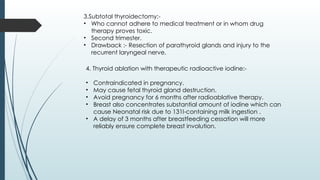
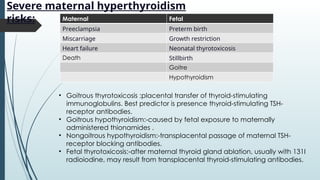
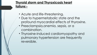
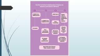
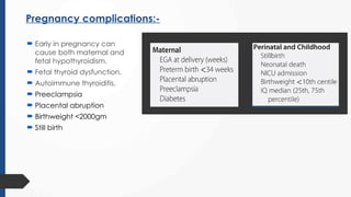
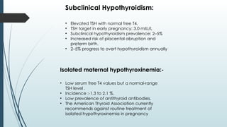
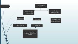
THYROID DISORDERS IN
PREGNANCY
PRESENTED BY :- DR SUNNY BHALADHARE
ANATOMICAL CHANGES DURING
PREGNANCY:-
1.Glandular hyperplasia
2.Increased vascularity
3.Slight enlargement of gland.
4.Enlargement begins in 4th
moth and
continues till term and regress within 2-3
weeks after delivery.
5.This hyperplasia occurs due to
stimulatory effect of hCG
PHYSIOLOGICAL CHANGES during
pregnancy:-
1.Synthesis of Thyroid binding globulin is
increased due to increased estrogen .
2.Total serum T3 and T4 increases.
3.Free T3 and T4 remain unaltered so they are
more accurate in pregnancy .
4. Throughout pregnancy, maternal thyroxine
is transferred to the fetus.
5. Maternal thyroxine is important for normal
fetal brain development.
6. The fetal gland begins concentrating
iodine and synthesizing thyroid hormone after
12 weeks’ gestation.
Influence of B-hCG on
thyroid physiology:
• Exhibits thyroid-stimulating properties
that increase soon after fertilization.
• Reaches its highest level around the
10th week of pregnancy, then
gradually falls by the 20th week then
plateaus.
• Stimulates TSH receptors while partially
suppressing pituitary gland activity.
• Levels dip between weeks 8 and 14,
reflecting the surge in hCG.
• Approximately one in five women
experience TSH levels dropping below
the normal range during this phase.
AUTOIMMUNITY AND THYROID DISEASE
1. Most Thyroid disorders are linked to autoantibodies.
2. These antibodies variably stimulate thyroid function,
block function, or cause thyroid inflammation that
may lead to follicular cell destruction.
3. Thyroid-stimulating autoantibodies, also called thyroid-
stimulating immunoglobulins (TSIs), bind and activate
the TSH receptor to cause thyroid hyperfunction and
growth ,identified in patients with classic graves
disease.
4. Thyroid peroxidase antibodies are directed against
TPO, associated with early pregnancy loss and
preterm birth,placental abruption, high risk for
postpartum thyroid dysfunction and at lifelong risk for
permanent thyroid failure.
Fetal microchimerism:-
• Fetal cells are known to enter maternal circulation
during pregnancy.
• Stem cell interchange can lead to engraftment in
several maternal tissues and is termed fetal
microchimerism
Normal TSH Levels by
Trimester:
Test 1st Trimester 2nd Trimester 3rd Trimester
TSH 0.1 – 2.5 mIU/L 0.2 – 3.0 mIU/L 0.3 – 3.0 mIU/L
Free T4 (FT4) 0.8 – 1.8 ng/dL
(10 – 23 pmol/L)
0.6 – 1.4 ng/dL
(8 – 18 pmol/L)
0.5 – 1.3 ng/dL
(7 – 17 pmol/L)
Total T4 ↑ ~1.5×
nonpregnant
(≈ 7 – 16 µg/dL)
↑ ~1.5×
(≈ 9 – 18 µg/dL)
↑ ~1.5×
(≈ 9 – 18 µg/dL)
Total T3 ↑ ~1.5×
nonpregnant
(≈ 120 – 250 ng/dL)
↑ ~1.5×
(≈ 140 – 280 ng/dL)
↑ ~1.5×
(≈ 140 – 280 ng/dL)
HYPERTHYROIDISM:-
• Incidence- 0.4 and 1.7 percent .
• TSH is low, free T4 high.
Clinical features:-
• Tachycardia
• Heat intolerance
• Hand tremor
• Thyromegaly
• Exophthalmos
• Failure to gain weight despite adequate food
intake.
• TSH levels are markedly depressed, while serum free
T4 (fT4) levels are elevated.
• Rarely, hyperthyroidism is caused by abnormally
high serum triiodothyronine (T3) levels and called T3-
toxicosis.
•Activity levels vary throughout pregnancy,
characterized by:
• A peak in symptoms during the first trimester
• A steady improvement as pregnancy progresses
into the later stages.
• An exacerabation of symptoms shortly after
childbirth.
GRAVES DISEASE:-
Diagnosis:-
• Diagnosis is difficult due to pregnancy-like symptoms.
• Thyroid exam shows goiter, low TSH, and high freeT4.
• Complications depend on duration and control of
hyperthyroidism.
• Autoantibodies mimicking TSH can cross placenta,
causing neonatal Graves’ disease.
•Testing for thyrotropin receptor antibodies) helps
diagnose Graves' disease during pregnancy.
Treatment:-
1. Thionamide drugs- Propylthiouracil (PTU), it partially inhibits the
conversion of T4 to T3.
• Crosses the placenta less readily than methimazole.
• Dose:- 50-150mg 3 times daily or 300-450 mg daily in three divided
doses. (Maximum 600mg) in first trimester.
• Transition to methimazole after the first trimester, with dose of 20:1
PTU to methimazole thoughout pregnancy is recommended.
2. Carbimazole and Methimazole:-
Dose:- Initial higher daily dose of 10 to 20 mg orally followed
by a lower maintenance dose of 5 to 10 mg.
Methimazole has sideeffects like esophageal or choanal
atresia and aplasia cutis, a congenital skin defects, fetal
malformation.
Side effects:-
• Liver toxicity.
• Transient leucopenia
• Agranulocytosis.
Follow up every 4-6 weeks.
3.Subtotal thyroidectomy:-
• Who cannot adhere to medical treatment or in whom drug
therapy proves toxic.
• Second trimester.
• Drawback :- Resection of parathyroid glands and injury to the
recurrent laryngeal nerve.
4. Thyroid ablation with therapeutic radioactive iodine:-
• Contraindicated in pregnancy.
• May cause fetal thyroid gland destruction.
• Avoid pregnancy for 6 months after radioablative therapy.
• Breast also concentrates substantial amount of iodine which can
cause Neonatal risk due to 131I-containing milk ingestion .
• A delay of 3 months after breastfeeding cessation will more
reliably ensure complete breast involution.
Severe maternal hyperthyroidism
risks: Maternal Fetal
Preeclampsia Preterm birth
Miscarriage Growth restriction
Heart failure Neonatal thyrotoxicosis
Death Stillbirth
Goitre
Hypothyroidism
• Goitrous thyrotoxicosis :placental transfer of thyroid-stimulating
immunoglobulins. Best predictor is presence thyroid-stimulating TSH-
receptor antibodies.
• Goitrous hypothyroidism:-caused by fetal exposure to maternally
administered thionamides .
• Nongoitrous hypothyroidism:-transplacental passage of maternal TSH-
receptor blocking antibodies.
• Fetal thyrotoxicosis:-after maternal thyroid gland ablation, usually with 131I
radioiodine, may result from transplacental thyroid-stimulating antibodies.
Thyroid storm and Thyroxicosis heart
failure:-
• Acute and life-threatening.
• Due to hypermetabolic state and the
profound myocardial effects of thyroxine.
• Preeclampsia,anemia, sepsis, or a
combination.
• Thyroxine-induced cardiomyopathy and
pulmonary hypertension are frequently
reversible.
HYPEREMESIS GRAVIDARUM:-
Gestational Transient Thyrotoxicosis.:-
• Severe nausea and vomiting
• High serum T4 levels and low TSH levels
due to TSH-receptor stimulation from
hCG.
• Normalisation of T4 mid gestation.
• Treatment is supportive care
Gestational trophoblastic disease:-
• Abnormally high hCG levels lead to overstimulation of the
TSH receptor and hence T4 level rises.
• With molar evacuation, serum free T4 levels usually drop
to normal levels rapidly in parallel with declining hCG
concentrations.
Sub clinical hyperthyroidism:-
• Abnormally low serum TSH with normal T4
hormone levels.
• Associated with osteoporosis , cardiac
morbidity,overt thyrotoxicosis and thyroid failure.
• Does not warrant treatment.
HYPOTHYROIDISM:-
• Hypothyroidism is the most common
thyroid disorder in pregnancy, affecting
0.2-1.2 percent of pregnancy.
Symptoms:-
• Fatigue
• Constipation
• Cold intolerance
• Muscle cramps
• Weight gain.
Clinical or Overt Hypothyroidism:-
• Elevated TSH with low free T4
• Most common cause of hypothyroidism in pregnancy
Hashimoto thyroiditis
Anti-TPO antibodies
Postablative Graves disease.
• Severe hypothyroidism during pregnancy is uncommon.
Treatment:-
Levothyroxine in doses of 1 to 2 μg/kg/d or approximately 100 μg daily.
The levothyroxine dose is adjusted by 25- to 50-μg increments until TSH values
approximate 2.5 mU/L.
Higher dose requirements begin as early as 5 weeks’ gestation.
Surveillance:- TSH levels measured at 4-6 week intervals in the first half of
pregnancy and at least once in the third trimester.
• If patient was previously taking levothyroxine then increase dose by 30%
• All overt hypothyroid with TSH>2.5 should be treated
• Target TSH<2.5
Pregnancy complications:-
 Early in pregnancy can
cause both maternal and
fetal hypothyroidism.
 Fetal thyroid dysfunction.
 Autoimmune thyroiditis.
 Preeclampsia
 Placental abruption
 Birthweight <2000gm
 Still birth
Subclinical Hypothyroidism:
• Elevated TSH with normal free T4.
• TSH target in early pregnancy: 3.0 mIU/L
• Subclinical hypothyroidism prevalence: 2–5%
• Increased risk of placental abruption and
preterm birth.
• 2–5% progress to overt hypothyroidism annually
Isolated maternal hypothyroxinemia:-
• Low serum free T4 values but a normal-range
TSH level .
• Incidence :-1.3 to 2.1 %.
• Low prevalence of antithyroid antibodies.
• The American Thyroid Association currently
recommends against routine treatment of
isolated hypothyroxinemia in pregnancy
Postpartum
Previously not
hypothyroid
<50mcg/d
>50mcg/d
Discontinue Continue
Repeat TSH after 6
weeks
Previously
hypothyroid
Revert to non
pregnant dose
Euthyroid Autoimmune Thyroid Disease
• Autoantibodies to TPO and thyroglobulin.
• Most who test positive for such antibodies, however, are
euthyroid.
• Presence of thyroid antibodies has also been associated
with Preterm birth, Placental abruption
Iodine Deficiency
• Decreasing iodide fortification led to occasional iodide deficiency.
• Dietary iodine requirements are higher during pregnancy.
• The American Thyroid Association recommends an average iodine
intake of 250 μg/d in all pregnant women.
• The American Thyroid Association advises against exceeding twice
the daily recommended intake of iodine, which is 500 μg/d.
Congenital Hypothyroidism
• May be caused by hereditary defects in thyroid hormone
production.
• Early and aggressive thyroxine replacement is critical for
affected newborns
Postpartum thyroiditis:-
• Autoimmune disorder with a self-limited hyperthyroid or hypothyroid phase
within one year after childbirth.
• 50% women who have thyroid antibodies in the first trimester will develop
postpartum thyroiditis.
• Presentations include:
Transient hyperthyroidism only
Transient hypothyroidism only
Transient hyperthyroidism followed by hypothyroidism, then recovery
• Occurs in 3–16% of cases after spontaneous or induced abortion.
• Small ,painless goitre with fatigue and palpitations.
• Antithyroid drugs are not effective.
• Beta blockers may b given.
• They might require thyroxine replacement therapy.
• Levothyroxine replacement at doses of 25 to 75 μg/d is typically given for 6 to
12 months.
Nodular
thyroid
disease:-
Thyroid tumors are the most common endocrine neoplasms.
Thyroid cancer represents 1% of all cancers, predominantly affecting
women, especially during reproductive years.
Diagnosis :-
Biopsy
• Serum TSH
• Free T4 levels
• Ultrasonography
• Fine needle aspiration.
Radionuclide scanning is contraindicated during pregnancy.
Surgery is recommended promptly for malignant or suspicious papillary
cancer cases.
Patients are maintained on thyroid replacement therapy post-treatment.
National
Iodine
Deficiency
Disorders
Control
Programme
(NIDDCP)
Objectives:-
• Surveys to assess the magnitude of
Iodine Deficiency Disorders in the
districts.
• Supply of iodated salt in place of
common salt.
• Resurveys to assess iodine deficiency
disorders and the impact of iodated
salt after every 5 years in the districts.
• Laboratory monitoring of iodated salt
and urinary iodine excretion.
• Health Education and Publicity.
Thank you.

THYROID DISORDERS IN PREGNANCY .pptx

  • 1.
  • 2.
    ANATOMICAL CHANGES DURING PREGNANCY:- 1.Glandularhyperplasia 2.Increased vascularity 3.Slight enlargement of gland. 4.Enlargement begins in 4th moth and continues till term and regress within 2-3 weeks after delivery. 5.This hyperplasia occurs due to stimulatory effect of hCG
  • 3.
    PHYSIOLOGICAL CHANGES during pregnancy:- 1.Synthesisof Thyroid binding globulin is increased due to increased estrogen . 2.Total serum T3 and T4 increases. 3.Free T3 and T4 remain unaltered so they are more accurate in pregnancy . 4. Throughout pregnancy, maternal thyroxine is transferred to the fetus. 5. Maternal thyroxine is important for normal fetal brain development. 6. The fetal gland begins concentrating iodine and synthesizing thyroid hormone after 12 weeks’ gestation.
  • 4.
    Influence of B-hCGon thyroid physiology: • Exhibits thyroid-stimulating properties that increase soon after fertilization. • Reaches its highest level around the 10th week of pregnancy, then gradually falls by the 20th week then plateaus. • Stimulates TSH receptors while partially suppressing pituitary gland activity. • Levels dip between weeks 8 and 14, reflecting the surge in hCG. • Approximately one in five women experience TSH levels dropping below the normal range during this phase.
  • 5.
    AUTOIMMUNITY AND THYROIDDISEASE 1. Most Thyroid disorders are linked to autoantibodies. 2. These antibodies variably stimulate thyroid function, block function, or cause thyroid inflammation that may lead to follicular cell destruction. 3. Thyroid-stimulating autoantibodies, also called thyroid- stimulating immunoglobulins (TSIs), bind and activate the TSH receptor to cause thyroid hyperfunction and growth ,identified in patients with classic graves disease. 4. Thyroid peroxidase antibodies are directed against TPO, associated with early pregnancy loss and preterm birth,placental abruption, high risk for postpartum thyroid dysfunction and at lifelong risk for permanent thyroid failure. Fetal microchimerism:- • Fetal cells are known to enter maternal circulation during pregnancy. • Stem cell interchange can lead to engraftment in several maternal tissues and is termed fetal microchimerism
  • 6.
    Normal TSH Levelsby Trimester: Test 1st Trimester 2nd Trimester 3rd Trimester TSH 0.1 – 2.5 mIU/L 0.2 – 3.0 mIU/L 0.3 – 3.0 mIU/L Free T4 (FT4) 0.8 – 1.8 ng/dL (10 – 23 pmol/L) 0.6 – 1.4 ng/dL (8 – 18 pmol/L) 0.5 – 1.3 ng/dL (7 – 17 pmol/L) Total T4 ↑ ~1.5× nonpregnant (≈ 7 – 16 µg/dL) ↑ ~1.5× (≈ 9 – 18 µg/dL) ↑ ~1.5× (≈ 9 – 18 µg/dL) Total T3 ↑ ~1.5× nonpregnant (≈ 120 – 250 ng/dL) ↑ ~1.5× (≈ 140 – 280 ng/dL) ↑ ~1.5× (≈ 140 – 280 ng/dL)
  • 7.
    HYPERTHYROIDISM:- • Incidence- 0.4and 1.7 percent . • TSH is low, free T4 high. Clinical features:- • Tachycardia • Heat intolerance • Hand tremor • Thyromegaly • Exophthalmos • Failure to gain weight despite adequate food intake. • TSH levels are markedly depressed, while serum free T4 (fT4) levels are elevated. • Rarely, hyperthyroidism is caused by abnormally high serum triiodothyronine (T3) levels and called T3- toxicosis.
  • 8.
    •Activity levels varythroughout pregnancy, characterized by: • A peak in symptoms during the first trimester • A steady improvement as pregnancy progresses into the later stages. • An exacerabation of symptoms shortly after childbirth. GRAVES DISEASE:- Diagnosis:- • Diagnosis is difficult due to pregnancy-like symptoms. • Thyroid exam shows goiter, low TSH, and high freeT4. • Complications depend on duration and control of hyperthyroidism. • Autoantibodies mimicking TSH can cross placenta, causing neonatal Graves’ disease. •Testing for thyrotropin receptor antibodies) helps diagnose Graves' disease during pregnancy.
  • 9.
    Treatment:- 1. Thionamide drugs-Propylthiouracil (PTU), it partially inhibits the conversion of T4 to T3. • Crosses the placenta less readily than methimazole. • Dose:- 50-150mg 3 times daily or 300-450 mg daily in three divided doses. (Maximum 600mg) in first trimester. • Transition to methimazole after the first trimester, with dose of 20:1 PTU to methimazole thoughout pregnancy is recommended. 2. Carbimazole and Methimazole:- Dose:- Initial higher daily dose of 10 to 20 mg orally followed by a lower maintenance dose of 5 to 10 mg. Methimazole has sideeffects like esophageal or choanal atresia and aplasia cutis, a congenital skin defects, fetal malformation. Side effects:- • Liver toxicity. • Transient leucopenia • Agranulocytosis. Follow up every 4-6 weeks.
  • 10.
    3.Subtotal thyroidectomy:- • Whocannot adhere to medical treatment or in whom drug therapy proves toxic. • Second trimester. • Drawback :- Resection of parathyroid glands and injury to the recurrent laryngeal nerve. 4. Thyroid ablation with therapeutic radioactive iodine:- • Contraindicated in pregnancy. • May cause fetal thyroid gland destruction. • Avoid pregnancy for 6 months after radioablative therapy. • Breast also concentrates substantial amount of iodine which can cause Neonatal risk due to 131I-containing milk ingestion . • A delay of 3 months after breastfeeding cessation will more reliably ensure complete breast involution.
  • 11.
    Severe maternal hyperthyroidism risks:Maternal Fetal Preeclampsia Preterm birth Miscarriage Growth restriction Heart failure Neonatal thyrotoxicosis Death Stillbirth Goitre Hypothyroidism • Goitrous thyrotoxicosis :placental transfer of thyroid-stimulating immunoglobulins. Best predictor is presence thyroid-stimulating TSH- receptor antibodies. • Goitrous hypothyroidism:-caused by fetal exposure to maternally administered thionamides . • Nongoitrous hypothyroidism:-transplacental passage of maternal TSH- receptor blocking antibodies. • Fetal thyrotoxicosis:-after maternal thyroid gland ablation, usually with 131I radioiodine, may result from transplacental thyroid-stimulating antibodies.
  • 12.
    Thyroid storm andThyroxicosis heart failure:- • Acute and life-threatening. • Due to hypermetabolic state and the profound myocardial effects of thyroxine. • Preeclampsia,anemia, sepsis, or a combination. • Thyroxine-induced cardiomyopathy and pulmonary hypertension are frequently reversible.
  • 14.
    HYPEREMESIS GRAVIDARUM:- Gestational TransientThyrotoxicosis.:- • Severe nausea and vomiting • High serum T4 levels and low TSH levels due to TSH-receptor stimulation from hCG. • Normalisation of T4 mid gestation. • Treatment is supportive care Gestational trophoblastic disease:- • Abnormally high hCG levels lead to overstimulation of the TSH receptor and hence T4 level rises. • With molar evacuation, serum free T4 levels usually drop to normal levels rapidly in parallel with declining hCG concentrations. Sub clinical hyperthyroidism:- • Abnormally low serum TSH with normal T4 hormone levels. • Associated with osteoporosis , cardiac morbidity,overt thyrotoxicosis and thyroid failure. • Does not warrant treatment.
  • 15.
    HYPOTHYROIDISM:- • Hypothyroidism isthe most common thyroid disorder in pregnancy, affecting 0.2-1.2 percent of pregnancy. Symptoms:- • Fatigue • Constipation • Cold intolerance • Muscle cramps • Weight gain.
  • 16.
    Clinical or OvertHypothyroidism:- • Elevated TSH with low free T4 • Most common cause of hypothyroidism in pregnancy Hashimoto thyroiditis Anti-TPO antibodies Postablative Graves disease. • Severe hypothyroidism during pregnancy is uncommon.
  • 17.
    Treatment:- Levothyroxine in dosesof 1 to 2 μg/kg/d or approximately 100 μg daily. The levothyroxine dose is adjusted by 25- to 50-μg increments until TSH values approximate 2.5 mU/L. Higher dose requirements begin as early as 5 weeks’ gestation. Surveillance:- TSH levels measured at 4-6 week intervals in the first half of pregnancy and at least once in the third trimester. • If patient was previously taking levothyroxine then increase dose by 30% • All overt hypothyroid with TSH>2.5 should be treated • Target TSH<2.5
  • 19.
    Pregnancy complications:-  Earlyin pregnancy can cause both maternal and fetal hypothyroidism.  Fetal thyroid dysfunction.  Autoimmune thyroiditis.  Preeclampsia  Placental abruption  Birthweight <2000gm  Still birth
  • 20.
    Subclinical Hypothyroidism: • ElevatedTSH with normal free T4. • TSH target in early pregnancy: 3.0 mIU/L • Subclinical hypothyroidism prevalence: 2–5% • Increased risk of placental abruption and preterm birth. • 2–5% progress to overt hypothyroidism annually Isolated maternal hypothyroxinemia:- • Low serum free T4 values but a normal-range TSH level . • Incidence :-1.3 to 2.1 %. • Low prevalence of antithyroid antibodies. • The American Thyroid Association currently recommends against routine treatment of isolated hypothyroxinemia in pregnancy
  • 21.
    Postpartum Previously not hypothyroid <50mcg/d >50mcg/d Discontinue Continue RepeatTSH after 6 weeks Previously hypothyroid Revert to non pregnant dose
  • 22.
    Euthyroid Autoimmune ThyroidDisease • Autoantibodies to TPO and thyroglobulin. • Most who test positive for such antibodies, however, are euthyroid. • Presence of thyroid antibodies has also been associated with Preterm birth, Placental abruption Iodine Deficiency • Decreasing iodide fortification led to occasional iodide deficiency. • Dietary iodine requirements are higher during pregnancy. • The American Thyroid Association recommends an average iodine intake of 250 μg/d in all pregnant women. • The American Thyroid Association advises against exceeding twice the daily recommended intake of iodine, which is 500 μg/d. Congenital Hypothyroidism • May be caused by hereditary defects in thyroid hormone production. • Early and aggressive thyroxine replacement is critical for affected newborns
  • 23.
    Postpartum thyroiditis:- • Autoimmunedisorder with a self-limited hyperthyroid or hypothyroid phase within one year after childbirth. • 50% women who have thyroid antibodies in the first trimester will develop postpartum thyroiditis. • Presentations include: Transient hyperthyroidism only Transient hypothyroidism only Transient hyperthyroidism followed by hypothyroidism, then recovery • Occurs in 3–16% of cases after spontaneous or induced abortion. • Small ,painless goitre with fatigue and palpitations. • Antithyroid drugs are not effective. • Beta blockers may b given. • They might require thyroxine replacement therapy. • Levothyroxine replacement at doses of 25 to 75 μg/d is typically given for 6 to 12 months.
  • 24.
    Nodular thyroid disease:- Thyroid tumors arethe most common endocrine neoplasms. Thyroid cancer represents 1% of all cancers, predominantly affecting women, especially during reproductive years. Diagnosis :- Biopsy • Serum TSH • Free T4 levels • Ultrasonography • Fine needle aspiration. Radionuclide scanning is contraindicated during pregnancy. Surgery is recommended promptly for malignant or suspicious papillary cancer cases. Patients are maintained on thyroid replacement therapy post-treatment.
  • 25.
    National Iodine Deficiency Disorders Control Programme (NIDDCP) Objectives:- • Surveys toassess the magnitude of Iodine Deficiency Disorders in the districts. • Supply of iodated salt in place of common salt. • Resurveys to assess iodine deficiency disorders and the impact of iodated salt after every 5 years in the districts. • Laboratory monitoring of iodated salt and urinary iodine excretion. • Health Education and Publicity.
  • 26.