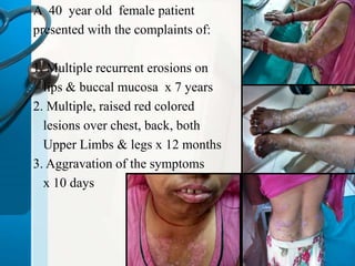
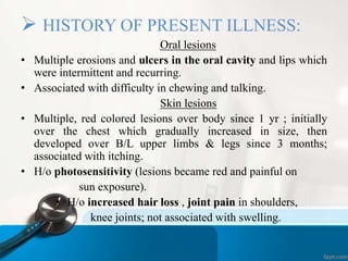
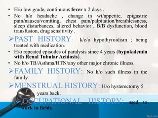
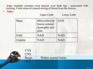
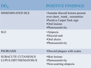
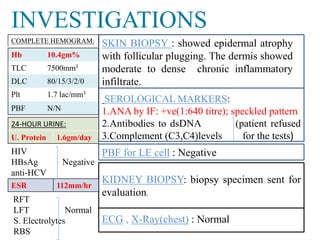
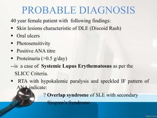
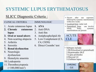
CLINICAL CASE 
VAISHALI 
MBBS 2012
A 40 year old female patient 
presented with the complaints of: 
1. Multiple recurrent erosions on 
lips & buccal mucosa x 7 years 
2. Multiple, raised red colored 
lesions over chest, back, both 
Upper Limbs & legs x 12 months 
3. Aggravation of the symptoms 
x 10 days
 HISTORY OF PRESENT ILLNESS: 
Oral lesions 
• Multiple erosions and ulcers in the oral cavity and lips which 
were intermittent and recurring. 
• Associated with difficulty in chewing and talking. 
Skin lesions 
• Multiple, red colored lesions over body since 1 yr ; initially 
over the chest which gradually increased in size, then 
developed over B/L upper limbs & legs since 3 months; 
associated with itching. 
• H/o photosensitivity (lesions became red and painful on 
sun exposure). 
• H/o increased hair loss , joint pain in shoulders, 
knee joints; not associated with swelling.
• H/o low grade, continuous fever x 2 days . 
• No h/o headache , change in wt/appetite, epigastric 
pain/nausea/vomiting, chest pain/palpitation/breathlessness, 
sleep disturbances, altered behavior , B/B dysfunction, blood 
transfusion, drug sensitivity . 
PAST HISTORY: k/c/o hypothyroidism ; being 
treated with medication. 
• H/o repeated episodes of paralysis since 4 years (hypokalemia 
with Renal Tubular Acidosis). 
• No h/o TB/Asthma/HTN/any other major chronic illness. 
FAMILY HISTORY: No h/o such illness in the 
family. 
MENSTRUAL HISTORY: H/o hysterectomy 5 
years back. 
OCCUPATIONAL HISTORY: used to 
work in fields.
EXAMINATION 
• GPE: conscious, co-operative, oriented to TPP, febrile 
• PR: 80/min, regular, good volume, vessel wall not palpable 
BP: 124/80 mmHg RR: 18/min 
• O/E: multiple, erythematous, P- I- Cy- Cl- JVPNR LAP- PE-discoid 
plaques with adherent scales 
and well-defined hyperpigmented 
border present over chest , lower 
back, B/L upper limbs & legs.Carpet 
Tack sign was positive. Grattage test 
and Auspitz sign were negative. 
• Oral mucosa: multiple ulcerations 
and superficial erosions were present 
on buccal mucosa and palate.
• Lips: multiple erosions were present over both lips ; associated with 
crusting. Crust removal caused oozing of blood from the lesions. 
• Nails : 
Upper Limb Lower Limb 
Plate Mild yellowish 
brown colored 
dystrophic nail 
plate 
NAD 
Fold NAD NAD 
Cuticle NAD NAD 
CVS 
CNS 
Respi. Within normal limits 
P/A
DDX POSITIVE FINDINGS 
DISSEMINATED DLE •Annular discoid lesions present 
over chest , trunk , extremities 
•Positive Carpet Tack sign 
•Oral lesions 
•Photosensitivity 
SLE •Alopecia 
•Discoid rash 
•Oral ulcers 
•Photosensitivity 
PSORIASIS •Discoid plaques with scales 
SUBACUTE CUTANEOUS 
LUPUS ERYTHEMATOSUS 
•Skin lesions 
•Photosensitivity 
•Non-scarring alopecia
INVESTIGATIONS 
KIDNEY BIOPSY: biopsy specimen sent for 
evaluation. 
ECG , X-Ray(chest) : Normal 
COMPLETE HEMOGRAM: 
Hb 10.4gm% 
TLC 7500mm3 
DLC 80/15/3/2/0 
Plt 1.7 lac/mm3 
PBF N/N 
24-HOUR URINE: 
U. Protein 1.6gm/day 
HIV 
HBsAg Negative 
anti-HCV 
ESR 112mm/hr 
SKIN BIOPSY : showed epidermal atrophy 
with follicular plugging. The dermis showed 
moderate to dense chronic inflammatory 
infiltrate. 
SEROLOGICALMARKERS: 
1.ANA by IF: +ve(1:640 titre); speckled pattern 
2.Antibodies to dsDNA (patient refused 
3.Complement (C3,C4)levels for the tests) 
PBF for LE cell : Negative 
RFT 
LFT Normal 
S. Electrolytes 
RBS
PROBABLE DIAGNOSIS 
40 year female patient with following findings: 
 Skin lesions characteristic of DLE (Discoid Rash) 
 Oral ulcers 
 Photosensitivity 
 Positive ANA titre 
 Proteinuria (>0.5 g/day) 
--is a case of Systemic Lupus Erythematosus as per the 
SLICC Criteria. 
 RTA with hypokalemic paralysis and speckled IF pattern of 
ANA indicate: 
? Overlap syndrome of SLE with secondary 
Sjogren’s Syndrome
SYSTEMIC LUPUS ERYTHEMATOSUS 
CHRONIC 
CLE 
SLICC Diagnostic Criteria : 
CLINICAL CRITERIA IMMUNOLOGIC 
CRITERIA 
1. Acute cutaneous lupus 
2. Chronic cutaneous 
lupus 
3. Oral or nasal ulcers 
4. Non-scarring alopecia 
5. Arthritis 
6. Serositis 
7. Renal dysfunction 
8. Neurologic 
dysfunction 
9. Hemolytic anaemia 
10.Leukopenia 
11.Thrombocytopenia 
(<100,000/mm3) 
1. ANA 
2. Anti-DNA 
3. Anti-Sm 
4. Antiphospholipid Ab 
5. Low Complement (C3, 
C4, CH50) 
6. Direct Coombs’ test 
• Occurs after sun exposure; 
followed by systemic manifestatons 
within few weeks 
• Localised form: malar rash 
• Generalised form: can involve 
whole body; systemic 
manifestations are present 
ACUTE 
CLE 
• Subtypes include: 
1.DLE(localised or generalised) 
2.Hypertrophic DLE 
3.Lupus profundus 
4.Mucosal LE 
5.Chilblain lupus
TREATMENT 
CUTANEOUS 
MANIFESTATIONS 
~Avoid sun exposure 
and use sunscreens 
with high SPF. 
~Cosmetic camouflage 
can be used. 
~Balanced diet low in 
fat. 
~ Topical steroids 
~ Intralesional steroids 
~Antimalarials 
(hydroxychloroquine) 
~ Oral steroids 
SYSTEMIC 
MANIFESTATIONS 
~Conservative treatment 
+ low dose 
corticosteroids 
~If organ threatening 
SLE, high dose 
corticosteroids with 
immunosuppressive 
drugs.
Case of SLE

Case of SLE

  • 1.
  • 2.
    A 40 yearold female patient presented with the complaints of: 1. Multiple recurrent erosions on lips & buccal mucosa x 7 years 2. Multiple, raised red colored lesions over chest, back, both Upper Limbs & legs x 12 months 3. Aggravation of the symptoms x 10 days
  • 3.
     HISTORY OFPRESENT ILLNESS: Oral lesions • Multiple erosions and ulcers in the oral cavity and lips which were intermittent and recurring. • Associated with difficulty in chewing and talking. Skin lesions • Multiple, red colored lesions over body since 1 yr ; initially over the chest which gradually increased in size, then developed over B/L upper limbs & legs since 3 months; associated with itching. • H/o photosensitivity (lesions became red and painful on sun exposure). • H/o increased hair loss , joint pain in shoulders, knee joints; not associated with swelling.
  • 4.
    • H/o lowgrade, continuous fever x 2 days . • No h/o headache , change in wt/appetite, epigastric pain/nausea/vomiting, chest pain/palpitation/breathlessness, sleep disturbances, altered behavior , B/B dysfunction, blood transfusion, drug sensitivity . PAST HISTORY: k/c/o hypothyroidism ; being treated with medication. • H/o repeated episodes of paralysis since 4 years (hypokalemia with Renal Tubular Acidosis). • No h/o TB/Asthma/HTN/any other major chronic illness. FAMILY HISTORY: No h/o such illness in the family. MENSTRUAL HISTORY: H/o hysterectomy 5 years back. OCCUPATIONAL HISTORY: used to work in fields.
  • 5.
    EXAMINATION • GPE:conscious, co-operative, oriented to TPP, febrile • PR: 80/min, regular, good volume, vessel wall not palpable BP: 124/80 mmHg RR: 18/min • O/E: multiple, erythematous, P- I- Cy- Cl- JVPNR LAP- PE-discoid plaques with adherent scales and well-defined hyperpigmented border present over chest , lower back, B/L upper limbs & legs.Carpet Tack sign was positive. Grattage test and Auspitz sign were negative. • Oral mucosa: multiple ulcerations and superficial erosions were present on buccal mucosa and palate.
  • 6.
    • Lips: multipleerosions were present over both lips ; associated with crusting. Crust removal caused oozing of blood from the lesions. • Nails : Upper Limb Lower Limb Plate Mild yellowish brown colored dystrophic nail plate NAD Fold NAD NAD Cuticle NAD NAD CVS CNS Respi. Within normal limits P/A
  • 7.
    DDX POSITIVE FINDINGS DISSEMINATED DLE •Annular discoid lesions present over chest , trunk , extremities •Positive Carpet Tack sign •Oral lesions •Photosensitivity SLE •Alopecia •Discoid rash •Oral ulcers •Photosensitivity PSORIASIS •Discoid plaques with scales SUBACUTE CUTANEOUS LUPUS ERYTHEMATOSUS •Skin lesions •Photosensitivity •Non-scarring alopecia
  • 8.
    INVESTIGATIONS KIDNEY BIOPSY:biopsy specimen sent for evaluation. ECG , X-Ray(chest) : Normal COMPLETE HEMOGRAM: Hb 10.4gm% TLC 7500mm3 DLC 80/15/3/2/0 Plt 1.7 lac/mm3 PBF N/N 24-HOUR URINE: U. Protein 1.6gm/day HIV HBsAg Negative anti-HCV ESR 112mm/hr SKIN BIOPSY : showed epidermal atrophy with follicular plugging. The dermis showed moderate to dense chronic inflammatory infiltrate. SEROLOGICALMARKERS: 1.ANA by IF: +ve(1:640 titre); speckled pattern 2.Antibodies to dsDNA (patient refused 3.Complement (C3,C4)levels for the tests) PBF for LE cell : Negative RFT LFT Normal S. Electrolytes RBS
  • 9.
    PROBABLE DIAGNOSIS 40year female patient with following findings:  Skin lesions characteristic of DLE (Discoid Rash)  Oral ulcers  Photosensitivity  Positive ANA titre  Proteinuria (>0.5 g/day) --is a case of Systemic Lupus Erythematosus as per the SLICC Criteria.  RTA with hypokalemic paralysis and speckled IF pattern of ANA indicate: ? Overlap syndrome of SLE with secondary Sjogren’s Syndrome
  • 10.
    SYSTEMIC LUPUS ERYTHEMATOSUS CHRONIC CLE SLICC Diagnostic Criteria : CLINICAL CRITERIA IMMUNOLOGIC CRITERIA 1. Acute cutaneous lupus 2. Chronic cutaneous lupus 3. Oral or nasal ulcers 4. Non-scarring alopecia 5. Arthritis 6. Serositis 7. Renal dysfunction 8. Neurologic dysfunction 9. Hemolytic anaemia 10.Leukopenia 11.Thrombocytopenia (<100,000/mm3) 1. ANA 2. Anti-DNA 3. Anti-Sm 4. Antiphospholipid Ab 5. Low Complement (C3, C4, CH50) 6. Direct Coombs’ test • Occurs after sun exposure; followed by systemic manifestatons within few weeks • Localised form: malar rash • Generalised form: can involve whole body; systemic manifestations are present ACUTE CLE • Subtypes include: 1.DLE(localised or generalised) 2.Hypertrophic DLE 3.Lupus profundus 4.Mucosal LE 5.Chilblain lupus
  • 11.
    TREATMENT CUTANEOUS MANIFESTATIONS ~Avoid sun exposure and use sunscreens with high SPF. ~Cosmetic camouflage can be used. ~Balanced diet low in fat. ~ Topical steroids ~ Intralesional steroids ~Antimalarials (hydroxychloroquine) ~ Oral steroids SYSTEMIC MANIFESTATIONS ~Conservative treatment + low dose corticosteroids ~If organ threatening SLE, high dose corticosteroids with immunosuppressive drugs.