Downloaded 51 times

![104 Winter 2014 • Volume 29 • Number 4
Introduction
Excellence will never be achieved by chance; rather,
it comes from a consistent, systematic approach to
diagnosis, communication, treatment planning, and
implementation. The incorporation of protocols and
checklists1-7
for quality control and information man-
agement help to guarantee that every critical point is
performed effectively, is double-checked, and is com-
municated correctly.
To obtain predictable and consistent outcomes, the
practitioner should define the design of the restorative
treatment at an early stage. The data must guide the
succeeding phases of the rehabilitation,8
scientifically
integrating all of the patient’s needs and desires and
the patient’s functional, structural, and biological is-
sues into the esthetic treatment design. The data serve
as a frame of reference for the treatment that will be
performed.9,10
However, many of these pieces of infor-
mation may not be taken into consideration if their
real meaning is not transferred in an adequate way to
the design of the restorations.
Digital Smile Design is a multipurpose digital tool
with clinically relevant advantages. It can strengthen
esthetic diagnostic abilities, improve communication
among team members, create predictable systems
throughout the treatment phases, enhance patients’
education and motivation through visualization, and
increase the effectiveness of case presentation. Be-
cause using DSD can make diagnosis more effective
and treatment planning more consistent, the effort
required to implement it is worthwhile and will make
the treatment sequence more logical and straightfor-
ward, saving time and materials and reducing the cost
of treatment.
Clinically Relevant Advantages
The advantages of using DSD are as follows:
• esthetic diagnosis
• treatment planning and communication
• feedback
• patient care
• case presentation
• education.
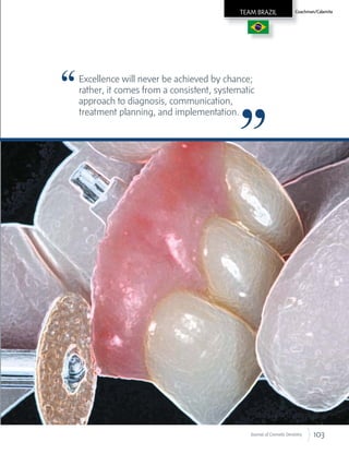
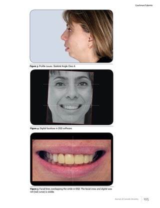
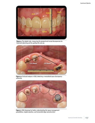
Esthetic Diagnosis
DSD allows a careful esthetic analysis of the patient’s
facial and dental features and a gradual discovery of
many critical factors that might have been overlooked
during the clinical, photographic, or study model
evaluation. Drawing reference lines and shapes over
extra- and intraoral digital photographs in presenta-
tion software (such as Keynote [Apple; Cupertino,
CA]; PowerPoint [Microsoft; Redmond, WA]; DSD software; or Smile De-
signer Pro [Tasty Tech; Toronto, Ontario, CA]), following a predetermined
sequence, helps widen the diagnostic vision. This visualization process
also helps the team to assess and understand limitations and risk fac-
tors such as asymmetries, disharmonies, and esthetic principle violations,
adding critical data to the process of treatment planning.1
Choosing the
appropriate technique is easier once problems have been identified and
the solution clearly visualized. The main steps related to diagnosis are
shown in Figures 1-11.
Figure 1: Preoperative extraoral view 20 years before first appointment
showing ankylosed teeth #21 and #22.
Figure 2: Preoperative extraoral view at first appointment.](https://image.slidesharecdn.com/virtualdesign-150102184118-conversion-gate02/85/V-irtual-design-3-320.jpg)
![116 Winter 2014 • Volume 29 • Number 4
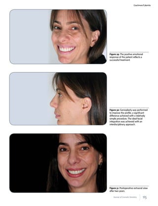
Summary
DSD is a practical multi-use tool with clinically rel-
evant advantages. It can strengthen esthetic diagnos-
tic abilities, improve communication among team
members, create predictable systems throughout the
treatment phases, enhance patients’ education and
motivation, and increase the effectiveness of case pre-
sentation. The drawing of reference lines and shapes
over the patient’s photograph, following a predeter-
mined sequence, allows the team to better evaluate
the esthetic relation among the teeth, the gingiva, the
smile, and the face.
Acknowledgment
The authors thank the restorative team—Dr. Marcos Pitta
(surgery and implants), Dr. Gustavo Giordani (perioplasty
and gingival leveling), Dr. Juliana Romanelli (orthodon-
tics), and Edson Silva (dental technician); all of São Pau-
lo, Brazil—for the high quality of treatment provided in the
case discussed in this article.
References
1. Coachman C, Van Dooren E, Gürel G, Landsberg CJ, Calamita
MA, Bichacho N. Digital smile design: from digital treatment
planning to clinical reality. In: Cohen M, editor. Interdisciplinary
treatment planning, Vol. II: comprehensive case studies. Hanover
Park (IL): Quintessence; 2011.
2. Goldstein RE. Esthetics in dentistry: principles, communication,
treatment methods. Hamilton (ONT): Decker; 1998.
3. Chiche GJ, Pinault A. Esthetics of anterior fixed prosthodontics.
Hanover Park (IL): Quintessence; 1996.
4. Magne P, Belser U. Bonded porcelain restorations in the anterior
dentition: a biomimetic approach. Hanover Park (IL): Quintes-
sence; 2002.
5. Fradeani M. Esthetic rehabilitation in fixed prosthodontics: es-
thetic analysis—a systematic approach to prosthetic treatment.
Hanover Park (IL): Quintessence; 2004.
6. Gürel G. The science and art of porcelain laminate veneers. Ber-
lin: Quintessence; 2003.
7. Rufenacht CR. Fundamentals of esthetics. Hanover Park (IL):
Quintessence; 1990.
8. Dawson PE. Functional occlusion: from TMJ to smile design. St.
Louis: Mosby; 2007.
9. Spear FM. The maxillary central incisor edge: a key to esthetic and functional treatment
planning. Compend Contin Educ Dent. 1999 Jun;20(6):512-6.
10. Kois JC. Diagnostically driven interdisciplinary treatment planning. Seattle Study Club J.
2002;6(4):28-34.
11. Paolucci B. Visagismo e Odontologia [Hair and makeup: and dentistry]. In: Hallawell
P, editor. Visagismo integrado: identidade, estilo, beleza [Hair and makeup: integrated:
identity, style, beauty]. São Paulo: Senac; 2009: p. 243-50. Portuguese.
12. Gürel G, Bichacho N. Permanent diagnostic provisional restorations for predictable re-
sults when redesigning smiles. Pract Proced Aesthet Dent. 2006 Jun;18(5):281-6.
13. Paolucci B, Hallawell P, Sauer C, Coachman C, Ricci A, Calamita M, Yoshinaga LG, Gurel
G. Visagismo: a arte de personalizar o desenho do sorriso [Hair and makeup: the art of
customizing the design of the smile]. São Paulo: VM Cultural; 2011. Portuguese. Available
from: http://www.fineartdental.com.br/downloads/visagismo_braulio-paolucci.pdf
14. Coachman C, Calamita MA. Digital Smile Design: a tool for treatment planning and com-
munication in esthetic dentistry. Hanover Park (IL): Quintessence; 2012. jCD
Dr. Calamita is an associate professor of prosthodontics, University of
Braz Cubas, São Paulo, Brazil, and University of Guarulhos, both in
São Paulo, Brazil. He maintains a private practice focusing on com-
prehensive restorative and implant dentistry in São Paulo.
Disclosure: Dr. Calamita did not report any disclosures.
Dr. Coachman is the owner of Well Clinic Esthetic Dentistry, Digital
Smile Design Center in São Paulo.
Disclosure: Dr. Coachman is the developer of Digital Smile Design.
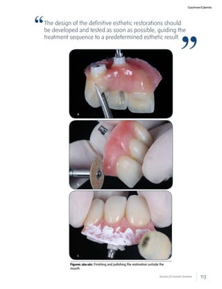
The drawing of reference lines and
shapes over the patient’s photograph,
following a predetermined sequence,
allows the team to better evaluate the
esthetic relation among the teeth, the
gingiva, the smile, and the face.](https://image.slidesharecdn.com/virtualdesign-150102184118-conversion-gate02/85/V-irtual-design-15-320.jpg)


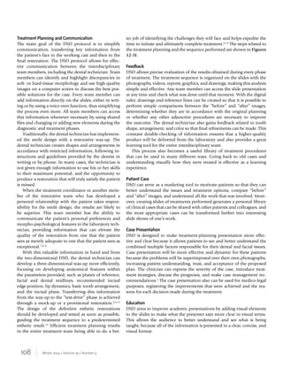
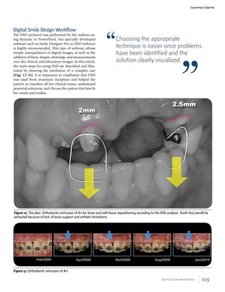
Digital Smile Design (DSD) is a digital tool that can strengthen esthetic diagnosis, improve communication between team members, and help organize treatment planning. It does this through a number of steps: 1) Careful analysis of facial and dental features through overlaying reference lines on digital photos to identify limitations and risk factors. 2) Allowing effective communication between team members who can discuss solutions by adding information directly to shared digital slides. 3) Allowing the dental technician to develop a three-dimensional wax-up more efficiently based on guidelines provided through the two-dimensional DSD.


