The document describes experiments to optimize the purification of serine palmitoyltransferase (SPT), a membrane protein involved in sphingolipid synthesis. Several factors were tested, including expression organism, induction time and cell density, salt concentrations during solubilization, and imidazole concentrations in wash buffers. Yeast expression was found to produce more functional protein than E. coli. Induction at an OD of 1 for 4.5 hours yielded the most protein. Purification was optimized using 0.5M salt during solubilization and 50mM imidazole in washes. The goal is to purify sufficient active SPT to crystallize and determine its structure, providing insight into related genetic diseases
Protein Protein Interactions Of Glycine Oxidase (Thi O)bturne
Project done as a final presentation for Experimental Biochemistry. The project was designed and proposed by me and performed by myself and Lauren Pioppo.
Effect of estradiol -17 β on arachidonic acid metabolism in sheep uterus: in ...iosrjce
The effect of estradiol-17 β on Arachidonic acid (AA) metabolism in non-pregnant sheep uterus was
studied under in vitro conditions. On incubation of uterine slices with estradiol-17β, the levels of prostaglandins
were altered but not Lipoxygenase (LOX) products. Based on their analysis on conventional TLC technique, the
Cyclooxygenase (COX) products PGF2α, 6-keto PGF1α and PGE2 were shown to be altered over an incubation
period of 0 to 120 minutes. The LOX products, HPETEs and HETEs did not show any change upon incubation
with estradiol-17β. This study gives a preliminary understanding of role of estradiol on AA metabolism.
Kinetic study of free and immobilized protease from Aspergillus sp.IOSR Journals
In the present investigation partially purified alkaline protease from Aspergillus sp. As#6 and As#7 strains were entrapped in calcium alginate beads and characterized using casein as a substrate. Temperature and pH maxima of protease from As#6 strain showed no changes before and after immobilization and remained stable at 450C and pH 9, respectively. However km value was slightly shifted from 4.5mg/ml to 5 mg/ml. Proteases from As#7 strain showed shifting in pH optima to a more alkaline range (10.0) as compared with free enzyme (9.0). Optimum temperature for protease from As#7 strain showed changes after immobilization and shifted from 650C to 850C. However there was no significant effect on Km value but Vmax of immobilized protease from As#7 strain was also shifted from 200U/ml to 370U/ml. Immobilized protease from As#6 strain was reused for 3 cycles with 22% loss in its activity whereas immobilize protease from As#7 strain was reused for 3 cycles with 17% loss in its activity. Protease from As#7 strain has a higher affinity for the substrate and higher proteolysis activity than protease from As#6 strain. The present work concludes that Aspergillus As#7 strain may be a good source of industrial protease
Bioconversion of Penicillin to CephalosporinIOSR Journals
Cephalosporins are known as 3rd generation broad spectrum Beta lactam antibiotics, which can also be produced synthetically. Commonly, chemical ring expansion followed by an enzymatic removal of the phenylacetyl side chain is commonly employed to convert penicillin G into 7-aminodeacetoxycephalosporanic acid, the precursor for the manufacture of semisynthetic cephalosporins. This process requires several steps, is expensive and highly polluting. Thus there is a need to device a simple biological route to replace the chemical process. A mutant of Streptomyces clavuligerus NP1 was reported to converts Penicillin G to Deacetoxycephalosporin G (DAOG;phenylacetyl-7-aminodeacetoxycephalosporanic acid) enzymatically[5,8] . This enzyme, deacetoxycephalosporin synthase has the potential for the large scale transformation of Penicillin G to deacetoxycephalosporin. The present work studies the conditions required for efficient transformation of Penicillin G to Deacetoxycephalosporin using the wild type strain Streptomyces clavuligerus . Detection of cephalosporin was carried out using various methods. Additionally succinic acid formation was also studied as it could be used as a commercially important by product of the transformation. Deacetoxycephalosporin synthase also extracted and partially purified and characterised.
The IOSR Journal of Pharmacy (IOSRPHR) is an open access online & offline peer reviewed international journal, which publishes innovative research papers, reviews, mini-reviews, short communications and notes dealing with Pharmaceutical Sciences( Pharmaceutical Technology, Pharmaceutics, Biopharmaceutics, Pharmacokinetics, Pharmaceutical/Medicinal Chemistry, Computational Chemistry and Molecular Drug Design, Pharmacognosy & Phytochemistry, Pharmacology, Pharmaceutical Analysis, Pharmacy Practice, Clinical and Hospital Pharmacy, Cell Biology, Genomics and Proteomics, Pharmacogenomics, Bioinformatics and Biotechnology of Pharmaceutical Interest........more details on Aim & Scope).
ABSTRACT- In this present study, the biotransformation of phenol to L-tyrosine was studied with resting cells of
Citrobacter freundii MTCC 2424 containing high tyrosine phenol lyase activity. Different process parameters leading to
synthesis of L-tyrosine were optimized. The L-tyrosine formed from biotransformation reactions was detected and
quantified by HPLC technique. The maximum L-tyrosine conversion 69% (6.49g/l) was obtained with ammonium
chloride 0.25M, phenol 0.1M, and sodium pyruvate 0.2M in borate buffer (0.1M) of pH 8.5 at 35°C temperature for
45min of incubation. Higher phenol concentration was found to be inhibitory for biotransformation due to phenol
inactivation of catalyst.
Key-words- Citrobacter freundii MTCC 2424, L-tyrosine, Tyrosine phenol lyase, Biotransformation
Expression Purification and Immunodetection of a fusion protein Glutathione S...iosrjce
Glutathione S Transferase(GST) is an enzyme of a multi gene family which is involved in reducing
oxidative damage to cells and detoxification of Xenobiotic compounds and plays critical role in life processes.
The entire work was completely qualitative and the objective of my work was to deal with the induction,
extraction and purification of the GST fusion protein from pGEX 3X vector.In order to achieve high degree of
transformed cells,the E.Coli BL21 host strain was made competent using 0.1M CaCl2 and adding of pGEX 3X
vector into host made it transformed.With the induction of GST protein by 0.1mM IPTG,the desired protein was
purified through glutathione Cl agarose column and was detected by immunoblotting method with the use of
anti GST HRP conjugate Ab which expressed the desired protein.
Dynamin I plays dual roles in the activity-dependent shift in exocytic mode i...Bryan Doreian
Under low stimulation, adrenal chromaffin cells release freely-soluble catecholamines through a restricted granule fusion pore while retaining the large neuropeptide-containing proteinacious granule core. Elevated activity causes dilation of the pore and release of all granule contents. Thus, physiological differential transmitter release is achieved through regulation of fusion pore dilation. We examined the mechanism for pore dilation utilizing a combined approach of peptide transfection, electrophysiology, electrochemistry and quantitative imaging techniques. We report that disruption of dynamin I function alters both fusion modes. Under low stimulation, interference with dynamin I does not affect granule fusion but blocks its re-internalization. In full collapse mode, disruption of dynamin I limits fusion pore dilation, but does not block membrane re-internalization. These data suggest that dynamin I is involved in both modes of exocytosis by regulating contraction or dilation of the fusion pore and thus contributes to activity-dependent differential transmitter release from the adrenal medulla.
Endothelins (ET) are 21-amino acid peptides that bind to membrane receptors to initiate a wide range of pathophysiological effects. ET binding to receptors has been shown to be almost irreversible because bound ET is difficult to dissociate. This report studies the dissociation characteristics of receptor antagonists and further examines the effects of ET's difficult-to-dissociate binding on the potency of antagonists. In membranes prepared from porcine cerebellum, [t25I]ET-1 binding was effectively blocked by ET-1 and ET-3 with similar IC5o values (0.08nM vs. 0.17 nM), suggesting that porcine cerebellum contains predominantly the ETB receptor subtype. [125I]ET-3 binding was inhibited by Ro46-2005 and PD142893, two non-selective antagonists, with IC5o values of 570 + 50 nM and 410 + 100 nM, respectively. Consistent with previous observations, bound [t25I]ET-1 in porcine cerebellum membranes was also difficult to dissociate. In contrast, bound Ro46-2005 or PD142893, but not bound ET-1, could be readily washed away from membranes, suggesting that antagonist binding was more reversible than ET-1 binding. Although Ro46-2005 or PD142893 at 0.5 laM inhibited 0.1 nM [125I]ET-1 binding by >80% after 15 min of incubation, the inhibitory effect decreased to approximately 50% after 3 h of incubation, and further decreased to <10% at 24 h. This decrease in antagonizing potency was further confirmed by the results that the IC50 values of the two antagonists against [125I]ET-3 binding increased with increasing incubation time. Control experiments indicate that the observed decrease in the potency of Ro46-2005 and PD142893 was not the result of ligand degradation. These results suggest that the potency of antagonists is critically dependent on the incubation time because antagonist binding is more reversible than ET binding.
Lipids Chemistry Structure & Function (More Detailed)hafizayyub
This presentation is for Medical students. It is more detailed explanation of Lipids including types and medical importance. It is made by Drs Charles Stephen and Dr Ayyub Patel
1. Optimization of Serine Palmitoyltransferase Purification
-Trishul Nagenalli
Dunn Lab, Department of Biochemistry
n
Background
Abstract
Background
Results
Methods
Conclusions
Future Research
Acknowledgments
Serine Palmitoyltransferase (abbreviated
SPT) is a membrane protein found in the endoplasmic reticulum
(ER). It catalyzes the reaction between the amino acid serine
and a fatty acid, palmitoyl CoA. The product of this reaction is 3-
ketosphinganine. 3-ketosphinganine is a precursor to many
sphingolipids, which are essential lipids in intercellular
communication. Metabolites of sphingolipid synthesis are used
as signaling molecules for a number of cellular function
including proliferation and senescence. Defects in sphingolipid
metabolism are generally caused by genetic diseases and can
have a large number of unwanted symptoms. Mutations in the
SPT gene are known to cause defects in the sphingolipid
pathway, and can cause hereditary sensory neuropathy.
Symptoms include permanently tingling nerve endings.
Despite its importance, the structure of SPT
is still unknown. A common way of determining structures of a
protein is X-Ray diffraction. This requires a purified sample of
the protein, which is later crystallized. The cell membrane is a
very complex organelle, and mimicking it in vitro can be very
difficult. For that reason, purifying membrane proteins is also a
difficult task. This experiment is aimed at determining the best
protocol for purifying SPT.
SPT is naturally encoded in three separate
peptides, two of which are required, and the last of which
tremendously increases the activity of the enzyme. For the
purpose of this experiment, however, we chose to fuse the
individual chains into one peptide with Factor Xa sites in
between. This allows formed SPT to be immediately functional.
We have also added GST and 10 Histidine tags to the front of
the coding sequence. The gene is driven by a copper-inducible
promoter. In our experiment we are testing various factors
including expression organism, induction time, cell density, salt
concentrations during solubilization and wash buffer imidazole
concentrations.
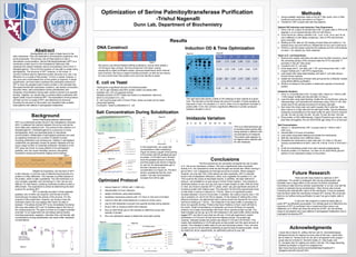
Induction OD & Time Optimization
Time 1 2 3 4 5
2:00 1.73 2.48 2.89 3.13 3.10
12:30 1.25 1.95 2.29 2.66 2.79
11:00 .80 1.39 1.73 2.09 2.17
9:30 .38 .69 .88 1.09 1.16
E. coli vs Yeast
Imidazole Variation
M S 20 30 40 50 60 70 80 100
There are still many factors to optimize in SPT
purification. The protein to detergent ratio during solubulization will
probably have a large effect on the amount of protein solubilized.
According to data found by another experimenter in our lab, only half the
protein is collected during solubulization. Other factors also include
mimicking the hydrophobic nature of the membrane. It may be possible
that the lipids inside the membrane increase SPT activity. We could add
lipids to our buffers during purification to see if we get a higher yield of
protein.
In the end, this research is aimed at being able to
purify SPT as efficiently as possible. Our ultimate goal is to determine the
structure of SPT by purifying it into a crystal and then using x-ray
diffraction on it. When we know the structure of SPT, we can develop
drugs to aid patients who have defects in sphingolipid metabolism due to
improperly functioning SPT.
The right hand side shows a table of OD readings of each sample at a given
time. The dot-blot on the left shows the amount of protein in these samples at
that point in time. For samples 3, 4, and 5, there is not a significant increase in
protein after 12:30. Nor is there a significant difference in protein produced
between samples 4 and 5.
This is an electrophoresis gel
of purified protein eluted after
being washed in different mM
concentrations of imidazole.
The protein yield appears to
be highest when washed with
50 mM imidazole.
Optimized Protocol
The first experiment we conducted compared the use of yeast
or E. coli as our expression medium. We had a significantly lower enzymatic
activity in E. coli compared to what we had thought we should see. When running a
gel on the E. coli, it appeared we had high amounts of protein. When assayed,
however, we only had 10% of the activity we were expecting. SPT is naturally
found in the Endoplasmic Reticulum, which is an organelle not present in E. coli.
This could be the cause of decreased activity. In addition, the best method for
lysing E. coli available to us was using a French Press machine. This machine is
extremely tedious to use, and can only lyse a small sample of cells. With all of this
in mind, we chose to express SPT in yeast, which can give significant amounts of
functional protein with relative ease. The induction OD and time experiments show
us that when a culture is induced at 1 OD/mL, there is no significant protein
increase between 3 and 4.5 hours later. In addition, the culture must be near 1 OD
(as in samples 3,4, and 5) for this to happen. In the interest of making this a time
efficient procedure, we determined that a culture should be induced for four and a
half hours starting at 1 OD/mL. The imidazole in the wash buffer is necessary to
block non-specific binding. Proteins that have Histidine in them could still bind to
the nickel. Small concentrations of imidazole can knock off these non-specific
proteins, but a large concentration will knock off our specific protein as well. The
data indicates we can go up to 50 mM imidazole in the wash buffer without eluting
tagged SPT, and that is now what we will use. In the salt experiment, protein
solubilized in 0.5 M and 0 M salt have the highest activity. The protein gel,
however, indicates double the protein was eluted in 0 M and 0.25 M NaCl. This
means that solubilizing in 0.5 M salt gives us less protein and the same amount of
activity. This indicates a better yield of pure and functional protein. Thus, we have
chosen to use 0.5 M salt while solubilizing to get the bets functional protein. Given
the data from all our experiments, an optimized protocol to your left.
Salt Concentration During Solubilization
0
0.05
0.1
0.15
0.2
0.25
9 14 19 24 29 34 39
mMCoA
Assay Time (minutes)
7/31 Elution Data
Series1
Series2
Series3
Series4
Series5
Series6
Elution 1
Elution 2
Elution 3
Elution 4
Elution 5
Elution 6
•Yeast gives a significant amount of functional protein.
•E. coli on gel indicates near 80% protein eluted, but assay data
indicates only 10% of expected activity.
•Multiple subunits of SPT folded and joined in endoplasmic reticulum,
which is not present in E. coli.
•E. coli must be lysed with a French Press, where as yeast can be lysed
using bead beating
Conclusion: Yeast is preferable to E. coli.
I would like to thank Dr. Jeffrey Harmon and Dr. Somashekarappa
Niranjana Kumari for helping me every step of the way. I would also
like to thank Dr. Teresa Dunn for her aid in all aspects of the project. I
would also lie to thank Dr. Kenneth Gable, Dr. Sita Gupta, and
Dr. Gongshe Han for helping me while in the lab. The image depicting
histidine purification in figure 8 is adapted from
http://www.biochem.arizona.edu/miesfeld/teaching/Bioc471-
2/pages/Lecture5/Lecture5.html
Sphingolipids are a class of lipids found in the
cells membrane. They are essential to all eukaryotic organisms, and
some prokaryotic. The primary role of these lipids is to aid in
intercellular communication. Serine Palmitoyltransferase (SPT) is a
membrane protein found in the Endoplasmic Reticulum that
catalyzes the reaction between serine and palmitoyl CoA to form 3-
ketosphinganine. This is the first reaction on the path to create many
sphingolipids. The structure of SPT, however, is not known. A
common method used to determine protein structure is to use x-ray
diffraction on a crystal of that protein. To form a crystal, however, a
very pure and concentrated form of the protein is required. In these
experiments, we have tried to optimize the purification protocol for
SPT to have the greatest yield of active protein in the shortest time.
We experimented with expression mediums, cell density at induction,
induction times, salt concentration during solubilization and
imidazole concentrations during wash cycles to prevent non-specific
binding. Together, our results help us achieve faster purification with
higher yields of pure functional SPT. The implications of this protocol
will help us form a crystal of SPT, and determine its structure.
Knowing the structure of this protein can hopefully help us better
treat patients with defects in sphingolipid metabolism.
DNA Construct
We want to use Nickel-Histidine affinity purification, and we have added a
10-His tag to help us there. We have fused the LCB 2a3a1 coding
sequences to make purification easier; previous experiments show minimal
loss of activity. We have a copper inducible promoter, so that we can induce
for only a short time if the protein turns out to be harmful to yeast.
1. Induce Yeast at 1 OD/mL with 1 mMCuSO4.
2. Harvest after 4.5 hour induction.
3. Isolate membrane using bead beating.
4. Solubilize membrane proteins with 1% Triton X-100 and 0.5 M NaCl
5. Leave to bind with nickel beads for a minimum of two hours.
6. Use 50 mM imidazole to prevent non-specific binding during washes.
7. Elute 5 250 uL fractions in500 mM Imidazole.
8. Run an SDS-PAGE gel on the samples to determine purity and
quantity of protein.
9. Run non-radioactive assay to determine enzymatic activity.
1. Using available restriction sites on the pET 28a vector, form a DNA
construct such as the one show in in Figure 1.
2. Transform LCB1 Δ yeast cells with this plasmid.
Optimal OD Induction and Induction Time Experiment:
1. Grow a 50 mL culture of transformed LCB 1 Δ yeast cells in YPD at 26
degrees C to an Optical Density (OD) of 0.85 OD/mL.
2. From the 50 mL culture, transfer 2 mL, 4 mL, 6 mL, 8 ml, and 10 mL
into 5 different 4-Liter flasks containing 1 liter of YPD and let them
grow overnight.
3. Starting at 9:30, take an OD reading of each flask and collect a 1 mL
sample every one and half hour. Repeat this for four and a half hours.
4. Perform a dot blot assay using the His antibody and the LCB antibody
on each 1 mL sample you have collected.
Yeast vs E. coli Experiment:
1. Remove the copper-inducible promoter from the DNA construct. Insert
the remaining portion of the construct after the IPTG inducible T7
promoter in the pET 28a vector.
2. Transform AG1 E. coli cells.
3. Grow yeast and E. coli cells until 1 OD, and induce them with 1 mM
Copper Sulfate and 1 mM IPTG respectively.
4. Lyse yeast cells using bead beating, and lyse E. coli cells using a
French Press machine.
5. Isolate the membranes of these cells and purify the LCB2a3a1 protein
using Nickel affinity purification.
6. Perform an assay on the protein to determine quantity of functional
protein.
Imidazole Variation:
1. Grow 1L of transformed LCB 1 Δ yeast cells. Induce at 1 OD/mL with
1 mM Copper Sulfate and harvest after 4.5 hours.
2. Lyse the cells using bead beating, isolate the cell membrane using an
ultracentrifuge, and solubilize the membrane using Triton X-100. Add
nickel resin to the sample and leave for binding overnight.
3. Spin down the nickel resin and split it equally into 8 columns. Wash
each column in a high salt and low salt wash buffer. Use the following
imidazole concentrations in the wash buffer for each of the 8 columns:
20 mM, 30 mM, 40 mM, 50 mM, 60 mM, 70 mM, 80 mM, 100 mM
4. Elute protein in 500 mMImidazole. Collect 6 fractions per column, and
pool them all together. Run an SDS-PAGE gel using 10 uL of the pool.
Salt Concentration:
1. Induce 1 L oftransformed LCB 1 Δ yeast cells at 1 OD/mL with 1
mM CuSO4.
2. Harvest after 4.5 hours of induction
3. Lyse yeast using bead beating and isolate the membranes
4. Split the membrane into 5 different tubes
5. Solubilize the membrane in each tube with 1% Triton X-100 and
varying concentrations of NaCl. Use 0 M, 0.25 M, 0.5 M, 0.75 M and 1
M.
6. Purify the solubilized protein from each sample independently.
7. Elute the protein in 5 fractions, run them on an SDS-PAGE gel and
perform a non-radioactive assay on the protein.
Figure 1
Figure 2a Figure 2b
Figure 4
Figure 5a
Figure 5b
Figure 6
Figure 7
Figure 8
0
0.2
0.4
0.6
0.8
1
1.2
1.4
1.6
0 2 4 6 8 10nmolCoAgeneratedbySPT
Time (min)
0 M y=0.15
.25 M y=.14
.5 M y=.16
.75 M y=.09
1 M y=.07
-0.01
0
0.01
0.02
0.03
0.04
0.05
0 2 4 6 8 10
0M NaCl Basline
Elution 3
Elution 4
Elution 5
Linear (Elution 3)
Linear (Elution 4)
Linear (Elution 5)
-0.02
-0.01
0
0.01
0.02
0.03
0.04
0.05
0 2 4 6 8 10
-CoA .5M .5M
NaCl Baseline
3 Elution
4 Elution
5 Elution
Linear (3 Elution )
In this experiment, we varied salt
concentrations while solubilizing.
Figure 3b shows a graph of the activity
in the elution with the greatest amount
of protein. 0.5 M NaCl and 0 M NaCl
have the greatest amount of activity,
and their graphs are shown in figures
3c and 3d. An electrophoresis gel of
the first 3 elutions for each
concentration is in figure 3a. The third
elution consistently has the most
protein. The salt concentrations
increase from left to right.
Figure 3a
Figure 3b
Figure 3c
Figure 3d