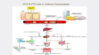
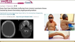
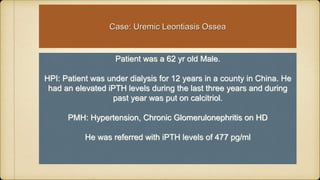
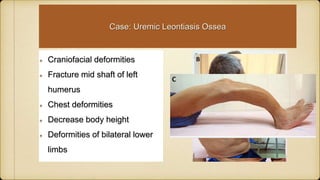
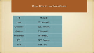
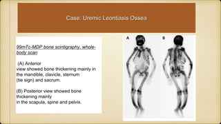
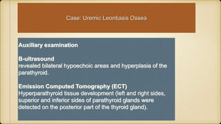
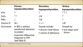
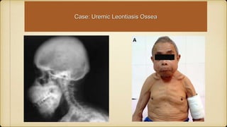
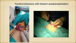
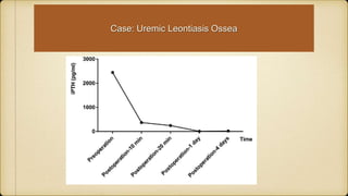
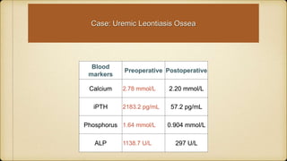
1
Dr Yogendranath Purrunsing
Uremic Leontiasis Ossea
Leontiasis ossea, also known as leontiasis or lion
face, is a form of severe bone remodeling that
prevails in patients with chronic kidney disease
(CKD) and secondary hyperparathyroidism
(SHPT), renal osteodystrophy, Paget diseases,
and fibrous dysplasia, characterized by
craniofacial, ribs, long bones, and spine
deformations.
The famed Rudolf Ludwig Carl Virchow
(1821-1902) a German physician,
anthropologist, pathologist and statesman,
first coined the term leontiasis ossea in
1864.
It is a historical term used to describe a
number of conditions that result in the
affected patient's face resembling that of
a lion. Although it is most frequently
associated withcraniofacial fibrous
dysplasia and has a broader meaning
encompassing other lesions that have
similar appearance.
Robert Kienböck (1871 –1953) was an Austrian
radiologist first mentioned symptomatic forms of
Leotiasis ossea as Osteitis Deformans (Paget’s
Disease) and Osteitis Fibrosa generalisata
(overproduction of Parathyroid glands causing bone
disorders)
In 1953
Cohen et al first described the symptoms
of secondary hyperparathyroidism (SHPT)
in chronic renal failure (CRF) as uremic
leontiasis ossea (ULO), which was
characterized by disfiguring facial
deformity due to progressive bony
overgrowth in the craniomaxillofacial
region, resulting in a lion-like face.
Secondary Hyperparathyroidism
(SHPT)
Secondary hyperparathyroidism (SHPT) is a long-term complication of chronic kidney
disease–mineral and bone disorder (CKD-MBD).
SHPT is characterized by hyperplasia of the parathyroid glands and abnormal secretion
of parathyroid hormones (PTH), calcium and phosphorous metabolic disorders, renal
osteodystrophy, vascular and soft tissue calcification, malnutrition, and other multiple
system complications, which can seriously affect the quality of life of the patient and
increase the risk of cardiovascular disease and mortality rate.
Uremic leontiasis ossea (ULO) is a medical condition only rarely encountered clinically.
Chronic Kidney Disease
CKD is defined as abnormalities of kidney structure or function, present for
>3 months, with implications for health.
CKD requires one of two criteria documented or inferred for >3 months:
either GFR <60 ml/min/1.73 m2 or markers of kidney damage, including
albuminuria, urinary sediment abnormalities.
Pathogenesis of Secondary Hyperparathyroidism
Definition, evaluation, and classification of renal
osteodystrophy: A position statement from Kidney Disease:
Improving Global Outcomes (KDIGO)
It is recommended that
(1) the term renal osteodystrophy be used exclusively to define alterations in bone
morphology associated with CKD, which can be further assessed by histomorphometry
and the results reported based on a unified classification system that includes
parameters of turnover, mineralization and volume.
(2) the term CKD-Mineral and Bone Disorder (CKD-MBD) be used to describe a
broader clinical syndrome that develops as a systemic disorder of mineral and bone
metabolism due to CKD, which is manifested by abnormalities in bone and mineral
metabolism or extra-skeletal calcification. The international adoption of these
recommendations will greatly enhance communication, facilitate clinical decision-
making and promote the evolution of evidence-based clinical practice guidelines
worldwide.
Vit D & PTH role in Calcium homeostasis
SHPT & CKD-MBD
The overproduction of parathyroid
hormone are secondary to
hypocalcemia (due to low levels of
Vit D/CKD) cause hyperplaisia of
parathyroid glands leading to
Secondary Hyperparathyroidism.
Sign and symptoms:
Joint/Bone pain
Deformed limbs
Increase IPTH
Loss of Calcium from bones
Vascular/CVD calcification
Neurological symptoms
Muscle weakness
Depression
Psychomotor depression
Patient was a 62 yr old Male.
HPI: Patient was under dialysis for 12 years in a county in China. He
had an elevated iPTH levels during the last three years and during
past year was put on calcitriol.
PMH: Hypertension, Chronic Glomerulonephritis on HD
He was referred with iPTH levels of 477 pg/ml
Case: Uremic Leontiasis Ossea
Craniofacial deformities
Fracture mid shaft of left
humerus
Chest deformities
Decrease body height
Deformities of bilateral lower
limbs
Case: Uremic Leontiasis Ossea
Hb 11.8 g/dl
Urea 23.79 mmol/L
Creatinine 606.1 mmol/L
Calcium 2.78 mmol/L
Phosphate 1.64mmol/L
iPTH 2183.2 pg/mL
ALP 1138.7 U/L
Case: Uremic Leontiasis Ossea
99mTc-MDP bone scintigraphy, whole-
body scan
(A) Anterior
view showed bone thickening mainly in
the mandible, clavicle, sternum
(tie sign) and sacrum.
(B) Posterior view showed bone
thickening mainly
in the scapula, spine and pelvis.
Case: Uremic Leontiasis Ossea
Auxillary examination
B-ultrasound
revealed bilateral hypoechoic areas and hyperplasia of the
parathyroid.
Emission Computed Tomography (ECT)
Hyperparathyroid tissue development (left and right sides,
superior and inferior sides of parathyroid glands were
detected on the posterior part of the thyroid gland).
What are parathyroid glands?
Parathyroid(PT) glands are 4 small glands
that are located near the thyroid gland in
your neck (size of a pea).
The glands release parathyroid hormone
from chief cells. PT hormones
regulates Ca2+ levels in blood:
A.Bones- PT stimulates release of calcium
from bone into bloodstream.
B.Kidneys- PT hormones reduces loss of
Ca2+ in urine. PT stimulates production of
active Vit D.
C.Intestine- Increase Calcium absorption
from food via Vit D metabolism.
Secondary Hyperparathyroidism
(SHPT)
It was postulated that a retention in phosphate in the extracellular space due to
decrease in GFR and decrease plasma Calcium was the primary event for
pathogenesis of SHPT.
However this hypothesis became less attracted when it was demonstrated that
phosphate level was rarely elevated in early stage of CKD.
Its was found that FGF23 a circulating peptide from bone osteocyte controlling
phosphate levels
Both FGF23 and PTH secretion could correct the high plasma phosphate.
Fibroblast Growth Factor-23 (FGF23)
produced by osteocytes and
osteoblasts:
•Decrease tubular reabsorption of
phosphate.
•Is independent of PTH and inhibits
synthesis of PTH
•Decrease renal synthesis of Calcitriol
Drüeke TB. Hyperparathyroidism in
Chronic Kidney Disease.
Published in Australian family physician
Case: Uremic Leontiasis Ossea
KDIGO 2017 Clinical Practice Guideline Update for the Diagnosis,
Evaluation, Prevention, and Treatment of Chronic Kidney Disease–Mineral
and Bone Disorder (CKD-MBD)
Guidelines
In patients with CKD G3a–G5D:
• Treatments of CKD-MBD should be based on serial assessments of phosphate,
calcium, and PTH levels and individualized to patients.
• We suggest avoidance of hypercalcaemia, maintaining serum calcium below the
upper limit of the reference range.
KDIGO 2017 Clinical Practice Guideline Update for the Diagnosis,
Evaluation, Prevention, and Treatment of Chronic Kidney Disease–Mineral
and Bone Disorder (CKD-MBD)
Guidelines Phosphate
In patients with CKD G3a–G5D:
• A pragmatic and individualized approach to lowering elevated phosphate
levels toward the normal range.
• Decisions about phosphate-lowering treatment should be based on
progressively or persistently elevated serum phosphate.
• We suggest limiting dietary phosphate intake in the treatment of
hyperphosphatemia alone or in combination with other treatments. It is
reasonable to consider phosphate source (e.g., animal, vegetable, additives)
in making dietary recommendations.
KDIGO 2017 Clinical Practice Guideline Update for the Diagnosis,
Evaluation, Prevention, and Treatment of Chronic Kidney Disease–Mineral
and Bone Disorder (CKD-MBD)
Guidelines PTH
In patients with CKD G3a–G5:
1. Patients with levels of intact PTH progressively rising or persistently above
the upper normal limit for the assay be evaluated for modifiable factors,
including hyperphosphatemia, hypocalcemia, high phosphate intake, and
vitamin D deficiency .
2. We suggest that calcitriol and vitamin D analogs not be routinely used. It is
reasonable to reserve the use of calcitriol and vitamin D analogs for patients
with CKD G4–G5 with severe and progressive hyperparathyroidism.
KDIGO 2017 Clinical Practice Guideline Update for the Diagnosis,
Evaluation, Prevention, and Treatment of Chronic Kidney Disease–Mineral
and Bone Disorder (CKD-MBD)
Guidelines PTH
3. In patients with CKD G5D requiring PTH-lowering therapy, we suggest
calcimimetics, calcitriol, or vitamin D analogs, or a combination of
calcimimetics with calcitriol or vitamin D analogs.
4. In patients with CKD G3a–G5D with severe hyperparathyroidism (HPT) who
fail to respond to medical or pharmacological therapy, we suggest
parathyroidectomy.
Parathyroidectomy with forearm autotransplantation
Case: Uremic Leontiasis Ossea
Blood
markers
Preoperative Postoperative
Calcium 2.78 mmol/L 2.20 mmol/L
iPTH 2183.2 pg/mL 57.2 pg/mL
Phosphorus 1.64 mmol/L 0.904 mmol/L
ALP 1138.7 U/L 297 U/L
Uremic patients often also experience cardiopulmonary insufficiency, vascular
calcification, malnutrition, bleeding during the intraoperative and perioperative periods,
infection, arrhythmia, circulatory disorders, and other complications.
In order to improve the safety and effectiveness of TPTX surgery. Preoperative
parathyroid localization tests include ultrasound and ECT is necessary.
Most people have four parathyroid glands. This patient had five, meaning that he had
one supernumerary parathyroid gland.
The reported rate of supernumerary parathyroid glands is around 2.5% to 13%.
Studies have shown that CKD - SHPT patients undergoing TPTX had an incidence of
ectopic parathyroid glands of about 15%, and these glands are mainly distributed
around the thymus, esophageal tissue, carotid sheath, and mediastinum.
Case: Uremic Leontiasis Ossea
ULO is the most dramatic pattern of hyperparathyroidism. Parathyroidectomy is
recommended for patients with severe SHPT refractory to drug therapy.
Presently, guidelines suggest surgical intervention if severe SHPT is persistently
elevated (serum iPTH>800 pg/mL) with hypercalcemia and hyperphosphatemia, and if
the case is resistant to drug treatment.
In conclusion, PTX with autotransplantation is an effective way to treat SHPT caused
by chronic renal failure. It relieves patients from mineral bone disorders, alleviates
vascular calcification, improves bone health and quality of life. However, abnormal
stature caused by vertebral fractures cannot be reversed in cases with excessive
skeletal malformation.
Case: Uremic Leontiasis Ossea
Uremic Leontiaisis Ossea.ppt

Uremic Leontiaisis Ossea.ppt

  • 1.
  • 2.
    Uremic Leontiasis Ossea Leontiasisossea, also known as leontiasis or lion face, is a form of severe bone remodeling that prevails in patients with chronic kidney disease (CKD) and secondary hyperparathyroidism (SHPT), renal osteodystrophy, Paget diseases, and fibrous dysplasia, characterized by craniofacial, ribs, long bones, and spine deformations.
  • 3.
    The famed RudolfLudwig Carl Virchow (1821-1902) a German physician, anthropologist, pathologist and statesman, first coined the term leontiasis ossea in 1864. It is a historical term used to describe a number of conditions that result in the affected patient's face resembling that of a lion. Although it is most frequently associated withcraniofacial fibrous dysplasia and has a broader meaning encompassing other lesions that have similar appearance.
  • 4.
    Robert Kienböck (1871–1953) was an Austrian radiologist first mentioned symptomatic forms of Leotiasis ossea as Osteitis Deformans (Paget’s Disease) and Osteitis Fibrosa generalisata (overproduction of Parathyroid glands causing bone disorders)
  • 5.
    In 1953 Cohen etal first described the symptoms of secondary hyperparathyroidism (SHPT) in chronic renal failure (CRF) as uremic leontiasis ossea (ULO), which was characterized by disfiguring facial deformity due to progressive bony overgrowth in the craniomaxillofacial region, resulting in a lion-like face.
  • 6.
    Secondary Hyperparathyroidism (SHPT) Secondary hyperparathyroidism(SHPT) is a long-term complication of chronic kidney disease–mineral and bone disorder (CKD-MBD). SHPT is characterized by hyperplasia of the parathyroid glands and abnormal secretion of parathyroid hormones (PTH), calcium and phosphorous metabolic disorders, renal osteodystrophy, vascular and soft tissue calcification, malnutrition, and other multiple system complications, which can seriously affect the quality of life of the patient and increase the risk of cardiovascular disease and mortality rate. Uremic leontiasis ossea (ULO) is a medical condition only rarely encountered clinically.
  • 7.
    Chronic Kidney Disease CKDis defined as abnormalities of kidney structure or function, present for >3 months, with implications for health. CKD requires one of two criteria documented or inferred for >3 months: either GFR <60 ml/min/1.73 m2 or markers of kidney damage, including albuminuria, urinary sediment abnormalities.
  • 10.
    Pathogenesis of SecondaryHyperparathyroidism
  • 11.
    Definition, evaluation, andclassification of renal osteodystrophy: A position statement from Kidney Disease: Improving Global Outcomes (KDIGO) It is recommended that (1) the term renal osteodystrophy be used exclusively to define alterations in bone morphology associated with CKD, which can be further assessed by histomorphometry and the results reported based on a unified classification system that includes parameters of turnover, mineralization and volume. (2) the term CKD-Mineral and Bone Disorder (CKD-MBD) be used to describe a broader clinical syndrome that develops as a systemic disorder of mineral and bone metabolism due to CKD, which is manifested by abnormalities in bone and mineral metabolism or extra-skeletal calcification. The international adoption of these recommendations will greatly enhance communication, facilitate clinical decision- making and promote the evolution of evidence-based clinical practice guidelines worldwide.
  • 13.
    Vit D &PTH role in Calcium homeostasis
  • 15.
    SHPT & CKD-MBD Theoverproduction of parathyroid hormone are secondary to hypocalcemia (due to low levels of Vit D/CKD) cause hyperplaisia of parathyroid glands leading to Secondary Hyperparathyroidism. Sign and symptoms: Joint/Bone pain Deformed limbs Increase IPTH Loss of Calcium from bones Vascular/CVD calcification Neurological symptoms Muscle weakness Depression Psychomotor depression
  • 17.
    Patient was a62 yr old Male. HPI: Patient was under dialysis for 12 years in a county in China. He had an elevated iPTH levels during the last three years and during past year was put on calcitriol. PMH: Hypertension, Chronic Glomerulonephritis on HD He was referred with iPTH levels of 477 pg/ml Case: Uremic Leontiasis Ossea
  • 18.
    Craniofacial deformities Fracture midshaft of left humerus Chest deformities Decrease body height Deformities of bilateral lower limbs Case: Uremic Leontiasis Ossea
  • 19.
    Hb 11.8 g/dl Urea23.79 mmol/L Creatinine 606.1 mmol/L Calcium 2.78 mmol/L Phosphate 1.64mmol/L iPTH 2183.2 pg/mL ALP 1138.7 U/L Case: Uremic Leontiasis Ossea
  • 20.
    Case: Uremic LeontiasisOssea Case: Uremic Leontiasis Ossea
  • 21.
  • 22.
  • 23.
    99mTc-MDP bone scintigraphy,whole- body scan (A) Anterior view showed bone thickening mainly in the mandible, clavicle, sternum (tie sign) and sacrum. (B) Posterior view showed bone thickening mainly in the scapula, spine and pelvis. Case: Uremic Leontiasis Ossea
  • 24.
    Case: Uremic LeontiasisOssea Auxillary examination B-ultrasound revealed bilateral hypoechoic areas and hyperplasia of the parathyroid. Emission Computed Tomography (ECT) Hyperparathyroid tissue development (left and right sides, superior and inferior sides of parathyroid glands were detected on the posterior part of the thyroid gland).
  • 25.
    What are parathyroidglands? Parathyroid(PT) glands are 4 small glands that are located near the thyroid gland in your neck (size of a pea). The glands release parathyroid hormone from chief cells. PT hormones regulates Ca2+ levels in blood: A.Bones- PT stimulates release of calcium from bone into bloodstream. B.Kidneys- PT hormones reduces loss of Ca2+ in urine. PT stimulates production of active Vit D. C.Intestine- Increase Calcium absorption from food via Vit D metabolism.
  • 26.
    Secondary Hyperparathyroidism (SHPT) It waspostulated that a retention in phosphate in the extracellular space due to decrease in GFR and decrease plasma Calcium was the primary event for pathogenesis of SHPT. However this hypothesis became less attracted when it was demonstrated that phosphate level was rarely elevated in early stage of CKD. Its was found that FGF23 a circulating peptide from bone osteocyte controlling phosphate levels Both FGF23 and PTH secretion could correct the high plasma phosphate.
  • 27.
    Fibroblast Growth Factor-23(FGF23) Fibroblast Growth Factor-23 (FGF23) produced by osteocytes and osteoblasts: •Decrease tubular reabsorption of phosphate. •Is independent of PTH and inhibits synthesis of PTH •Decrease renal synthesis of Calcitriol Drüeke TB. Hyperparathyroidism in Chronic Kidney Disease.
  • 29.
    Published in Australianfamily physician
  • 30.
  • 31.
    KDIGO 2017 ClinicalPractice Guideline Update for the Diagnosis, Evaluation, Prevention, and Treatment of Chronic Kidney Disease–Mineral and Bone Disorder (CKD-MBD) Guidelines In patients with CKD G3a–G5D: • Treatments of CKD-MBD should be based on serial assessments of phosphate, calcium, and PTH levels and individualized to patients. • We suggest avoidance of hypercalcaemia, maintaining serum calcium below the upper limit of the reference range.
  • 32.
    KDIGO 2017 ClinicalPractice Guideline Update for the Diagnosis, Evaluation, Prevention, and Treatment of Chronic Kidney Disease–Mineral and Bone Disorder (CKD-MBD) Guidelines Phosphate In patients with CKD G3a–G5D: • A pragmatic and individualized approach to lowering elevated phosphate levels toward the normal range. • Decisions about phosphate-lowering treatment should be based on progressively or persistently elevated serum phosphate. • We suggest limiting dietary phosphate intake in the treatment of hyperphosphatemia alone or in combination with other treatments. It is reasonable to consider phosphate source (e.g., animal, vegetable, additives) in making dietary recommendations.
  • 33.
    KDIGO 2017 ClinicalPractice Guideline Update for the Diagnosis, Evaluation, Prevention, and Treatment of Chronic Kidney Disease–Mineral and Bone Disorder (CKD-MBD) Guidelines PTH In patients with CKD G3a–G5: 1. Patients with levels of intact PTH progressively rising or persistently above the upper normal limit for the assay be evaluated for modifiable factors, including hyperphosphatemia, hypocalcemia, high phosphate intake, and vitamin D deficiency . 2. We suggest that calcitriol and vitamin D analogs not be routinely used. It is reasonable to reserve the use of calcitriol and vitamin D analogs for patients with CKD G4–G5 with severe and progressive hyperparathyroidism.
  • 34.
    KDIGO 2017 ClinicalPractice Guideline Update for the Diagnosis, Evaluation, Prevention, and Treatment of Chronic Kidney Disease–Mineral and Bone Disorder (CKD-MBD) Guidelines PTH 3. In patients with CKD G5D requiring PTH-lowering therapy, we suggest calcimimetics, calcitriol, or vitamin D analogs, or a combination of calcimimetics with calcitriol or vitamin D analogs. 4. In patients with CKD G3a–G5D with severe hyperparathyroidism (HPT) who fail to respond to medical or pharmacological therapy, we suggest parathyroidectomy.
  • 35.
    Parathyroidectomy with forearmautotransplantation
  • 36.
  • 37.
    Case: Uremic LeontiasisOssea Blood markers Preoperative Postoperative Calcium 2.78 mmol/L 2.20 mmol/L iPTH 2183.2 pg/mL 57.2 pg/mL Phosphorus 1.64 mmol/L 0.904 mmol/L ALP 1138.7 U/L 297 U/L
  • 38.
    Uremic patients oftenalso experience cardiopulmonary insufficiency, vascular calcification, malnutrition, bleeding during the intraoperative and perioperative periods, infection, arrhythmia, circulatory disorders, and other complications. In order to improve the safety and effectiveness of TPTX surgery. Preoperative parathyroid localization tests include ultrasound and ECT is necessary. Most people have four parathyroid glands. This patient had five, meaning that he had one supernumerary parathyroid gland. The reported rate of supernumerary parathyroid glands is around 2.5% to 13%. Studies have shown that CKD - SHPT patients undergoing TPTX had an incidence of ectopic parathyroid glands of about 15%, and these glands are mainly distributed around the thymus, esophageal tissue, carotid sheath, and mediastinum. Case: Uremic Leontiasis Ossea
  • 39.
    ULO is themost dramatic pattern of hyperparathyroidism. Parathyroidectomy is recommended for patients with severe SHPT refractory to drug therapy. Presently, guidelines suggest surgical intervention if severe SHPT is persistently elevated (serum iPTH>800 pg/mL) with hypercalcemia and hyperphosphatemia, and if the case is resistant to drug treatment. In conclusion, PTX with autotransplantation is an effective way to treat SHPT caused by chronic renal failure. It relieves patients from mineral bone disorders, alleviates vascular calcification, improves bone health and quality of life. However, abnormal stature caused by vertebral fractures cannot be reversed in cases with excessive skeletal malformation. Case: Uremic Leontiasis Ossea

Editor's Notes

  • #6 The scout film demonstrates overgrowth of the jaw, in particular the maxilla, with classical salt and pepper sign (arrow); found as multiple tiny well-defined calvarial lucencies in background of sclerotic ground-glass appearance associated with indistinct inner and outer tables
  • #9 The KDIGO classification for CKD is new. "CKD is classified based on cause, GFR category, and albuminuria category (CGA)." Previously CKD was classified into 5 stages according to GFR alone, but guidelines for evaluation and treatment had specifically recommended ascertaining the cause of disease and level of albuminuria or proteinuria. • GFR category 3 (30-59 ml/min/1.73 m2) is subdivided into 3a (45-59 ml/min/1.73 m2) and 3b (30-44 ml/min/1.73 m2) because of the large number of people in this category (6.2% of US adults, 12.4 million), and the generally worse prognosis of patients in category 3b vs. 3a. Previous guidelines directed toward patients with CKD stages 1-5 remain applicable for patients with GFR categories 1-5. • The inclusion of albuminuria for all GFR categories recognizes the importance of albuminuria in defining risk and individualizing treatment. The three categories correspond to albumin-to-creatinine ratio (ACR) <30, 30-300, and >300 mg/g, corresponding to what was previously referred to normoalbuminuria, microalbuminuria and macroalbuminuria (these terms are no longer used).
  • #11 In CKD , phosphate levels increase and patient become hypocalcemia. Each of these input on PT gland increase PTH production iPTH is secreted by chief cells in PT. As it progresses (CKD) , both Vitamin D and Ca sensing receptor system are down regulated and PT glands become enlarged.
  • #13 The term ‘renal osteodystrophy’ has recently been replaced by ‘CKD-mineral and bone disorder (CKD-MBD)’, which includes abnormalities in bone and mineral metabolism and vascular calcification. Disturbances in mineral and bone metabolism have a critical role in the pathogenesis of cardiovascular complications in patients with chronic kidney disease (CKD).
  • #15 Abnormalities of mineral metabolism occur in early ckd Blood Calcium/phosphorus/PTH/ Vit D was drawn There was a relative stability of Serum Ca2+/PO/IPHT until Gfr <20 ml/min/1.73 m2 Decrease in 1,25 OH2 D3 at a GFR level < 30 ml/min/1.73m2
  • #18 Pico gram/ml
  • #25 Uncontrolled SHPT causes complications such as bone remodeling, skeletal turnover, cardiovascular disturbances,metabolic imbalances. Tertiary hyperparathyroidism-(THPT), first described in 1963, is classically defined as persistent hyperparathyroidism with hypercalcemia due to either prolonged SHPT or autonomous hypersecretion of one or more parathyroid glands after kidney transplantation. Therefore, the term THPT can also be used to describe this patient. In this case, the patient experienced facial and whole-body remodeling with fracture of the long bone occurring without any traumatic events because of severe SHPT.
  • #26 Parathyroid hormone regulates calcium levels in the blood, largely by increasing the levels when they are too low. It does this through its actions on the kidneys, bones and intestine.
  • #29 As CKD progresses, high phosphate levels/ low calcium/calcitriol/increase uremia contribute to synthesis of PTH. CKD FGF23’s effect is partially or even completely abolished owing to downregulation of the expression of its receptor and co-receptor Klotho. This figure shows the progression from polyclonal to monoclonal parathyroid hyperplasia and corresponds to sonographic features Alpha-klotho is found in tubular epithelial cells in DCT and together with FGF23 they decrease reabsorption of phosphate.
  • #31 This patient had all the complications of uncontrolled SHPT complications such as whole body bone remodeling, skeletal turnover with fractures of long bone without any traumatic event, cardiovascular disturbances, metabolic imbalances. Tertiary hyperparathyroidism-(THPT), first described in 1963, is classically defined as persistent hyperparathyroidism with hypercalcemia due to either prolonged SHPT or autonomous hyper secretion of one or more parathyroid glands after kidney transplantation. Therefore, the term THPT can also be used to describe this patient.
  • #36 Researchers found that individuals who were under hemodialysis for 10 and 20 years had a prevalence of parathyroisdectomy with forearm autotransplantation of 10% and 30%.
  • #37 The patient was evaluated and PTX was proposed according to the Kidney Disease Improving Global Outcomes (KDIGO) guidelines; when a CKD patient with SHPT is experiencing biochemical, radiological, and cardiovascular irregularities and does not respond to medical/pharmacological therapy, parathyroidectomy is recommended. Blood iPTH was measure pre and postoperatively
  • #38 iPTH ;10-55 pg/ml
  • #39 All of these glands must be removed from the neck to ensure the efficacy of the surgery.