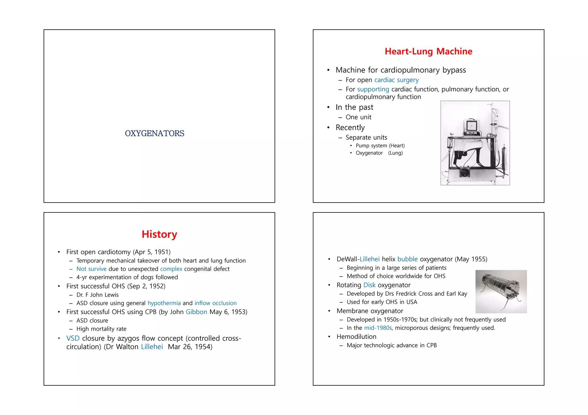
OXYGENATORS
Heart-Lung Machine
• Machine for cardiopulmonary bypass
– For open cardiac surgery
– For supporting cardiac function, pulmonary function, or
cardiopulmonary function
• In the past
– One unit
• Recently
– Separate units
• Pump system (Heart)
• Oxygenator (Lung)
History
• First open cardiotomy (Apr 5, 1951)
– Temporary mechanical takeover of both heart and lung function
– Not survive due to unexpected complex congenital defect
– 4-yr experimentation of dogs followed
• First successful OHS (Sep 2, 1952)
– Dr. F John Lewis
– ASD closure using general hypothermia and inflow occlusion
• First successful OHS using CPB (by John Gibbon May 6, 1953)
– ASD closure
– High mortality rate
• VSD closure by azygos flow concept (controlled cross-
circulation) (Dr Walton Lillehei Mar 26, 1954)
• DeWall-Lillehei helix bubble oxygenator (May 1955)
– Beginning in a large series of patients
– Method of choice worldwide for OHS
• Rotating Disk oxygenator
– Developed by Drs Fredrick Cross and Earl Kay
– Used for early OHS in USA
• Membrane oxygenator
– Developed in 1950s-1970s; but clinically not frequently used
– In the mid-1980s, microporous designs; frequently used.
• Hemodilution
– Major technologic advance in CPB
Cardiopulmonary Bypass
• Goals
1. Still, bloodless heart for cardiac surgery
2. Replacement of cardiac and pulmonary function
Functions of CPB
• Respiration
• Ventilation
• Oxygenation
• Circulation
• Venous drainage (by gravity, centrifugal pump, or negative
pressure)
• Arterial inflow
• Temperature regulation (hypothermia)
• Low blood flow -> decreased blood trauma
• Decreased body metabolism
Components of CPB
• Total CPB
• Partial CPB
• Integral Components of Extracorporeal Circuit
– Pumps
– Oxygenator
– Heat exchanger
– Arterial filter
– Cardioplegic delivery system
– Cannulae (aortic; arterial; vena caval)
– Suction and vent
Basic CPB circuit with oxygenator and
centrifugal pump
Typical CPB Circuit Pumps
• Two principle types
– Displacement pumps
• Roller pump
• Non occlusive roller pumps
– Rotatory pumps
• Radial (centrifugal) pumps
• Axial pumps (Archimedes’ screw)
• Diagonal pumps
Roller Pumps
• Most commonly used
• Volume Displacement
• Non pulsatile blood flow
• Used for
• Forward flow
• Cardioplegic delivery
• LV vent suction
Roller Pumps
• Flow determined
– Tubing diameter, roller RPM, length of tubing in contact with rollers
• Proper set occlusion for minimal hemolysis
• 100% occlusion in cardioplegia and vent pumps
– Full occlusion -> hemolysis
• Larger tubing and lesser rotations cause minimal hemolysis.
– High RPM and fully occlusive setting -> hemolysis
• Tubing spallation cause microemboli
• Easily pump air
• Resistance = resistance of tubing + oxygenator + heat exchanger
+ filter + aortic cannula + SVR
• Line pressure depends on SVR and pump flow rate
• Pressure limit = 150-350 mmHg ( >250 mmHg seldom accepted)
Nonocclusive Roller Pumps
• Flat compliant tubing placed over the rollers
• Positive pressure at the inlet to fill the tubing
• Unlikely microair emboli
• Require use of an in-line flowmeter
Radial (Centrifugal) Pumps
• Impeller spinning within a rigid housing
– Creates regions of lower and higher pressure
– Blood moved from inlet to outlet
• No spallation with rigid housing
• Very dependent on afterload
• Nonocclusive
– Permit back-bleeding
– Require occlusive device
• Reqiure use of in-line flow meter
Axial / Diagonal Pumps
• Axial pumps
– Low internal volume, high-velocity axial impeller
– Currently best suited for ventricular assist application
• Diagonal pumps
– Very similar to centrifugal pump in design and application
Differences of Rotatory Pumps
Alternate Classification of Pumps
a. Roller pumps
b. Impeller pumps (Impeller >)
c. Centrifugal pumps (Cone >)
Centrifugal pumps > Roller pumps
• Long-term CPB
• In high-risk angioplasty patients
• Ventricular assistance
• Neonatal ECMO
• Centrifugal pumps
– Biomedicus Biopump (Medtronic Inc)
– Sarns/3M centrifugal pump (Terumo)
– Levitronix CentriMag blood pump
• LVAD, RVAD, Bi VAD
• BiVAD + oxygenator in RVAD = ECMO
Pulsatile Perfusion
• Significant physiologic advantages
• Diastolic run-off
• Stimulation of the endothelium
• Problem
• Noncompliant high resistance CPB circuit
• High flow with resultant shear stress
• Hemolysis
• Possible with roller pump and diagonal pump, but not with
centrifugal pump
• Requires larger bore arterial cannulas
• Alternative method for generating pulsatile flow in high-risk
patients
• Use of IABP during CPB
• Additional cost and invasiveness
Oxygenator
• Limited reserve for gas transfer vs. natural lung
• Much smaller surface
• Limited by diffusion
• Types of oxygenator
• Disk oxygenator
• Bubble oxygenator
• Membrane oxygenator
• Maximum oxygen transfer
• Less than 25% that of normal lung
• Proportional to partial pressure difference and surface area,
inversely to diffusion distance
Disk or bubble oxygenator
• Direct contact oxygenators
• Bubbles in direct contact with blood
• Increasing cellular trauma
Bubble oxygenator
• Bubble oxygenator
• Larger bubbles improve removal of CO2
• Smaller bubbles are very efficient at oxygenation but poor in CO2
removal
• Larger the No. of bubbles, Greater the efficiency of the oxygenator
Deforming Chamber of Bubble Oxygenator
• Deforming the frothy blood
• Large surface area coated with silicone
– Increased surface tension of bubbles -> causing them to burst
Bubble Oxygenator
• Advantage
– Easy to assemble
– Relatively small priming volume
– Deforming the frothy blood
– Low cost
• Disadvantages
– Micro emboli
– Blood cell trauma
– Destruction of plasma protein
– Excessive removal of CO2
– Deforming capacity exhausted
Membrane Oxygenator
• Charateristics
– Gas exchange across a thin membrane
– No need in direct contact with blood and no need for
deformer; so more physiologic
– Minimal blood damage
• Two types
– Solid type (Silicone)
– Microporous type (polypropylene)
• 0.3-0.8-um pores
• Most popular design = hollow fibers (120-200 um)
Membrane Oxygenator
• Microporous / Hollow fibers
Microporous (Polypropylene) Membrane
Oxygenator
• Currently predominant design used for CPB
• Micropores
– Less than 1.0 um in diameter
– Initially porous, but plasma protein coating the membrane-gas
interface
– Surface tension of blood prevent gas leakage into the blood
phase
– Conduit for O2 and CO2 exchange
• Problems
– Plasma leakage and membrane wet at use of period > 24
hours
Silicone Membrane Oxygenator
• True membrane oxygenator
• Silicone polymer
– Improved biocompatibility -> long-term support
– The 1980s-the mid-1990s
– Still the membrane of choice for long-term procedures
• ECLS/ECMO
• Problems
– Gas exchnage inferior to that of polypropylene (microporous)
oxygenator
• Need greater surface area and larger prime volume
– Difficult in manufacturing and quality control
New Generation Membrane Oxygenator
• Silicone polymer
• A continuous sheet of silicone membrane rolled into a
coil
– Manufactured by Medtronic Cardiopulmonary Inc.
– Membrane surface area + 0.6-4.5 M2
– Most common use for ECLS/ECMO
Heat Exchanger
• Intergrated into oxygenator for warming and cooling of the blood
stream
• Exchange surface made of
– Stainless steel, aluminium, or polypropylene
• Counter-current mechanism
• Temperature difference between waterside and blood side
– Historic reports : maximum difference of 10 °C
– Recent recommendation : 6 °C and longer rewarming times
• To improve neurocognitive outcome
• Hyperthermic circulatory temperature
– Blood damage (protein denaturation
– Limit absolute maximum temperature (42 °C) in blood
Circuits
• Venous drainage by gravity into oxygenator
– Height difference between venae cavae and oxygenator > 20-30 cm
• Mechanical suction Not desirable
– Entrain air
– Suck the vena cava walls against the cannula orifices
• Arterial blood return to the systemic circulation under pressure
Size of cannula
Adult Children
• SVC (1/3 of total flow) 28 24
• IVC (2/3 of total flow) 36 28
• Example: 1.8 m2 patient
– Total flow 5.4 l/min
– SVC 1.8 l/min, IVC 3.6 l/min
– SVC > 30 Fr, IVC > 34 Fr : Single cannula > 38 Fr
– 36-51 Fr cannula required.
Arterial Return
• Ascending aorta just proximal to inniminate artery
• Femoral artery access in
• Dissecting aortic aneurysm (0.2-3%)
• Reoperation
• Emergency
• Problems of femoral cannulation (more than ascending aorta
cannulation)
• Sepsis
• Formation of false aneurysm
• Development of lymphatic fistula
• Arterial cannula
• The narrowest part of CPB circuit
• Should be as short as possible
• As large as the diameter of vessel permits
• < 100 mmHg in full CBP flow
Arterial Cannula
• Long or diffuse-tipped cannula
• Minimize risk of dislodgement of atheroma in the ascending or transverse
aorta
• Axillary –subclavian artery, innimonate artery, LV apex
• In special circumstances
• Limitations and more complications
• Dissection of aorta
• All sites of arterial cannulation
• Prompt recognition and surgical correction
• TEE helpful for diagnosis
Other circuits
• Tubing sizes and lengths and connectors
• Should minmize blood velocity and priming volume
• Search for better biomaterials
• Cardiotomy suction
• Major source of microemboli and activated blood (humeral and cellular)
• Minimize amount, substition by cell salvage
• Cell processed blood may pose hazards
• Hemoconcentrator
• During and after CPB
• Removal of plasma and raising of Hct
• More cost effective than cell salvage and washing devices
Prime Fluid
• Ideally close to ECF
• Whole blood not used
• Homologous blood syndrome
• Postperfusion bleeding diathesis
• Incompatibility reaction
• Demand on blood banks
• Advantages of hemodilution
• Lower blood viscosity
• Improve microcirculation
• Counteract the increased viscosity by hypothermia
• Risk of hemodilution
• Decreased viscosity : SVR decreased
• Low oncotic pressure
• O2 carrying
• Coagulation factor
Composition of Prime
• Average 1,500-2,000ml
• Hct 20- 25%
• Example
• Balanced salt sol. RL 1250 ml
• Osmotically active agent (Mannitol, Dextran 40, Hexastarch)
100 ml
• NaHCO3 50 ml
• KCL 10 ml
• Heparin 1 ml
• CPB for cardiac surgery
• ECMO for ECLS
• ECMO for supporting cardio/pulmonary function
• VAD for supporting cardiac function
– RVAD; LVAD; Bi VAD
– BiVAD + oxygenator in RVAD = ECMO

Unit 1.pdf

  • 1.
    OXYGENATORS Heart-Lung Machine • Machinefor cardiopulmonary bypass – For open cardiac surgery – For supporting cardiac function, pulmonary function, or cardiopulmonary function • In the past – One unit • Recently – Separate units • Pump system (Heart) • Oxygenator (Lung) History • First open cardiotomy (Apr 5, 1951) – Temporary mechanical takeover of both heart and lung function – Not survive due to unexpected complex congenital defect – 4-yr experimentation of dogs followed • First successful OHS (Sep 2, 1952) – Dr. F John Lewis – ASD closure using general hypothermia and inflow occlusion • First successful OHS using CPB (by John Gibbon May 6, 1953) – ASD closure – High mortality rate • VSD closure by azygos flow concept (controlled cross- circulation) (Dr Walton Lillehei Mar 26, 1954) • DeWall-Lillehei helix bubble oxygenator (May 1955) – Beginning in a large series of patients – Method of choice worldwide for OHS • Rotating Disk oxygenator – Developed by Drs Fredrick Cross and Earl Kay – Used for early OHS in USA • Membrane oxygenator – Developed in 1950s-1970s; but clinically not frequently used – In the mid-1980s, microporous designs; frequently used. • Hemodilution – Major technologic advance in CPB
  • 2.
    Cardiopulmonary Bypass • Goals 1.Still, bloodless heart for cardiac surgery 2. Replacement of cardiac and pulmonary function Functions of CPB • Respiration • Ventilation • Oxygenation • Circulation • Venous drainage (by gravity, centrifugal pump, or negative pressure) • Arterial inflow • Temperature regulation (hypothermia) • Low blood flow -> decreased blood trauma • Decreased body metabolism Components of CPB • Total CPB • Partial CPB • Integral Components of Extracorporeal Circuit – Pumps – Oxygenator – Heat exchanger – Arterial filter – Cardioplegic delivery system – Cannulae (aortic; arterial; vena caval) – Suction and vent Basic CPB circuit with oxygenator and centrifugal pump
  • 3.
    Typical CPB CircuitPumps • Two principle types – Displacement pumps • Roller pump • Non occlusive roller pumps – Rotatory pumps • Radial (centrifugal) pumps • Axial pumps (Archimedes’ screw) • Diagonal pumps Roller Pumps • Most commonly used • Volume Displacement • Non pulsatile blood flow • Used for • Forward flow • Cardioplegic delivery • LV vent suction Roller Pumps • Flow determined – Tubing diameter, roller RPM, length of tubing in contact with rollers • Proper set occlusion for minimal hemolysis • 100% occlusion in cardioplegia and vent pumps – Full occlusion -> hemolysis • Larger tubing and lesser rotations cause minimal hemolysis. – High RPM and fully occlusive setting -> hemolysis • Tubing spallation cause microemboli • Easily pump air • Resistance = resistance of tubing + oxygenator + heat exchanger + filter + aortic cannula + SVR • Line pressure depends on SVR and pump flow rate • Pressure limit = 150-350 mmHg ( >250 mmHg seldom accepted)
  • 4.
    Nonocclusive Roller Pumps •Flat compliant tubing placed over the rollers • Positive pressure at the inlet to fill the tubing • Unlikely microair emboli • Require use of an in-line flowmeter Radial (Centrifugal) Pumps • Impeller spinning within a rigid housing – Creates regions of lower and higher pressure – Blood moved from inlet to outlet • No spallation with rigid housing • Very dependent on afterload • Nonocclusive – Permit back-bleeding – Require occlusive device • Reqiure use of in-line flow meter Axial / Diagonal Pumps • Axial pumps – Low internal volume, high-velocity axial impeller – Currently best suited for ventricular assist application • Diagonal pumps – Very similar to centrifugal pump in design and application Differences of Rotatory Pumps
  • 5.
    Alternate Classification ofPumps a. Roller pumps b. Impeller pumps (Impeller >) c. Centrifugal pumps (Cone >) Centrifugal pumps > Roller pumps • Long-term CPB • In high-risk angioplasty patients • Ventricular assistance • Neonatal ECMO • Centrifugal pumps – Biomedicus Biopump (Medtronic Inc) – Sarns/3M centrifugal pump (Terumo) – Levitronix CentriMag blood pump • LVAD, RVAD, Bi VAD • BiVAD + oxygenator in RVAD = ECMO Pulsatile Perfusion • Significant physiologic advantages • Diastolic run-off • Stimulation of the endothelium • Problem • Noncompliant high resistance CPB circuit • High flow with resultant shear stress • Hemolysis • Possible with roller pump and diagonal pump, but not with centrifugal pump • Requires larger bore arterial cannulas • Alternative method for generating pulsatile flow in high-risk patients • Use of IABP during CPB • Additional cost and invasiveness Oxygenator • Limited reserve for gas transfer vs. natural lung • Much smaller surface • Limited by diffusion • Types of oxygenator • Disk oxygenator • Bubble oxygenator • Membrane oxygenator • Maximum oxygen transfer • Less than 25% that of normal lung • Proportional to partial pressure difference and surface area, inversely to diffusion distance
  • 6.
    Disk or bubbleoxygenator • Direct contact oxygenators • Bubbles in direct contact with blood • Increasing cellular trauma Bubble oxygenator • Bubble oxygenator • Larger bubbles improve removal of CO2 • Smaller bubbles are very efficient at oxygenation but poor in CO2 removal • Larger the No. of bubbles, Greater the efficiency of the oxygenator
  • 7.
    Deforming Chamber ofBubble Oxygenator • Deforming the frothy blood • Large surface area coated with silicone – Increased surface tension of bubbles -> causing them to burst Bubble Oxygenator • Advantage – Easy to assemble – Relatively small priming volume – Deforming the frothy blood – Low cost • Disadvantages – Micro emboli – Blood cell trauma – Destruction of plasma protein – Excessive removal of CO2 – Deforming capacity exhausted Membrane Oxygenator • Charateristics – Gas exchange across a thin membrane – No need in direct contact with blood and no need for deformer; so more physiologic – Minimal blood damage • Two types – Solid type (Silicone) – Microporous type (polypropylene) • 0.3-0.8-um pores • Most popular design = hollow fibers (120-200 um)
  • 8.
    Membrane Oxygenator • Microporous/ Hollow fibers Microporous (Polypropylene) Membrane Oxygenator • Currently predominant design used for CPB • Micropores – Less than 1.0 um in diameter – Initially porous, but plasma protein coating the membrane-gas interface – Surface tension of blood prevent gas leakage into the blood phase – Conduit for O2 and CO2 exchange • Problems – Plasma leakage and membrane wet at use of period > 24 hours Silicone Membrane Oxygenator • True membrane oxygenator • Silicone polymer – Improved biocompatibility -> long-term support – The 1980s-the mid-1990s – Still the membrane of choice for long-term procedures • ECLS/ECMO • Problems – Gas exchnage inferior to that of polypropylene (microporous) oxygenator • Need greater surface area and larger prime volume – Difficult in manufacturing and quality control New Generation Membrane Oxygenator • Silicone polymer • A continuous sheet of silicone membrane rolled into a coil – Manufactured by Medtronic Cardiopulmonary Inc. – Membrane surface area + 0.6-4.5 M2 – Most common use for ECLS/ECMO
  • 9.
    Heat Exchanger • Intergratedinto oxygenator for warming and cooling of the blood stream • Exchange surface made of – Stainless steel, aluminium, or polypropylene • Counter-current mechanism • Temperature difference between waterside and blood side – Historic reports : maximum difference of 10 °C – Recent recommendation : 6 °C and longer rewarming times • To improve neurocognitive outcome • Hyperthermic circulatory temperature – Blood damage (protein denaturation – Limit absolute maximum temperature (42 °C) in blood Circuits • Venous drainage by gravity into oxygenator – Height difference between venae cavae and oxygenator > 20-30 cm • Mechanical suction Not desirable – Entrain air – Suck the vena cava walls against the cannula orifices • Arterial blood return to the systemic circulation under pressure Size of cannula Adult Children • SVC (1/3 of total flow) 28 24 • IVC (2/3 of total flow) 36 28 • Example: 1.8 m2 patient – Total flow 5.4 l/min – SVC 1.8 l/min, IVC 3.6 l/min – SVC > 30 Fr, IVC > 34 Fr : Single cannula > 38 Fr – 36-51 Fr cannula required. Arterial Return • Ascending aorta just proximal to inniminate artery • Femoral artery access in • Dissecting aortic aneurysm (0.2-3%) • Reoperation • Emergency • Problems of femoral cannulation (more than ascending aorta cannulation) • Sepsis • Formation of false aneurysm • Development of lymphatic fistula • Arterial cannula • The narrowest part of CPB circuit • Should be as short as possible • As large as the diameter of vessel permits • < 100 mmHg in full CBP flow
  • 10.
    Arterial Cannula • Longor diffuse-tipped cannula • Minimize risk of dislodgement of atheroma in the ascending or transverse aorta • Axillary –subclavian artery, innimonate artery, LV apex • In special circumstances • Limitations and more complications • Dissection of aorta • All sites of arterial cannulation • Prompt recognition and surgical correction • TEE helpful for diagnosis Other circuits • Tubing sizes and lengths and connectors • Should minmize blood velocity and priming volume • Search for better biomaterials • Cardiotomy suction • Major source of microemboli and activated blood (humeral and cellular) • Minimize amount, substition by cell salvage • Cell processed blood may pose hazards • Hemoconcentrator • During and after CPB • Removal of plasma and raising of Hct • More cost effective than cell salvage and washing devices Prime Fluid • Ideally close to ECF • Whole blood not used • Homologous blood syndrome • Postperfusion bleeding diathesis • Incompatibility reaction • Demand on blood banks • Advantages of hemodilution • Lower blood viscosity • Improve microcirculation • Counteract the increased viscosity by hypothermia • Risk of hemodilution • Decreased viscosity : SVR decreased • Low oncotic pressure • O2 carrying • Coagulation factor Composition of Prime • Average 1,500-2,000ml • Hct 20- 25% • Example • Balanced salt sol. RL 1250 ml • Osmotically active agent (Mannitol, Dextran 40, Hexastarch) 100 ml • NaHCO3 50 ml • KCL 10 ml • Heparin 1 ml
  • 11.
    • CPB forcardiac surgery • ECMO for ECLS • ECMO for supporting cardio/pulmonary function • VAD for supporting cardiac function – RVAD; LVAD; Bi VAD – BiVAD + oxygenator in RVAD = ECMO