

Recommended
Recommended
More Related Content
What's hot
What's hot (20)
Similar to Principles of CPB
Similar to Principles of CPB (20)
More from Dhanesh Bhardwaj
More from Dhanesh Bhardwaj (20)
Recently uploaded
Recently uploaded (20)
Principles of CPB
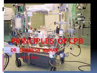
- 1. DR DHANESH KUMAR 19-03-2016 PRINCIPLES OF CPB
- 2. What is It ? Temporary mechanical circulatory support to the stationary heart and lungs Heart and Lungs are made “functionless temporarily” , in order to perform surgeries
- 3. History First open cardiotomy (Apr 5, 1951) First successfulOHS (Sep 2, 1952) Dr. F John Lewis ASD closure using hypothermia and inflow occlusion First successfulOHS using CPB (by JohnGibbon May 6, 1953) ASD closure High mortality rate VSD closure by azygos flow concept (controlled cross- circulation) (Dr C.Walton Lillehei Mar 26, 1954)
- 4. DeWall-Lillehei helix bubble oxygenator (May 1955) Beginning in a large series of patients Method of choice worldwide for OHS Rotating Disk oxygenator Developed by Dr Fredrick Cross and Earl Kay in 1962 Used for early OHS in USA Membrane oxygenator Developed in 1950s-1970s; but initially not frequently used In the mid-1980s, microporous designs; frequently used. Hemodilution Major technologic advance in CPB
- 5. Cardiopulmonary Bypass Goals 1. Still, bloodless heart for cardiac surgery 2. Replacement of cardiac and pulmonary function
- 6. Functions of CPB • Respiration Ventilation Oxygenation • Circulation Venous drainage (by gravity, centrifugal pump, or negative pressure) Arterial inflow • Temperature regulation (hypothermia) Low blood flow -> decreased blood trauma Decreased body metabolism
- 7. Components of CPB Total CPB Partial CPB Integral Components of Extracorporeal Circuit Pumps Oxygenator Heat exchanger Arterial filter Cardioplegic delivery system Cannulae (aortic; arterial; vena caval) Suction and vent
- 8. Basic CPB circuit with oxygenator and centrifugal pump
- 10. Pumps Two principal types Displacement pumps Roller pump Non occlusive roller pumps Rotatory pumps Radial (centrifugal) pumps Axial pumps (Archimedes’ screw) Diagonal pumps
- 11. Impeller PumpRoller Pump Centrifugal Pump Pumps
- 12. Centrifugal pumps > Roller pumps Long-term CPB Ventricular assistance Neonatal ECMO Centrifugal pumps Biomedicus Biopump (Medtronic Inc) Sarns/3M centrifugal pump (Terumo) Levitronix CentriMag blood pump LVAD, RVAD, BiVAD BiVAD + oxygenator in RVAD = ECMO
- 13. Pulsatile Perfusion Significant physiologic advantages • Diastolic run-off • Stimulation of the endothelium Problem • Noncompliant high resistance CPB circuit • High flow with resultant shear stress Hemolysis • Possible with roller pump and diagonal pump, but not with centrifugal pump • Requires larger bore arterial cannulas Alternative method for generating pulsatile flow in high-risk patients • Use of IABP during CPB • Additional cost and invasiveness
- 14. Oxygenator Limited reserve for gas transfer vs. natural lung • Much smaller surface • Limited by diffusion Types of oxygenator • Disk oxygenator • Bubble oxygenator • Membrane oxygenator Maximum oxygen transfer • Less than 25% that of normal lung • Proportional to pO2 difference and surface area, inversely to diffusion distance
- 15. Disk or bubble oxygenator Direct contact oxygenators • Bubbles in direct contact with blood • Increasing cellular trauma
- 16. Bubble oxygenator Bubble oxygenator • Larger bubbles improve removal of CO2 • Smaller bubbles are very efficient at oxygenation but poor in CO2 removal • Larger the No. of bubbles, Greater the efficiency of the oxygenator
- 17. Bubble Oxygenator Advantage Easy to assemble Relatively small priming volume Deforming the frothy blood Low cost Disadvantages Micro emboli Blood cell trauma Destruction of plasma protein Excessive removal of CO2 Deforming capacity exhausted
- 18. Membrane Oxygenator Characteristics Gas exchange across a thin membrane No direct contact with blood and no defoamer; more physiologic Minimal blood damage Two types Solid type (Silicone) Microporous type (polypropylene) 0.3-0.8-micron pores Most popular design = hollow fibers (120-200 microns)
- 19. Membrane Oxygenator Microporous / Hollow fibers
- 20. Microporous (Polypropylene) Membrane Oxygenator Currently predominant design used forCPB Micropores Less than 1.0 um in diameter Initially porous, but plasma protein coating the membrane-gas interface Surface tension of blood prevent gas leakage into the blood phase Conduit for O2 and CO2 exchange Problems Plasma leakage and membrane wet at use of period > 24 hours
- 21. Silicone Membrane Oxygenator True membrane oxygenator Silicone polymer Improved biocompatibility -> long-term support 1980s to mid-1990s Still the membrane of choice for long-term procedures ECMO Problems Gas exchange inferior to polypropylene (microporous) oxygenator Need greater surface area and larger prime volume Difficult in manufacturing and quality control
- 22. New Generation Membrane Oxygenator Silicone polymer A continuous sheet of silicone membrane rolled into a coil Manufactured by Medtronic Cardiopulmonary Inc. Membrane surface area + 0.6-4.5 sq.m Most common use for ECLS/ECMO
- 23. Heat Exchanger Integrated into oxygenator for warming and cooling Exchange surface made of Stainless steel, aluminum, or polypropylene Counter-current mechanism Temperature difference between waterside and blood side Historic reports : maximum difference of 10 °C Recent recommendation : 6 °C and longer rewarming times To improve neurocognitive outcome Hyperthermic circulatory temperature Blood damage (protein denaturation) Limit absolute maximum temperature (42 °C) in blood
- 24. Filters and Bubble Traps In the circuit, micro emboli are monitored by arterial line ultrasound or monitoring screen filtration pressure. Depth filters consist of porous foam, have a large, wetted surface and remove micro emboli by impaction and absorption Screen filters are usually made of woven polyester or nylon thread.
- 25. Tubing Medical grade Polyvinyl Chloride (PVC) tubing It is flexible, compatible with blood, inert, nontoxic, smooth, nonwettable, tough, transparent, resistant to kinking and collapse Can be heat sterilized The Duraflo II heparin coating ionically attaches heparin to a quaternary ammonium carrier (alkylbenzyl dimethyl - ammonium chloride), which binds to plastic surfaces.
- 26. Perfusion Monitors and Sensors A sensor with alarms on the venous reservoir and a bubble detector on the arterial line are desirable safety devices. Flow-through devices are available to continuously measure blood gases, hemoglobin/hematocrit , and some electrolytes Temperatures of the water entering heat exchangers
- 27. Circuits Venous drainage by gravity into oxygenator Height difference between venae cavae and oxygenator > 20- 30 cm Mechanical suction Not desirable Entrain air Suck the vena cava walls against the cannula orifices Arterial blood return to the systemic circulation under pressure
- 29. Arterial Return Ascending aorta just proximal to innominate A Femoral artery access in • Dissecting aortic aneurysm (0.2-3%) • Reoperation • Emergency • MICS Problems of femoral cannulation (more than ascending aorta cannulation) • Sepsis • Pseudoaneurysm • lymphatic fistula Arterial cannula • The narrowest part of CPB circuit • As short as possible • As large as the diameter of vessel permits < 100 mmHg in full flow
- 30. Arterial Cannula Straight/Angled cannula • Minimize risk of dislodgement of atheroma in the ascending Aorta or Arch Axillary –subclavian artery, innominate artery, LV apex • In special circumstances • Limitations and more complications Complication: Dissection of aorta • All sites of arterial cannulation • Prompt recognition and surgical correction • TEE helpful for diagnosis
- 31. Size of venous cannula Adult Children SVC (1/3 of total flow) 28 24 IVC (2/3 of total flow) 36 28 Example: 1.8 m2 patient Total flow 5.4 l/min SVC 1.8 l/min, IVC 3.6 l/min SVC > 30 Fr, IVC > 34 Fr : Single cannula > 38 Fr 36-51 Fr cannula required.
- 32. Prime Fluid Ideally close to ECF Whole blood not used • Homologous blood syndrome • Postperfusion bleeding diathesis • Incompatibility reaction • Demand on blood banks Advantages of hemodilution • Lower blood viscosity • Improve microcirculation • Counteract the increased viscosity by hypothermia Risk of hemodilution • Decreased viscosity : SVR decreased • Low oncotic pressure • O2 carrying • Coagulation factor
Editor's Notes
- University of Minesta hospital; Thomas Jefferson University hospital in Philadelphia; 18-yr-old woman; Oxford univesity, physics, architec;
- University of Minesta hospital; Thomas Jefferson University hospital in Philadelphia; 18-yr-old woman; Oxford univesity, physics, architec;