Ubiquitination of Activation-Induced Cytidine Deaminase by RNF126 2011
1. Ubiquitination of Activation-Induced Cytidine Deaminase by RING Finger 126
Identification of the Site of Ubiquitination
Chittampalli Yashaswini, Rebecca Delker, F. Nina Papavasiliou
Laboratory of Lymphocyte Biology, The Rockefeller University, New York, NY
INTRODUCTION:
Activation-induced cytidine deaminase (AID) is involved in antibody
diversification, which provides protection against a wide array of
pathogens. AID undergoes ubiquitination, a process in which ubiquitin
proteins are covalently bonded to a target protein with the help of an
enzymatic cascade which includes a protein ligase that provides
specificity for the target. The ubiquitin ligase specific for AID is RING
Finger Protein 126 (RNF126). Ubiquitination is known to occur on lysine
residues of proteins. AID has eight lysine residues itself, but the site of
RNF126-dependent ubiquitination remains unknown. By means of site-
specific mutagenesis, immunoprecipitation (IP), and immunoblotting (IB),
we have found that when AID is mutated to have no lysine residues (K0),
ubiquitination does not occur. We have shown that RNF126 and ubiquitin
must be present for ubiquitination of AID to occur. In addition, we have
found that lysine residue 22 (K22) of AID is the main site of ubiquitination
by RNF126, however this ligase is able to ubiquitinate AID at other
residues. The importance of each one of these ubiquitination events
remains to be determined.
METHODS :
•Nine different AID mutants were made using site-specific mutagenesis
on the pcDNA3 vector backbone. The first was K0, where all eight
lysine (K) residues of AID were mutated to arginine (R). The other eight
mutants were made using the K0 mutant as a template. Each mutant
had a different arginine residue mutated back into a lysine to make
single lysine mutants. Wild Type AID (WT AID) was also used.
•The DNA from eight of the AID mutants (K10, K16, K22, K34, K52,
K120, K142 and K160 )was cloned using bacterial transformation. The
DNA was purified and we confirmed by gel electrophoresis that the AID
gene was successfully incorporated into the bacterial plasmid (Figure
2).
•293T cells were used to conduct a ubiquitination assay. AID and AID
mutants were transfected in the presence of exogenous HA-tagged
RNF126 and FLAG-tagged ubiquitin to determine if they were able to
be ubiquitinated.
•Immunoprecipitation was used to isolate AID. Tubulin levels were
measured using immunoblotting to ensure that all samples had the
same protein levels. αAID and αFLAG antibodies were used to detect
the ubiquitination status of each of the mutants. αHA antibody was used
to ensure equal expression of RNF126.
Figure 2. Digestion of
the plasmid was done
with EcoR1 and Xba1
to cut the AID gene out
of the plasmid. The
lower band at 600 base
pairs is AID. The higher
band at 5,446 base
pairs is the plasmid.
Figure 5. This blot detected AID. The third lane was the only one to show
ubiquitination because it had a combination of AID, ubiquitin and RNF126.
Ubiquitination could not occur in the first lane because of the absence of
ubiquitin and RNF126 (the E3 ubiquitin ligase). Although the second lane had
AID and ubiquitin, ubiquitination still could not occur because of the absence of
RNF126. Ubiquitination could not occur in the fourth lane because of the
absence of AID (the substrate). The lower blot detected tubulin, to ensure that
each sample had the same amount of protein.
CONCLUSIONS:
•We have shown that RNF126 is able to ubiquitinate AID
• An AID construct lacking all lysines (K0) was not able to be ubiquitinated
by RNF126. This was the first suggestion that RNF126 ubiquitinates AID
on a lysine residue.
•The ubiquitination ladders visible for the K22 mutant suggests that the
22nd lysine residue is important for ubiquitination of AID by RNF126.
REFERENCES:
RESULTS:
10kb
1kb
K52-1
K52-2
K52-3
K52-4
Plasmid
AID
10,000 base pairs
1,000 base pairs
100 base pairs
ACKNOWLEDGMENTS:
I would like to thank my mentor, Rebecca Delker, for her guidance and
patience. I would also like to thank Dr. Nina Papavasiliou, Ted Scovell, Dr.
C. S Narayanan, and Dr. Ramon Bonfil.
RNF126 is required for ubiquitination of AID
Figure 4. WT AID has 8 lysines as shown above. K0 AID was an AID mutant in
which all lysine residues were mutated to arginine residues. K0 AID was used as a
template to create the other eight mutants. Each of the other eight mutants had
one different arginine residue mutated back into a lysine residue. The K10 mutant
is shown as an example. This is not to scale.
N CWT K
10
K
16
K
22 K160K142K120K52K34
N CK0 R
10
R
16
R
22
R
160
R
142
R
120
R
52
R
34
N CK10 K
10
R
16
R
22
R
160
R
142
R
120
R
52
R
34
Activation-induced Cytidine Deaminase (AID)
•AID regulates antibody diversification via somatic hypermutation (SHM)
and class-switch recombination (CSR) in B-cells.
•SHM is the insertion of point mutations (cytidine to uridine) in DNA at
the immunoglobulin (Ig) locus.
•CSR changes the constant region of an antibody, which is caused by
deletional mutations in the Ig locus.
•Deficiencies in AID or other proteins involved in CSR can result in Hyper
IgM syndrome, a disease in which only one type of antibody (IgM) is
made.
•Misregulation of AID can lead to an accumulation of mutations outside of
the immunoglobulin locus, which can cause cancer.
•The activity of AID is regulated at many levels, including transcription,
translation and the addition of post-translational modifications such as
phosphorylation.
Figure 1. The E1 enzyme binds to ubiquitin in an ATP-consuming process. The E1
passes the ubiquitin to the E2, which binds to RNF126 (the E3 ubiquitin ligase). The
ubiquitin is then covalently bound to AID (the substrate). This process can repeat
several times in order to build the desired ubiquitin chain.
RNF126
E1 E1
E1E2
E2
Ub + ATP AMP + PP
Ub
Ub
UbE2
RNF126
E2
AID
AID
AID Ub
Ub
Ub
Ub
Ub
Lysine residues of AID are necessary for RNF126
dependent ubiquitination
50
20
37
25
75
100
RNF126+ubiquitin
RNF126+ubiquitin+WTAID
RNF126+ubiquitin+KOAID
RNF126+ubiquitin
RNF126+ubiquitin+WTAID
RNF126+ubiquitin+KOAID
WCE
IP
Heavy Chain
Mono-Ub AID
Unmodified AID
αtubulin IB
αAID IP, αAID IB
Figure 6. This blot detected AID. Ubiquitination only occurred in the second
lanes because ubiquitin, RNF126, and AID with lysine residues were present.
Ubiquitination did not occur in the first lanes because of the absence of AID, and
did not occur in the third lanes because of the absence of lysine residues. The
lower blot detected tubulin, to ensure that each sample had the same amount of
protein.
Alberts, et al (2002). Molecular biology of the cell. New York, NY: Garland
Science.
Basu, Uttiya, et al (2005). The AID antibody diversification enzyme is
regulated by protein kinase A phosphorylation. Nature, 438 (24), 508-511.
Chen, Z.J., Sun, L.J. (2009). Nonproteolytic functions of ubiquitin in cell
signaling. Molecular Cell, 33, 275-286.
Delker, R.K. (2009). A coming of age story: activation-induced cytidine
deaminase turns 10. Nature Immunology,10, 1147-1153.
Komander, David (2009). The emerging complexity of protein ubiquitination.
Biochemical society, 37, 937-953.
McBride, K.M., et al (2006). Regulation of hypermutation by activation-
induced cytidine deaminase phosphorylation. PNAS, 103 (23), 8798-8803.
Successful mutagenesis provided usable AID mutants
Ampicillin
resistance gene
P CMV promoter
EcoR1 recognition site
Xba1
recognition
site
AID gene
pcDNA3
5.4 kb
Figure 3. The pcDNA3 plasmid
contains the AID gene, which is
expressed by the P CMV promoter.
It also contains an ampicillin
resistance gene used for drug
selection. It had EcoR1 and Xba1
restriction enzyme recognition sites
on either side of the AID gene,
which allowed us to cut out the AID
gene for gel electrophoresis.
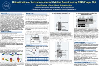
RNF126 favors lysine residue 22 (K22) as the main
ubiquitination site on AID
Figure 7A. The blot on the left panel detected AID. The band at 50 kD is the rat
heavy chain from the antibody we used for IP. The band at 25 kD is unmodified AID.
The band between 25 and 37 kD is likely mono-ubiquitinated AID. This band is most
prevalent in WTAID and K22AID. Another band between 37 and 50 kD is likely di-
ubiquitinated AID. This band is also most prevalent in WT AID and K22 AID. The blot
on the right panel detected ubiquitin proteins that were tagged with FLAG. The K16
lane looks unusually empty, which can be explained by the low expression of
RNF126 in that assay. Higher migrating bands which we detected with the anti-AID
antibody also appear using the αFLAG antibody, suggesting that these bands are in
fact ubiquitinated AID. Again, the mono-ubiquitination band is strongest in WT AID,
but also can be seen in the K22 mutant. Figure 7B. αtubulin antibody was used to
confirm equal loading of the protein in each IP and each gel. Figure 7C. αHA
antibody was used to show equal expression of HA-RNF126 in each 293T
ubiquitination assay. There is less RNF126 expression in the K16 lane, which
explains the emptiness in the αFLAG immunoblot.
Ubiquitination:
•is the covalent attachment of a ubiquitin protein to a substrate protein,
which occurs on lysine residues through isopeptide bonds.
•is carried out by E1 (ubiquitin activating enzyme), E2 (ubiquitin
conjugating enzyme), and E3 (ubiquitin ligase) enzymes.
•RNF126, an E3 ligase, was previously determined to ubiquitinate AID.
•has many roles, including protein degradation by proteasome, protein
regulation, autophagy, endocytosis, and DNA repair.
ABSTRACT:
αAID IP, αAID IB
WTAID
K0AID
K10AID
K16AID
K22AID
K34AID
K52AID
K120AID
K142AID
K160AID
50
20
37
25
75
100
Unmodified AID
Mono-Ub AID
Heavy Chain
Di-Ub AID
WTAID
K0AID
K10AID
K16AID
K22AID
K34AID
K52AID
K120AID
K142AID
K160AID
αAID IP, αFLAG IB
50
20
37
25
75
100
AID
AID+ubiquitin
AID+ubiquitin+RNF126
Ubiquitin+RNF126
Unmodified AID
Mono-Ub AID
Di-Ub AID
WCE αtubulin IB
αAID IP, αAID IB
C
B WCE, αtubulin IB
WCE, αHA IB
A