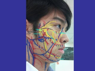
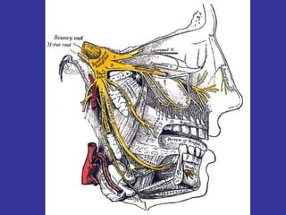
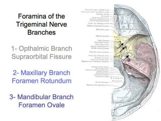
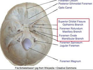
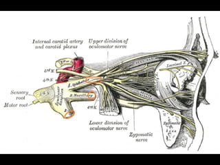
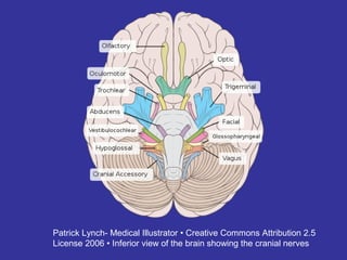
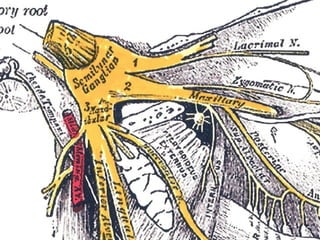
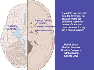
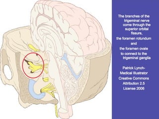
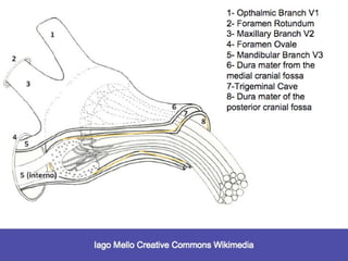
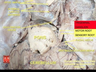
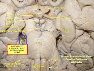
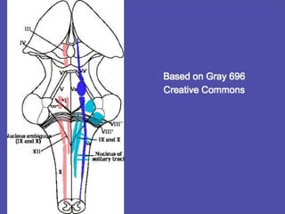
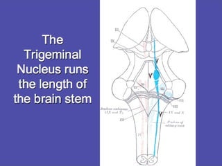
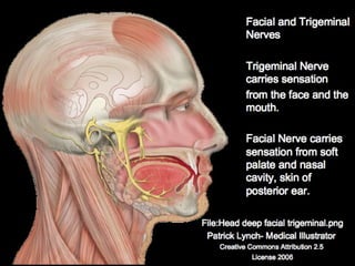
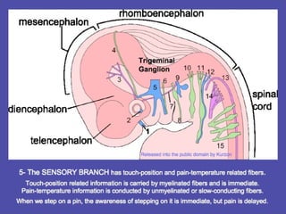
The trigeminal nerve is responsible for sensory innervation of the face and front parts of the head. It has three main branches that enter the skull through different openings and relay sensory information to the trigeminal ganglion and trigeminal nucleus in the brainstem. The trigeminal nucleus is a collection of nerve cells that receives all sensory information from the face, including touch, pain, and temperature. It contains a detailed map of the face and relates this sensory input to different areas of the brain. Disturbances in trigeminal nerve function can lead to pain conditions like trigeminal neuralgia.
This session works with the (1) local tissues, (2) the entrance through the foramen and into the dura, (3) the trigeminal ganglion, (4) the trigeminal nucleus and (5) higher brain facilitation.
trigeminal nerve and pathology/rotary endodontic courses by indian dental aca...Indian dental academy
Indian Dental Academy: will be one of the most relevant and exciting training center with best faculty and flexible training programs for dental professionals who wish to advance in their dental practice,Offers certified courses in Dental implants,Orthodontics,Endodontics,Cosmetic Dentistry, Prosthetic Dentistry, Periodontics and General Dentistry.
The Indian Dental Academy is the Leader in continuing dental education , training dentists in all aspects of dentistry and
offering a wide range of dental certified courses in different formats.for more details please visit
www.indiandentalacademy.com
This lecture was prepared for second year MBBS students of Sir Salimullah Medical College, Dhaka. Here six cranial nerves are described in short and other six are described in detail. You are requested to see the youtube videos for understanding course of the cranial nerves. The lecture was delivered by Dr. Zobayer Mahmud Khan, lecturer, Departmenyt of Anatomy, SSMC.
Anatomy of Cranial Nerve for BPT class.
Require 10 classes. Require help of brain specimen during the class. Testing of the nerves can also be taken together.
The Indian Dental Academy is the Leader in continuing dental education , training dentists in all aspects of dentistry and
offering a wide range of dental certified courses in different formats.
Trigeminal nerve / orthodontic courses by Indian dental academyIndian dental academy
The Indian Dental Academy is the Leader in continuing dental education , training dentists in all aspects of dentistry and
offering a wide range of dental certified courses in different formats.
How to Become a Thought Leader in Your NicheLeslie Samuel
Are bloggers thought leaders? Here are some tips on how you can become one. Provide great value, put awesome content out there on a regular basis, and help others.
Trigeminal nerve (V):
Responsible for sensation in the face and motor functions such as chewing. The trigeminal nerve has both sensory and Medial Motor roots that emerges from the pons and enlarge forming trigeminal ganglia.
The anatomy of the nerve supply of the head and neck has many significant applications in maxillofacial surgery. Understanding these important anatomic relations- variations enables surgeons to perform the surgical procedures safely. Knowledge of these concepts helps us to recognize the problems and complications as and when they occur and manage them accordingly.
Crispy seminar on trigeminal or dentists nerve
treatment of trigeminal neuralgia in detail
including gamma knife cryotherapy glycerol injections
radiofrequency lesioning
pretty useful for last minute brush ups at both undergraduate as well as masters level from both theory as well as practical point of view
This presentation was developed by me and another classmate to present some of the major features and characteristics of the nervous system as relating to orofacial structures. We also focused on learning how to make adjustments and adaptations for individuals with nervous system disorders.
These simplified slides by Dr. Sidra Arshad present an overview of the non-respiratory functions of the respiratory tract.
Learning objectives:
1. Enlist the non-respiratory functions of the respiratory tract
2. Briefly explain how these functions are carried out
3. Discuss the significance of dead space
4. Differentiate between minute ventilation and alveolar ventilation
5. Describe the cough and sneeze reflexes
Study Resources:
1. Chapter 39, Guyton and Hall Textbook of Medical Physiology, 14th edition
2. Chapter 34, Ganong’s Review of Medical Physiology, 26th edition
3. Chapter 17, Human Physiology by Lauralee Sherwood, 9th edition
4. Non-respiratory functions of the lungs https://academic.oup.com/bjaed/article/13/3/98/278874
Report Back from SGO 2024: What’s the Latest in Cervical Cancer?bkling
Are you curious about what’s new in cervical cancer research or unsure what the findings mean? Join Dr. Emily Ko, a gynecologic oncologist at Penn Medicine, to learn about the latest updates from the Society of Gynecologic Oncology (SGO) 2024 Annual Meeting on Women’s Cancer. Dr. Ko will discuss what the research presented at the conference means for you and answer your questions about the new developments.
MANAGEMENT OF ATRIOVENTRICULAR CONDUCTION BLOCK.pdfJim Jacob Roy
Cardiac conduction defects can occur due to various causes.
Atrioventricular conduction blocks ( AV blocks ) are classified into 3 types.
This document describes the acute management of AV block.
New Directions in Targeted Therapeutic Approaches for Older Adults With Mantl...i3 Health
i3 Health is pleased to make the speaker slides from this activity available for use as a non-accredited self-study or teaching resource.
This slide deck presented by Dr. Kami Maddocks, Professor-Clinical in the Division of Hematology and
Associate Division Director for Ambulatory Operations
The Ohio State University Comprehensive Cancer Center, will provide insight into new directions in targeted therapeutic approaches for older adults with mantle cell lymphoma.
STATEMENT OF NEED
Mantle cell lymphoma (MCL) is a rare, aggressive B-cell non-Hodgkin lymphoma (NHL) accounting for 5% to 7% of all lymphomas. Its prognosis ranges from indolent disease that does not require treatment for years to very aggressive disease, which is associated with poor survival (Silkenstedt et al, 2021). Typically, MCL is diagnosed at advanced stage and in older patients who cannot tolerate intensive therapy (NCCN, 2022). Although recent advances have slightly increased remission rates, recurrence and relapse remain very common, leading to a median overall survival between 3 and 6 years (LLS, 2021). Though there are several effective options, progress is still needed towards establishing an accepted frontline approach for MCL (Castellino et al, 2022). Treatment selection and management of MCL are complicated by the heterogeneity of prognosis, advanced age and comorbidities of patients, and lack of an established standard approach for treatment, making it vital that clinicians be familiar with the latest research and advances in this area. In this activity chaired by Michael Wang, MD, Professor in the Department of Lymphoma & Myeloma at MD Anderson Cancer Center, expert faculty will discuss prognostic factors informing treatment, the promising results of recent trials in new therapeutic approaches, and the implications of treatment resistance in therapeutic selection for MCL.
Target Audience
Hematology/oncology fellows, attending faculty, and other health care professionals involved in the treatment of patients with mantle cell lymphoma (MCL).
Learning Objectives
1.) Identify clinical and biological prognostic factors that can guide treatment decision making for older adults with MCL
2.) Evaluate emerging data on targeted therapeutic approaches for treatment-naive and relapsed/refractory MCL and their applicability to older adults
3.) Assess mechanisms of resistance to targeted therapies for MCL and their implications for treatment selection
New Drug Discovery and Development .....NEHA GUPTA
The "New Drug Discovery and Development" process involves the identification, design, testing, and manufacturing of novel pharmaceutical compounds with the aim of introducing new and improved treatments for various medical conditions. This comprehensive endeavor encompasses various stages, including target identification, preclinical studies, clinical trials, regulatory approval, and post-market surveillance. It involves multidisciplinary collaboration among scientists, researchers, clinicians, regulatory experts, and pharmaceutical companies to bring innovative therapies to market and address unmet medical needs.
- Video recording of this lecture in English language: https://youtu.be/lK81BzxMqdo
- Video recording of this lecture in Arabic language: https://youtu.be/Ve4P0COk9OI
- Link to download the book free: https://nephrotube.blogspot.com/p/nephrotube-nephrology-books.html
- Link to NephroTube website: www.NephroTube.com
- Link to NephroTube social media accounts: https://nephrotube.blogspot.com/p/join-nephrotube-on-social-media.html
Lung Cancer: Artificial Intelligence, Synergetics, Complex System Analysis, S...Oleg Kshivets
RESULTS: Overall life span (LS) was 2252.1±1742.5 days and cumulative 5-year survival (5YS) reached 73.2%, 10 years – 64.8%, 20 years – 42.5%. 513 LCP lived more than 5 years (LS=3124.6±1525.6 days), 148 LCP – more than 10 years (LS=5054.4±1504.1 days).199 LCP died because of LC (LS=562.7±374.5 days). 5YS of LCP after bi/lobectomies was significantly superior in comparison with LCP after pneumonectomies (78.1% vs.63.7%, P=0.00001 by log-rank test). AT significantly improved 5YS (66.3% vs. 34.8%) (P=0.00000 by log-rank test) only for LCP with N1-2. Cox modeling displayed that 5YS of LCP significantly depended on: phase transition (PT) early-invasive LC in terms of synergetics, PT N0—N12, cell ratio factors (ratio between cancer cells- CC and blood cells subpopulations), G1-3, histology, glucose, AT, blood cell circuit, prothrombin index, heparin tolerance, recalcification time (P=0.000-0.038). Neural networks, genetic algorithm selection and bootstrap simulation revealed relationships between 5YS and PT early-invasive LC (rank=1), PT N0—N12 (rank=2), thrombocytes/CC (3), erythrocytes/CC (4), eosinophils/CC (5), healthy cells/CC (6), lymphocytes/CC (7), segmented neutrophils/CC (8), stick neutrophils/CC (9), monocytes/CC (10); leucocytes/CC (11). Correct prediction of 5YS was 100% by neural networks computing (area under ROC curve=1.0; error=0.0).
CONCLUSIONS: 5YS of LCP after radical procedures significantly depended on: 1) PT early-invasive cancer; 2) PT N0--N12; 3) cell ratio factors; 4) blood cell circuit; 5) biochemical factors; 6) hemostasis system; 7) AT; 8) LC characteristics; 9) LC cell dynamics; 10) surgery type: lobectomy/pneumonectomy; 11) anthropometric data. Optimal diagnosis and treatment strategies for LC are: 1) screening and early detection of LC; 2) availability of experienced thoracic surgeons because of complexity of radical procedures; 3) aggressive en block surgery and adequate lymph node dissection for completeness; 4) precise prediction; 5) adjuvant chemoimmunoradiotherapy for LCP with unfavorable prognosis.
Explore natural remedies for syphilis treatment in Singapore. Discover alternative therapies, herbal remedies, and lifestyle changes that may complement conventional treatments. Learn about holistic approaches to managing syphilis symptoms and supporting overall health.
NVBDCP.pptx Nation vector borne disease control programSapna Thakur
NVBDCP was launched in 2003-2004 . Vector-Borne Disease: Disease that results from an infection transmitted to humans and other animals by blood-feeding arthropods, such as mosquitoes, ticks, and fleas. Examples of vector-borne diseases include Dengue fever, West Nile Virus, Lyme disease, and malaria.
Basavarajeeyam is an important text for ayurvedic physician belonging to andhra pradehs. It is a popular compendium in various parts of our country as well as in andhra pradesh. The content of the text was presented in sanskrit and telugu language (Bilingual). One of the most famous book in ayurvedic pharmaceutics and therapeutics. This book contains 25 chapters called as prakaranas. Many rasaoushadis were explained, pioneer of dhatu druti, nadi pareeksha, mutra pareeksha etc. Belongs to the period of 15-16 century. New diseases like upadamsha, phiranga rogas are explained.
9. The trigeminal nerve receives sensory
input from many areas including:
• oral cavity, teeth and TMJ
• nasopharynx and nasal cavity
• most of the face and the scalp
• eyeballs and conjunctiva
• ear pinae and external ear canal
• lacrimal glands(tears)
• meningeal membranes of the
anterior and middle cranial fossae
• portions of the superior tentorium
10. Dermatomes-
Sensory areas of
the head- the
trigeminal nerve
and cervical
plexus
from Henry Gray- Anatomy of the Human Body before 1858
11. Three branches of the trigeminal nerve enter
the skull through three different foramina in
the sphenoid bone:
The Mandibular Branch (V3) enters through
the foramen ovale.
The Maxillary Branch (V2) enters through
the foramen rotundum.
The Opthalmic Branch (V1) enters through
the superior orbital fissure.
12.
13.
14.
15. Midway between theMidway between the
peripheral nervesperipheral nerves
and trigeminal nucleiand trigeminal nuclei
in the brain stem,in the brain stem,
there is a ganglionthere is a ganglion
or junctionor junction
16. The trigeminal ganglion is locatedThe trigeminal ganglion is located
near the apex of the petrousnear the apex of the petrous
portion of the temporal boneportion of the temporal bone
and is surrounded byand is surrounded by
cerebrospinal fluidcerebrospinal fluid
and nested in anand nested in an
arachnoid poucharachnoid pouch
17. Patrick Lynch- Medical Illustrator • Creative Commons Attribution 2.5
License 2006 • Inferior view of the brain showing the cranial nerves
21. The Trigeminal Cave is alsoThe Trigeminal Cave is also
known as Meckel's Cave.known as Meckel's Cave.
It is an arachnoidal pouch containingIt is an arachnoidal pouch containing
cerebrospinal fluidcerebrospinal fluid
formed by two layers of dura materformed by two layers of dura mater
which are part of an evagination of thewhich are part of an evagination of the
tentorium cerebellitentorium cerebelli
22.
23. The Trigeminal CaveThe Trigeminal Cave
envelops theenvelops the
trigeminal ganglion.trigeminal ganglion.
It is named forIt is named for
Johann Friedrich Meckel the ElderJohann Friedrich Meckel the Elder..
31. There is about a 2 cmThere is about a 2 cm
connection from theconnection from the trigeminaltrigeminal
ganglionganglion
to the trigeminal nucleusto the trigeminal nucleus
37. The trigeminal nucleus is dividedThe trigeminal nucleus is divided
anatomically into three parts,anatomically into three parts,
visible in microscopic sections of the brainstem.visible in microscopic sections of the brainstem.
From caudal to rostralFrom caudal to rostral
going up from the medulla to the midbraingoing up from the medulla to the midbrain
they are:they are:
1- The spinal trigeminal nucleus1- The spinal trigeminal nucleus
2- The main trigeminal nucleus2- The main trigeminal nucleus
3- The mesencephalic trigeminal3- The mesencephalic trigeminal
nucleus.nucleus.
38. The three parts of the trigeminal nucleus receiveThe three parts of the trigeminal nucleus receive
different types of sensory information:different types of sensory information:
1- The spinal trigeminal nucleus (lowest)1- The spinal trigeminal nucleus (lowest)
receivesreceives pain/temperature fibers.pain/temperature fibers.
2- The main trigeminal nucleus (middle)2- The main trigeminal nucleus (middle)
receivesreceives touch/position fibers.touch/position fibers.
3- The mesencephalic nucleus (highest)3- The mesencephalic nucleus (highest)
receivesreceives proprioceptor andproprioceptor and
mechanoreceptormechanoreceptor
fibers from the jaws and teeth.fibers from the jaws and teeth.
39.
40. It is not widely appreciatedIt is not widely appreciated
that all sensory informationthat all sensory information
from the facefrom the face (all touch/position(all touch/position
information and allinformation and all
pain/temperature information)pain/temperature information) isis
sent to thesent to the
trigeminal nucleus.trigeminal nucleus.
41. In classical anatomy, most sensoryIn classical anatomy, most sensory
information from the face is carried by theinformation from the face is carried by the
fifth (trigeminal) nerve.fifth (trigeminal) nerve.
Sensation from certain parts of the mouth,Sensation from certain parts of the mouth,
certain parts of the ear and certain parts ofcertain parts of the ear and certain parts of
the meninges is carried bythe meninges is carried by
"general somatic afferent" (GSA)"general somatic afferent" (GSA)
fibers in cranial nervesfibers in cranial nerves
VII (the facial nerve)VII (the facial nerve)
IX (the glossopharyngeal nerve) andIX (the glossopharyngeal nerve) and
X (the vagus nerve).X (the vagus nerve).
42. Without exception, however,
all sensory fibers from these nerves (VII,
IX, X) terminate in the trigeminal nucleus.
On entering the brainstem,On entering the brainstem,
sensory fibers from V, VII, IX, and Xsensory fibers from V, VII, IX, and X
are sorted out and sent to theare sorted out and sent to the
trigeminal nucleus, which thus contains atrigeminal nucleus, which thus contains a
complete sensory map of the face and mouth.complete sensory map of the face and mouth.
43. The trigeminal nucleus extendsThe trigeminal nucleus extends
throughout the entire brainstem, from thethroughout the entire brainstem, from the
midbrain to the medulla, and continuesmidbrain to the medulla, and continues
into the cervical cord. It contains ainto the cervical cord. It contains a
sensory map of the face, mouth andsensory map of the face, mouth and
relates to cranial membranes.relates to cranial membranes.
The trigeminal nucleus merges with theThe trigeminal nucleus merges with the
dorsal horn cells of the spinal cord whichdorsal horn cells of the spinal cord which
contain a sensory map of the rest of thecontain a sensory map of the rest of the
body.body.
44. The coverings of the brain are called meningesThe coverings of the brain are called meninges
and consist of the dura, arachnoid and pia. Theand consist of the dura, arachnoid and pia. The
dura in particular has a lot of pain receptors anddura in particular has a lot of pain receptors and
may be responsible for many headaches. As amay be responsible for many headaches. As a
neurosurgeon I have seen this first hand duringneurosurgeon I have seen this first hand during
awake brain surgery when we open the dura.awake brain surgery when we open the dura.
The patient usually doesn't report any painThe patient usually doesn't report any pain
when you drill a hole in their skull. However, theywhen you drill a hole in their skull. However, they
start to report dull pain or headache whenstart to report dull pain or headache when
you stimulate or touch the dura.you stimulate or touch the dura.
Brain Surgeon responseBrain Surgeon response
45. Not only is the trigeminal nerve (V)Not only is the trigeminal nerve (V)
feeding informationfeeding information
into the trigeminal nucleus,into the trigeminal nucleus,
but also sensory informationbut also sensory information
related to cranial nerves VII, IX, and X.related to cranial nerves VII, IX, and X.
All four of these nerves are part ofAll four of these nerves are part of
Porges Polyvagal TheoryPorges Polyvagal Theory
46.
47.
48.
49. Pain related to thePain related to the
Trigeminal NerveTrigeminal Nerve
51. A headache or cephalalgia is pain anywhere inA headache or cephalalgia is pain anywhere in
the region of the head or neck. It can be athe region of the head or neck. It can be a
symptom of a number of different conditions ofsymptom of a number of different conditions of
the head and neck. The brain tissue itself is notthe head and neck. The brain tissue itself is not
sensitive to pain because it lacks pain receptors.sensitive to pain because it lacks pain receptors.
Rather, the pain is caused by disturbance of theRather, the pain is caused by disturbance of the
pain-sensitive structures around the brain.pain-sensitive structures around the brain.
52. Nine areas of the head and neck have these pain-Nine areas of the head and neck have these pain-
sensitive structures, which are thesensitive structures, which are the
•• Cranium (The periosteum of the skull)Cranium (The periosteum of the skull)
• MusclesMuscles
• NervesNerves
• Arteries and veinsArteries and veins
• Subcutaneous tissuesSubcutaneous tissues
• EyesEyes
• EarsEars
• Sinuses and mucous membranesSinuses and mucous membranes
53. The complex processing of pain-temperatureThe complex processing of pain-temperature
information in the thalamus and cortexinformation in the thalamus and cortex
reflects a phylogenetically olderreflects a phylogenetically older
and more primitive sensory systemand more primitive sensory system
relative to the simple processingrelative to the simple processing
of touch-position information.of touch-position information.
Pain is an individualized sensationPain is an individualized sensation
that varies among people andthat varies among people and
is conditionedis conditioned
by memory and emotion.by memory and emotion.
54. All information from touch-position andAll information from touch-position and
pain-temperature receptors is sent to thepain-temperature receptors is sent to the
primary somato-sensory cortex.primary somato-sensory cortex.
However, pain-temperature information isHowever, pain-temperature information is
communicated with more cortical centers thancommunicated with more cortical centers than
the touch-position fibers.the touch-position fibers.
It communicates with:It communicates with:
• the medial dorsal thalamic nucleus which• the medial dorsal thalamic nucleus which
projects to the anterior cingulate gyrus.projects to the anterior cingulate gyrus.
•• the ventromedial nucleus of the thalamusthe ventromedial nucleus of the thalamus
which is then sent to the insular cortex.which is then sent to the insular cortex.
• some fibers are sent from the• some fibers are sent from the
intralaminar nucleus of the thalamusintralaminar nucleus of the thalamus
via the reticular formation.via the reticular formation.
55. Trigeminal NeuralgiaTrigeminal Neuralgia
TN is a severe neuropathic chronic painTN is a severe neuropathic chronic pain
disorder affecting the trigeminal nerve.disorder affecting the trigeminal nerve.
There is intense pain along theThere is intense pain along the
trigeminal nerve divisions.trigeminal nerve divisions.
Evidence indicates it is causedEvidence indicates it is caused
by a loss of myelin from theby a loss of myelin from the
sensory fibers within thesensory fibers within the
nerve root itself.nerve root itself.
56. In type 1, it is characterized by sudden severeIn type 1, it is characterized by sudden severe
pain along the trigeminal nerve itself.pain along the trigeminal nerve itself.
Sometimes in type 2, there is constant painSometimes in type 2, there is constant pain
that varies from dull to excruciating.that varies from dull to excruciating.
Experts say this is one of the mostExperts say this is one of the most
painful medical conditions possible.painful medical conditions possible.
Dental work and herpes zosterDental work and herpes zoster
may be contributing factors.may be contributing factors.