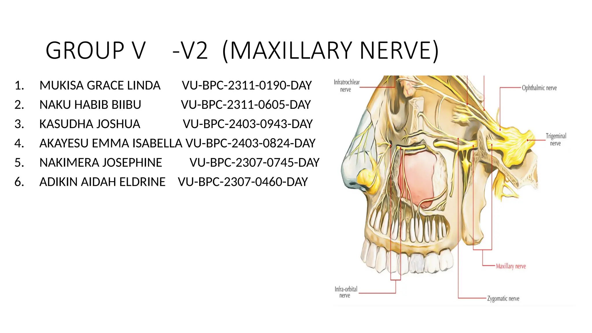
GROUP V -V2(MAXILLARY NERVE)
1. MUKISA GRACE LINDA VU-BPC-2311-0190-DAY
2. NAKU HABIB BIIBU VU-BPC-2311-0605-DAY
3. KASUDHA JOSHUA VU-BPC-2403-0943-DAY
4. AKAYESU EMMA ISABELLA VU-BPC-2403-0824-DAY
5. NAKIMERA JOSEPHINE VU-BPC-2307-0745-DAY
6. ADIKIN AIDAH ELDRINE VU-BPC-2307-0460-DAY
2.
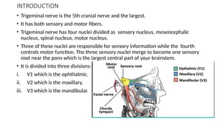
INTRODUCTION
• Trigeminal nerveis the 5th cranial nerve and the largest.
• It has both sensory and motor fibers.
• Trigeminal nerve has four nuclei divided as sensory nucleus, mesencephalic
nucleus, spinal nucleus, motor nucleus.
• Three of these nuclei are responsible for sensory information while the fourth
controls motor function. The three sensory nuclei merge to become one sensory
root near the pons which is the largest central part of your brainstem.
• It is divided into three divisions:
i. V1 which is the ophthalmic.
ii. V2 which is the maxillary.
iii. V3 which is the mandibular.
3.
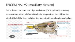
TRIGEMINAL V2 (maxillarydivision)
This is the second branch of trigeminal nerve (CN V), primarily a sensory
nerve carrying sensory information (pain, temperature, touch) from the
middle third of the face, including the upper teeth, nasal cavity, and palate.
4.
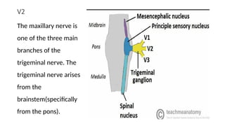
V2
The maxillary nerveis
one of the three main
branches of the
trigeminal nerve. The
trigeminal nerve arises
from the
brainstem(specifically
from the pons).
5.
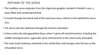
PATHWAY OF THENERVE
• The maxillary nerve originates from the trigeminal ganglion, located in Meckel's cave, a
space filled with cerebrospinal fluid.
• It travels through the lateral wall of the cavernous sinus, inferior to the ophthalmic nerve
(V1).
• The nerve exits the skull base through the foramen rotundum.
• It then enters the pterygopalatine fossa, where it gives off several branches, including the
middle meningeal nerve, zygomatic nerve and branches to the nasal cavity and palate.
• The main trunk continues anteriorly in the orbital floor and emerges onto the face as the
infraorbital nerve.
6.
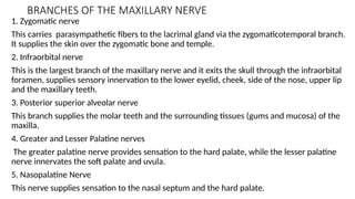
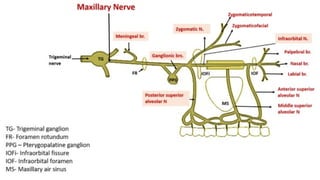
BRANCHES OF THEMAXILLARY NERVE
1. Zygomatic nerve
This carries parasympathetic fibers to the lacrimal gland via the zygomaticotemporal branch.
It supplies the skin over the zygomatic bone and temple.
2. Infraorbital nerve
This is the largest branch of the maxillary nerve and it exits the skull through the infraorbital
foramen, supplies sensory innervation to the lower eyelid, cheek, side of the nose, upper lip
and the maxillary teeth.
3. Posterior superior alveolar nerve
This branch supplies the molar teeth and the surrounding tissues (gums and mucosa) of the
maxilla.
4. Greater and Lesser Palatine nerves
The greater palatine nerve provides sensation to the hard palate, while the lesser palatine
nerve innervates the soft palate and uvula.
5. Nasopalatine Nerve
This nerve supplies sensation to the nasal septum and the hard palate.
8.
Areas supplied bythe maxillary nerve
1.Skin
It supplies the midface which includes the lower eyelid, upper lip,
lateral nose and cheeks.
2.Mucosa
It supplies the nasal cavity, nasopharynx and palate.
3.Teeth
It supplies the upper teeth via superior alveolar nerves.
4.Sinuses
It supplies the maxillary and part of ethmoid.
9.
CLINICAL IMPORTANCE
• Themaxillary nerve is one of the crucial nerves in dentistry as it provides
the sensation of the middle face which includes all of the upper teeth
and their supporting structures.
• In dental procedures, a maxillary nerve block may be performed to
anesthetize the upper teeth and surrounding areas and this is often done
using an injection near the pterygopalatine fossa or infraorbital foramen.
• The V2 carries postganglionic fibers from the pterygopalatine ganglion
which supply the lacrimal gland and mucous glands of the nasal mucosa.
• It carries parasympathetic fibers that are involved in the secretion of
tears from the lacrimal gland. These fibers travel with the zygomatic
nerve to reach the lacrimal gland.
• Facial trauma, V2 can be damaged in facial trauma leading to numbness,
tingling or pain in the mid-face region.
10.
CONCLUSION
• The maxillarynerve (V2) is a sensory branch of the trigeminal nerve
that provides innervation to the midfacial region and supplies areas
such as the skin, mucosa, upper teeth and sinuses.
• It passes through the foramen rotundum into the pterygopalatine fossa
and then branches into the infraorbital, zygomatic and palatine nerves.
• Understanding its course and connections helps in clinical settings like
dental anesthesia, sinus pathologies and facial pain syndromes.
11.
REFERENCES
1.Richard S. Snell,Clinical Neuroanatomy 7th Edition pg 361,362.
2.James D. Fix Neuroanatomy.
3.Grays Anatomy textbook.