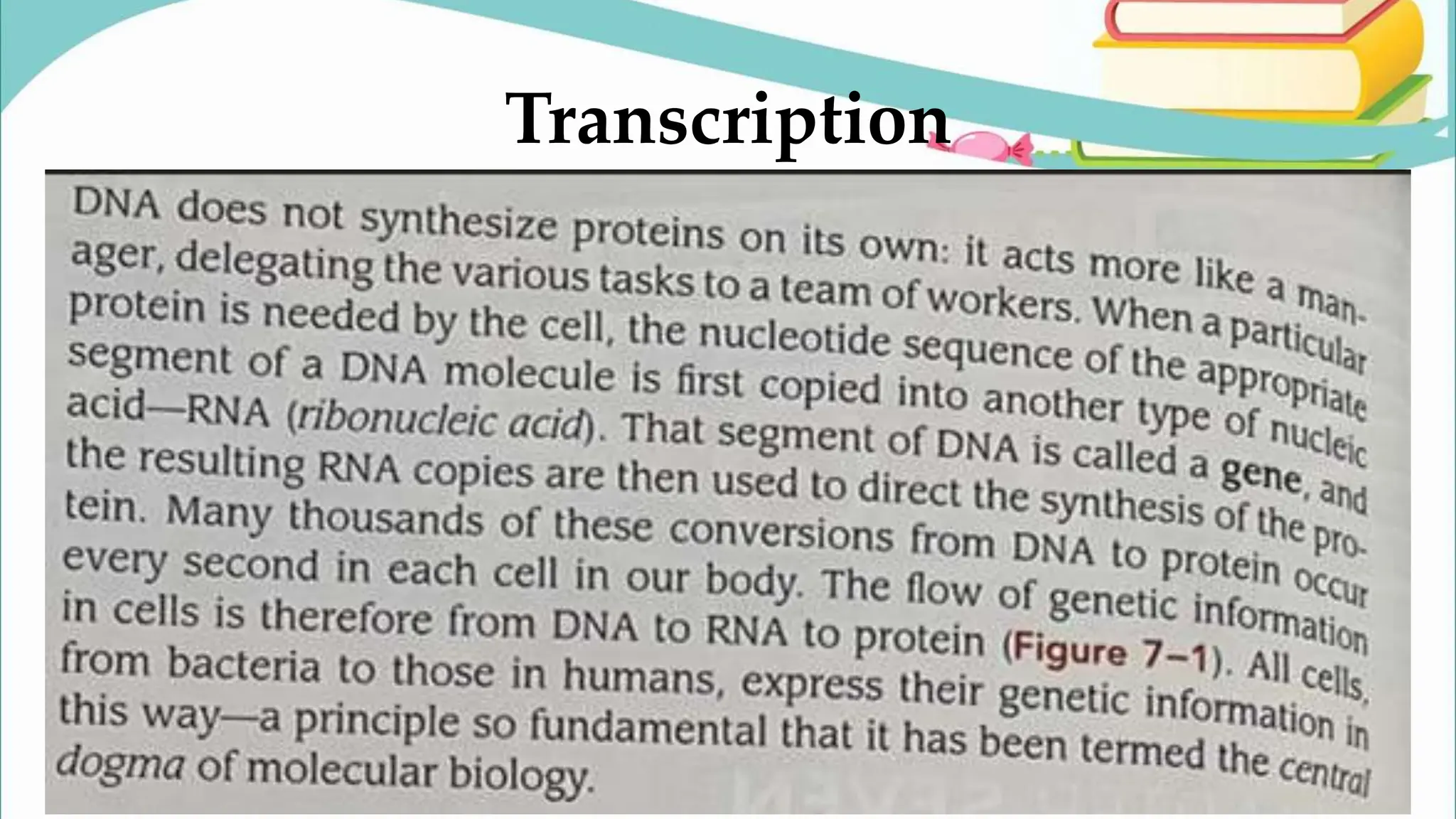
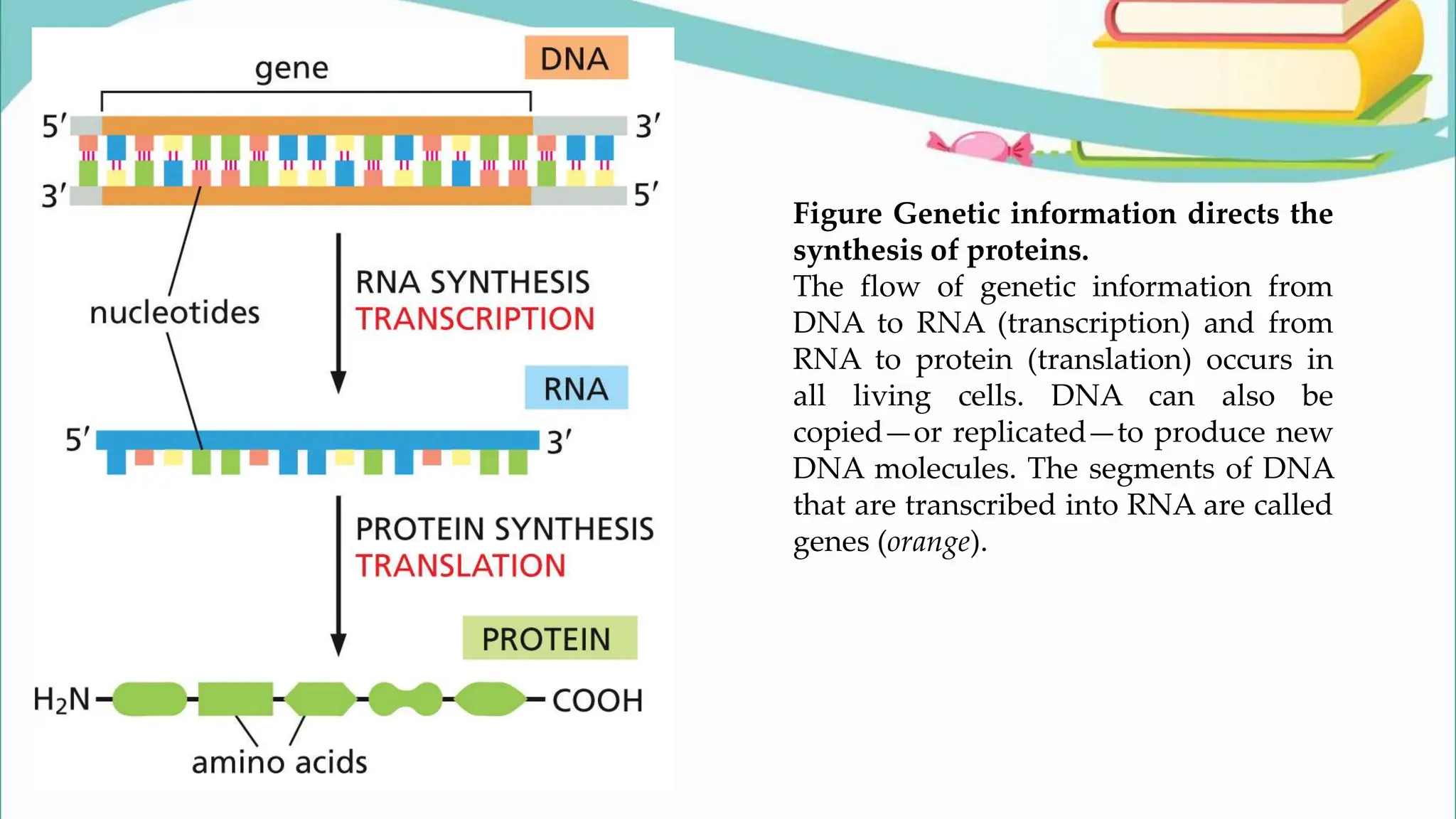
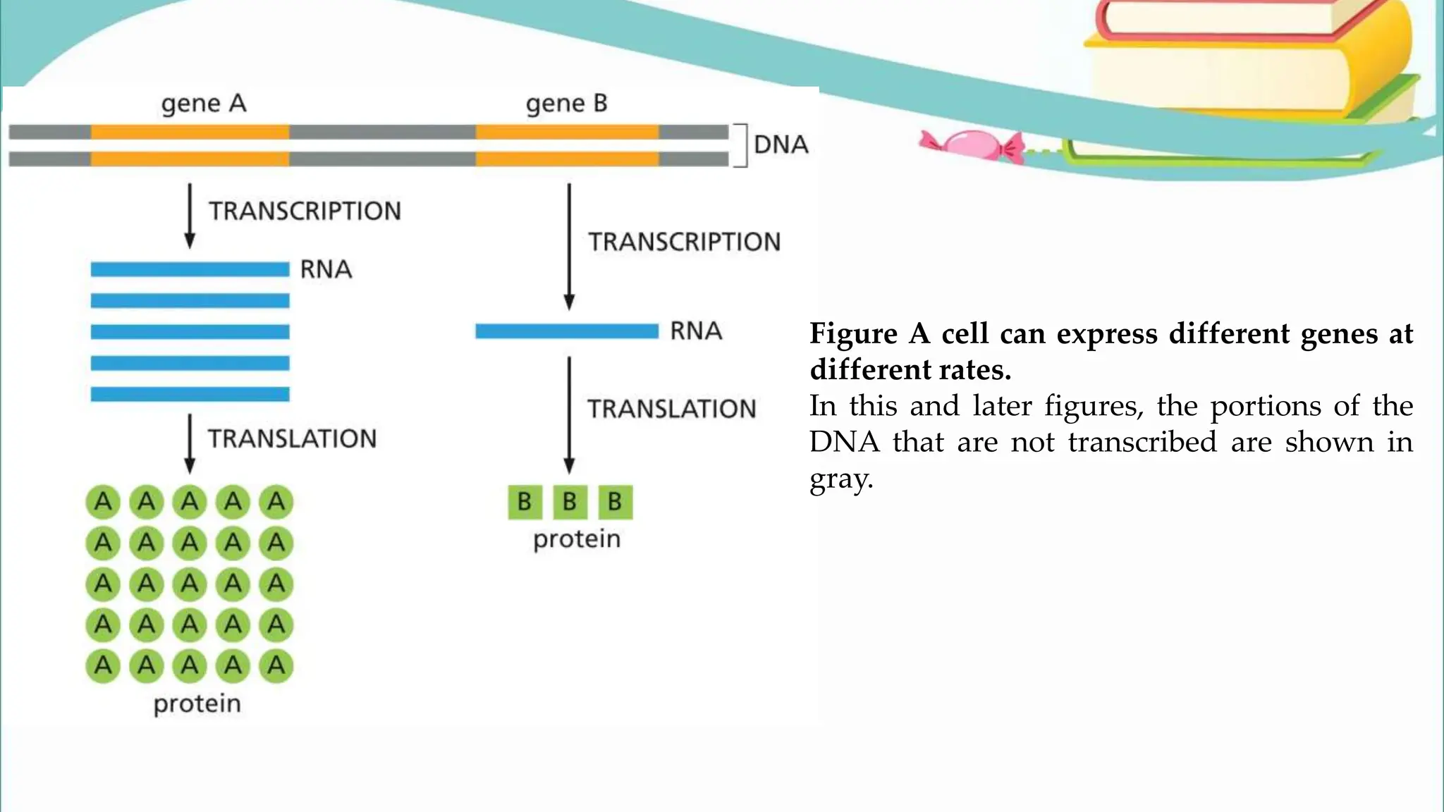
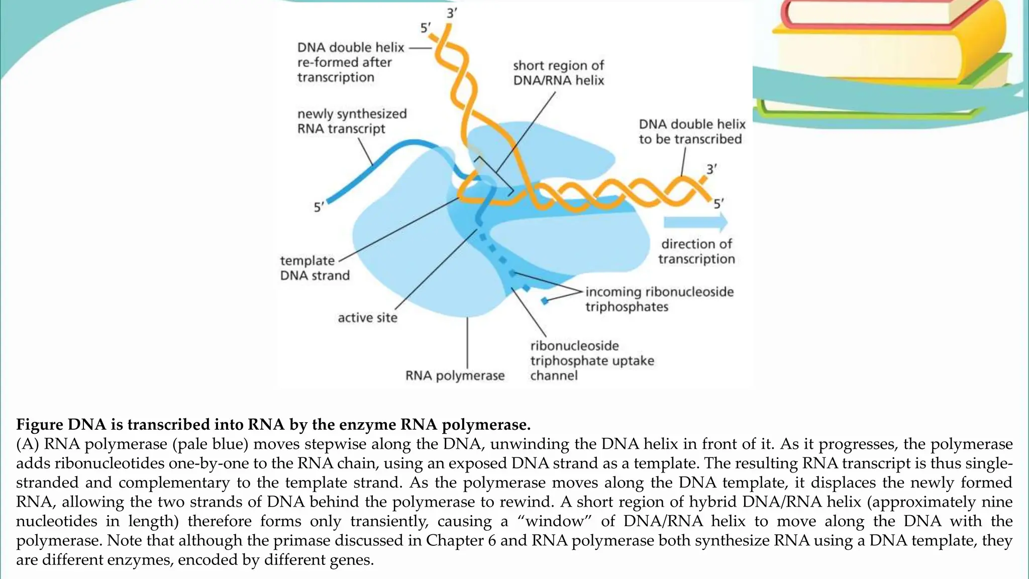
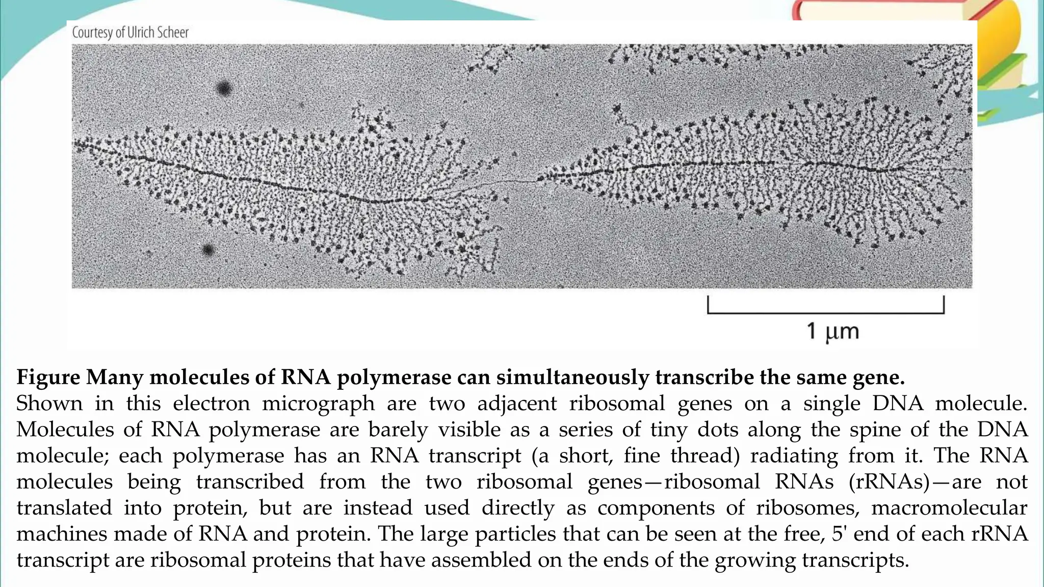
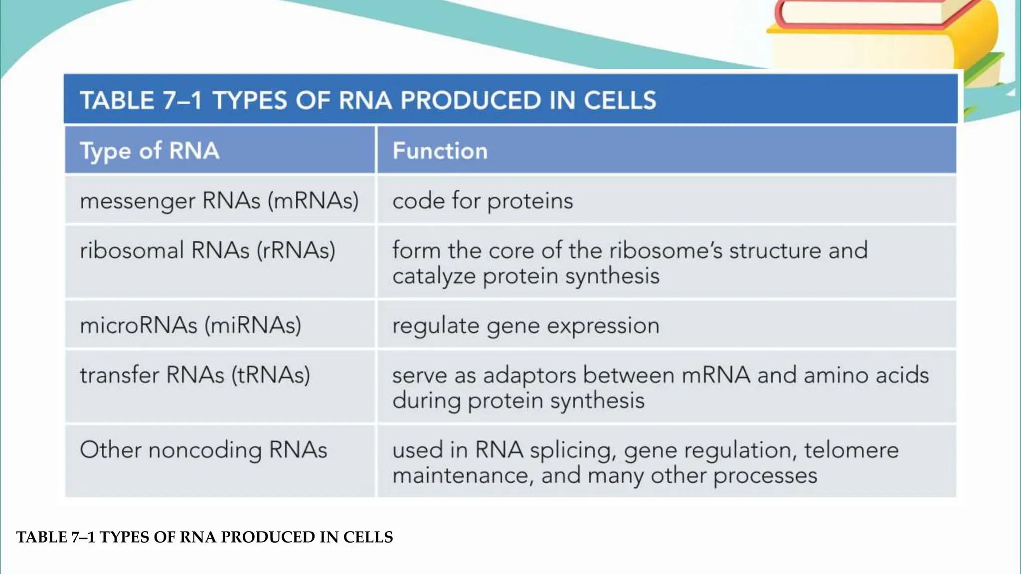
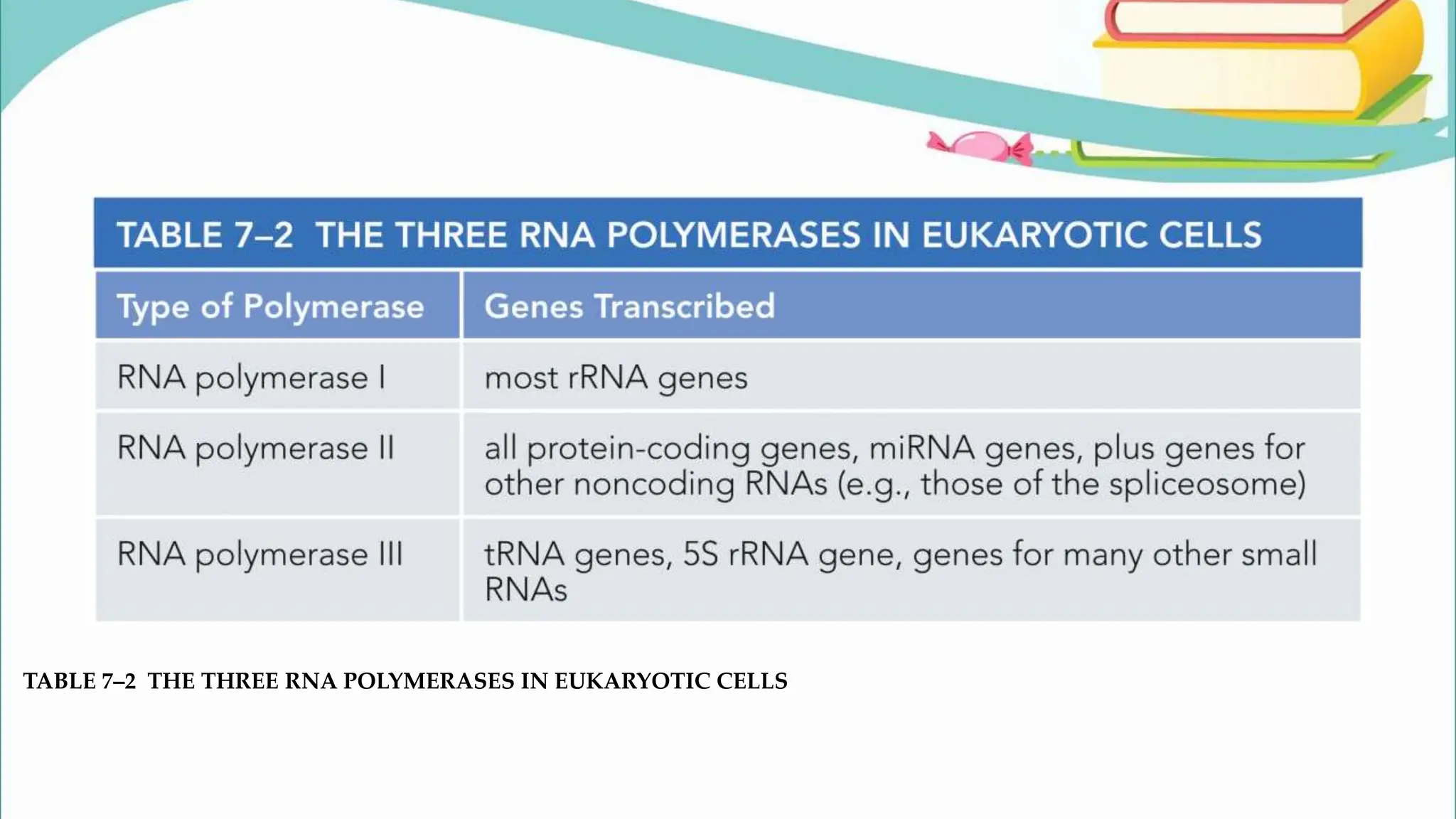
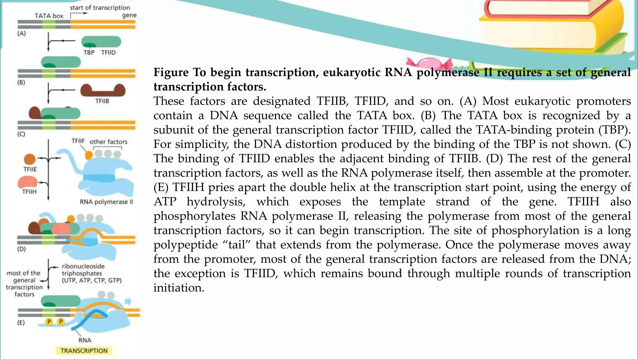
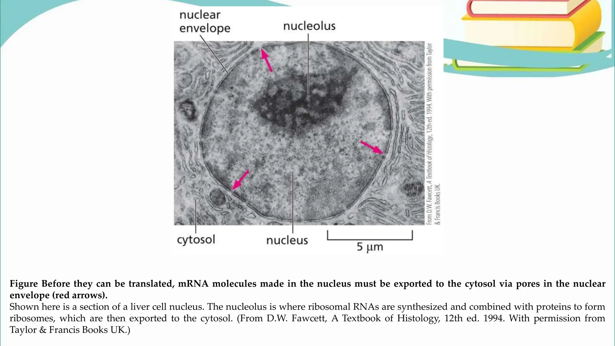
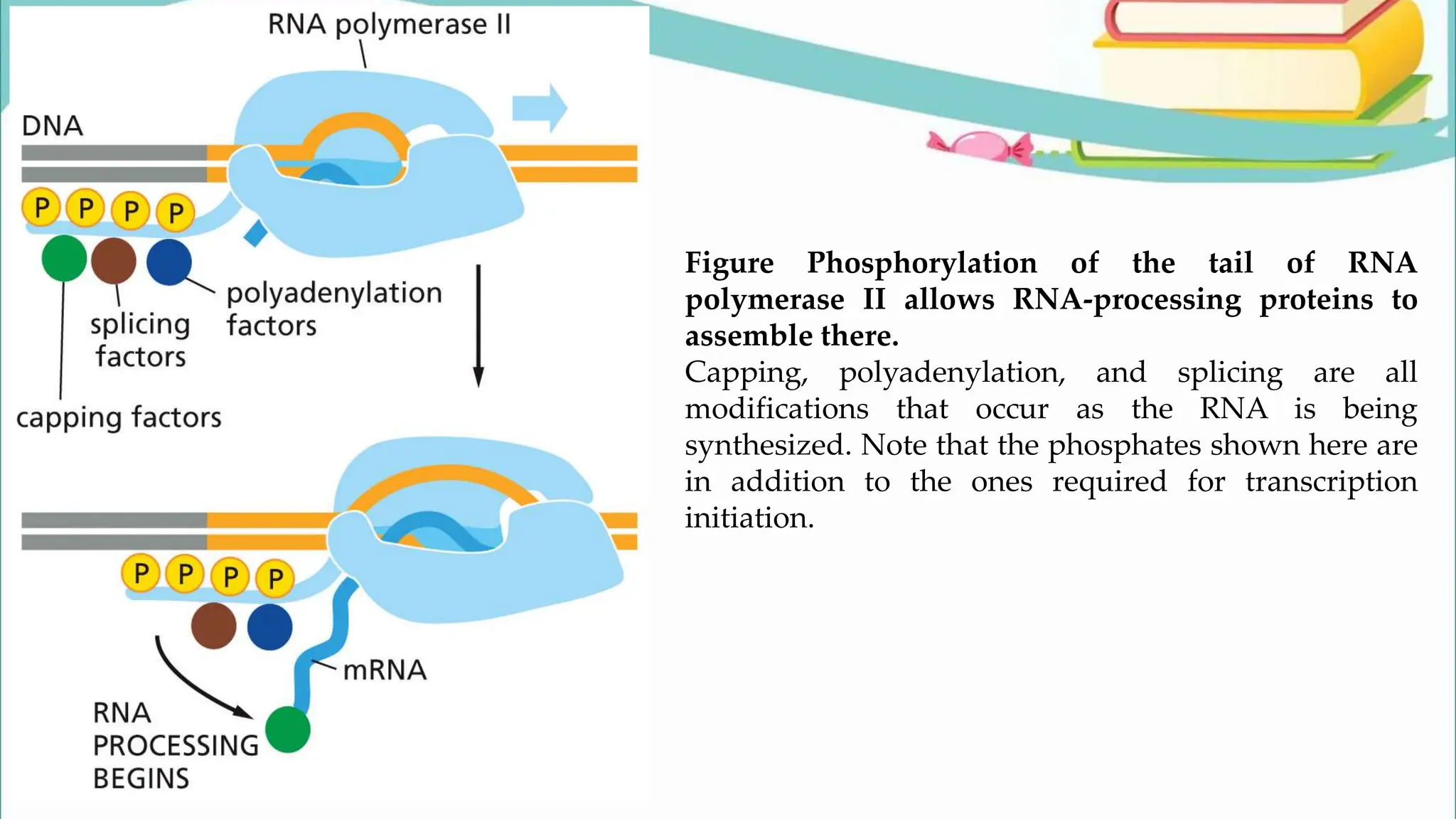
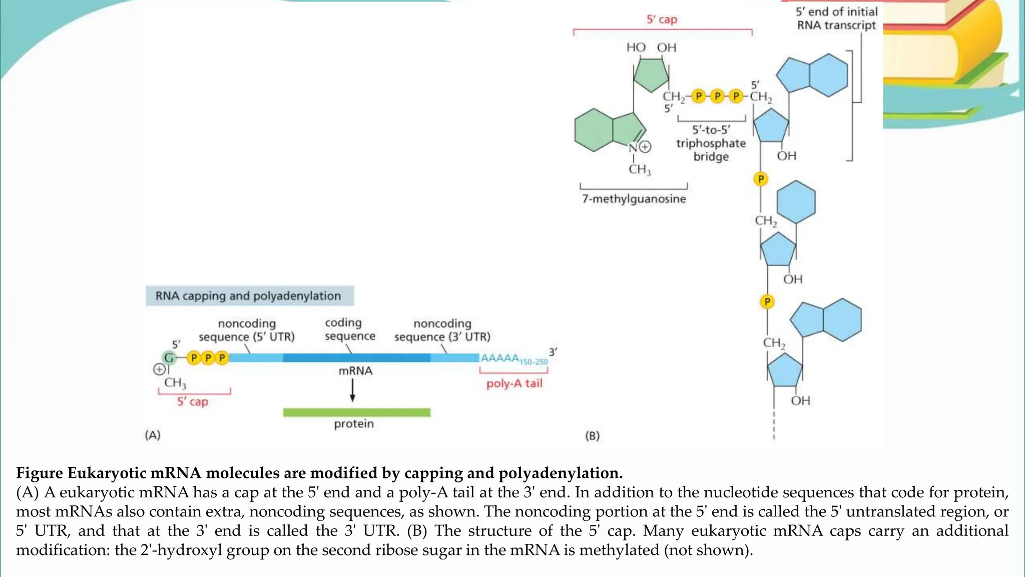
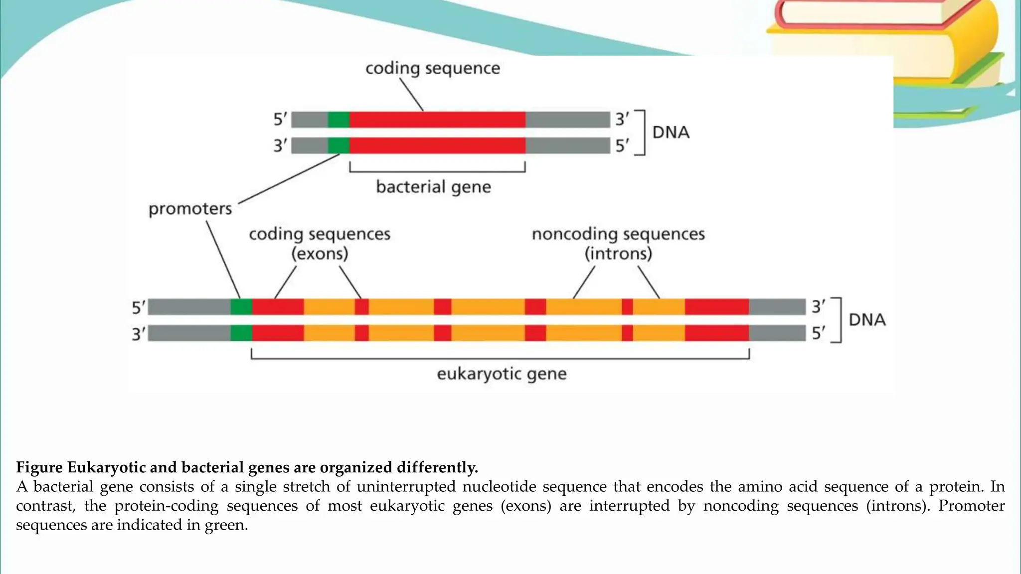
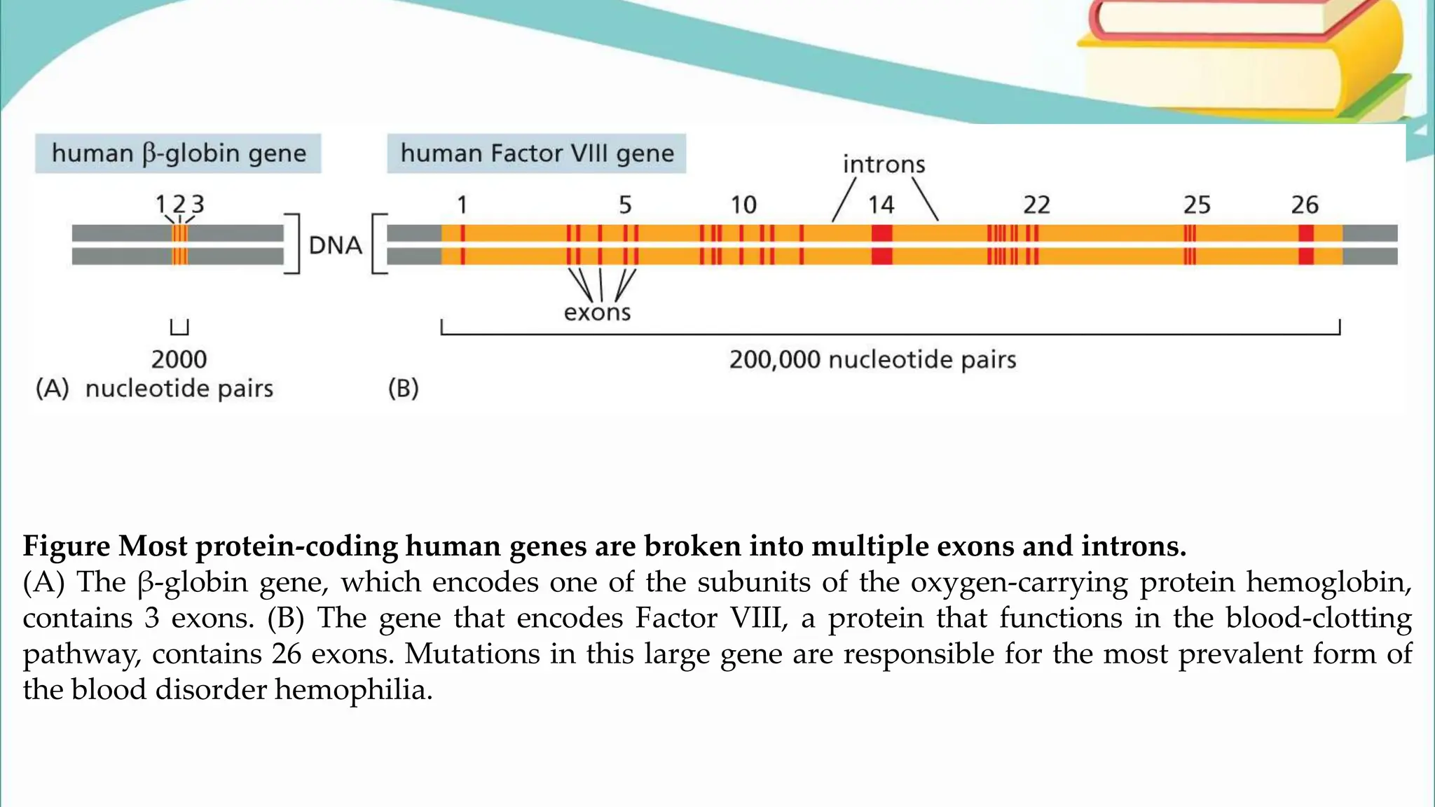
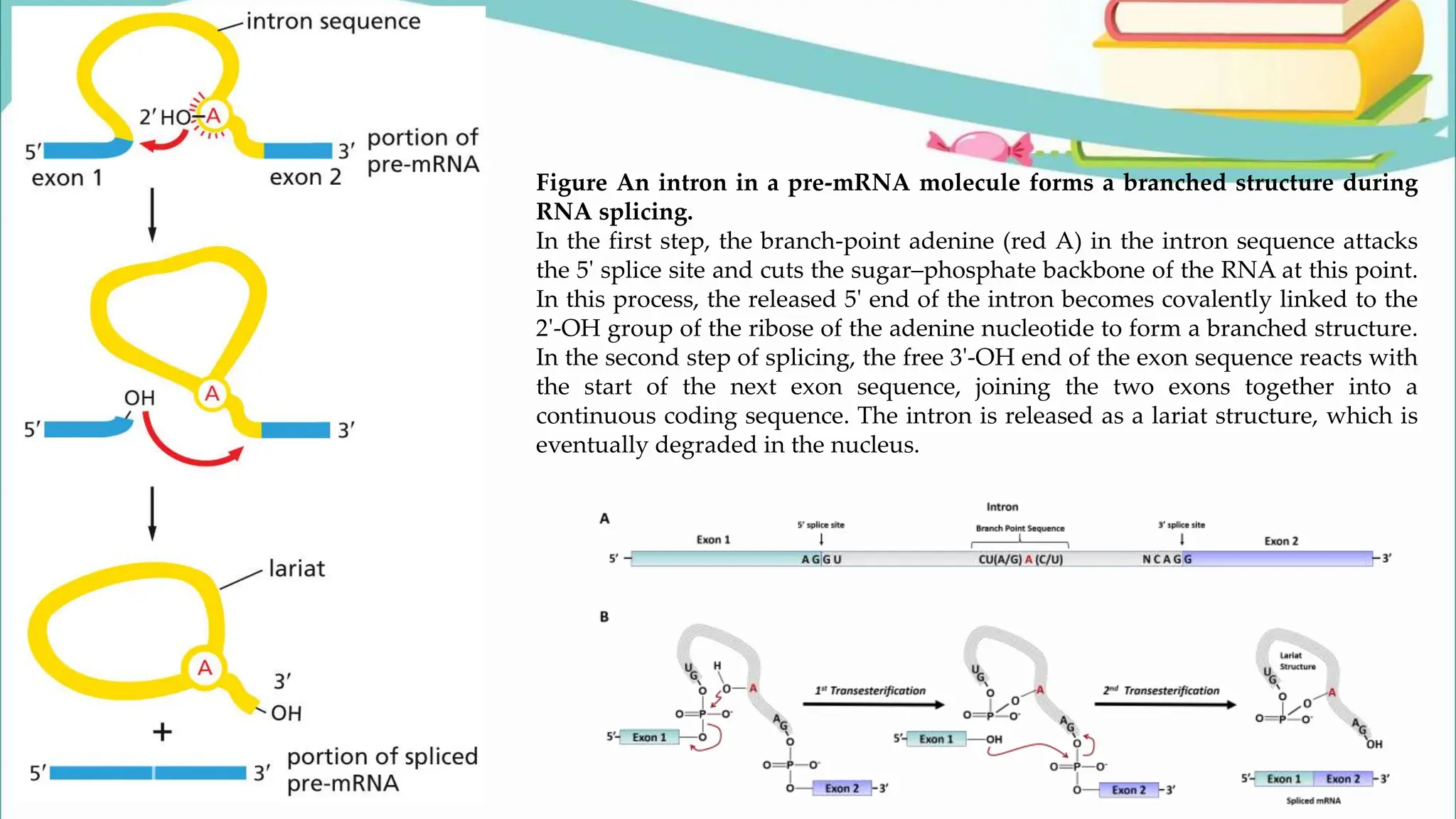
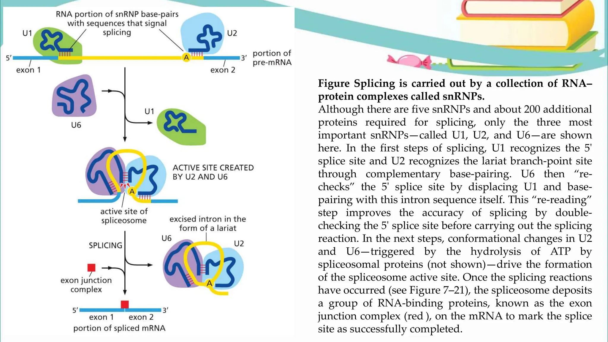
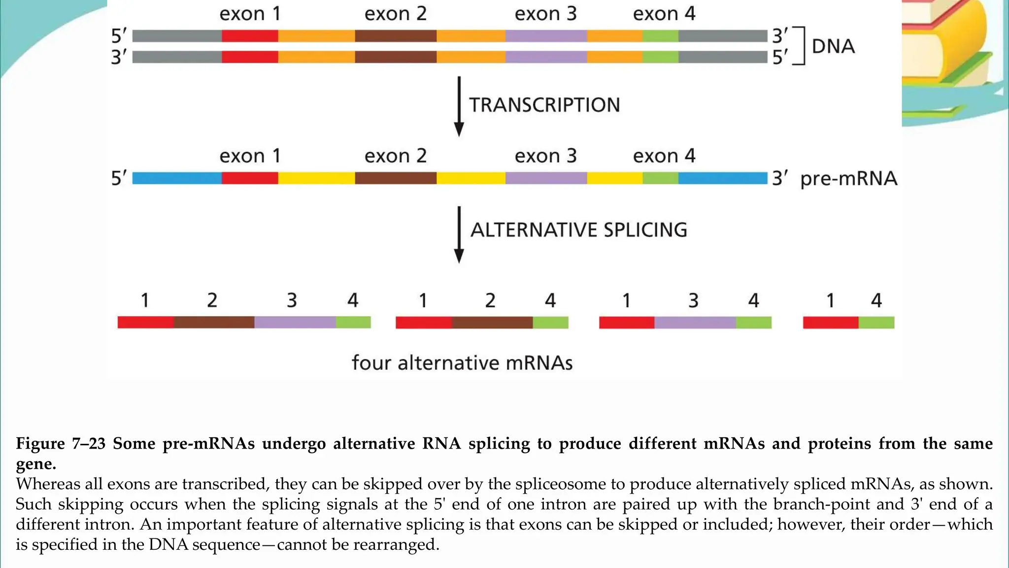
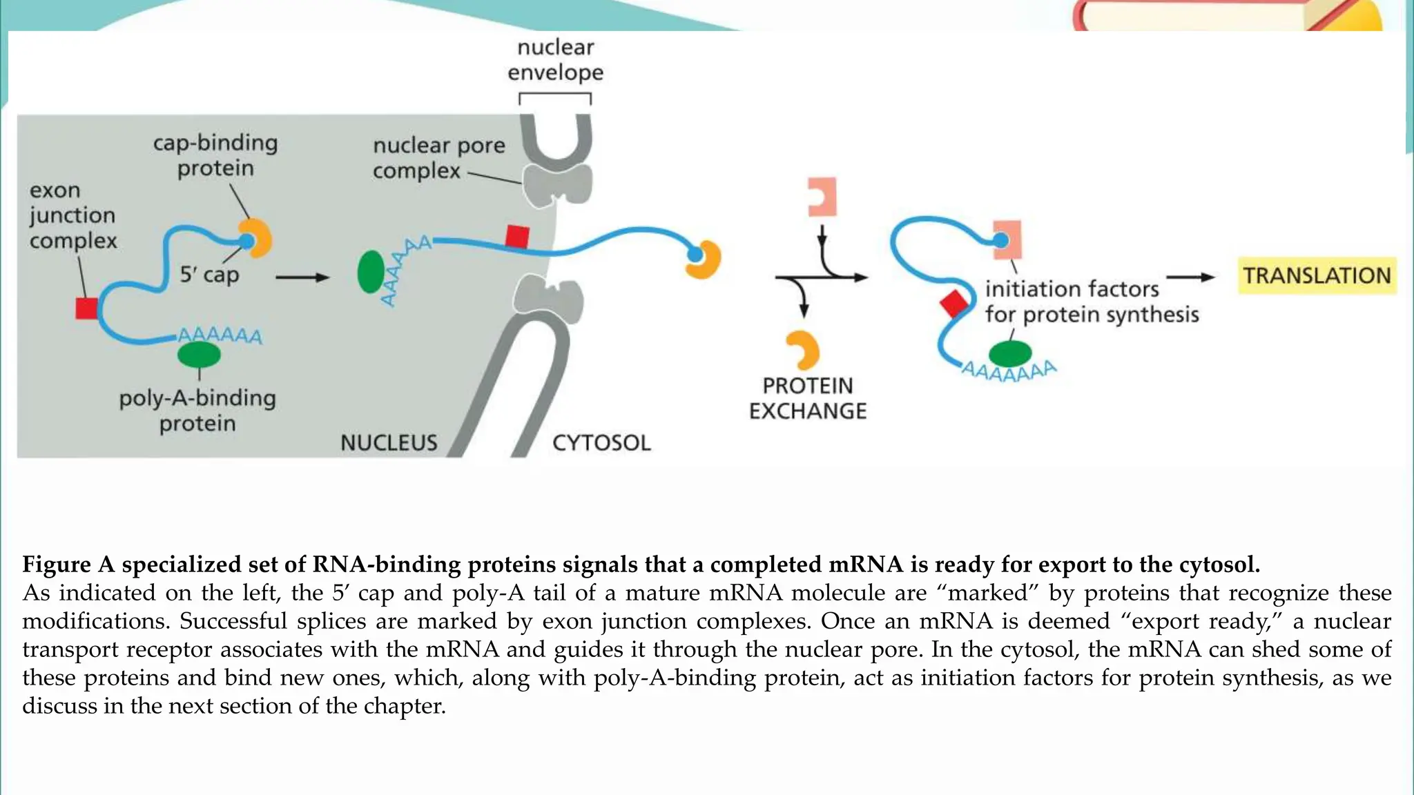
Dr. Salman Ul Islam
Unit#1 BASIC CELL FUNCTIONS
Topic: Transcription
Physiology-A
Transcription
Figure Genetic information directs the
synthesis of proteins.
The flow of genetic information from
DNA to RNA (transcription) and from
RNA to protein (translation) occurs in
all living cells. DNA can also be
copied—or replicated—to produce new
DNA molecules. The segments of DNA
that are transcribed into RNA are called
genes (orange).
Figure A cell can express different genes at
different rates.
In this and later figures, the portions of the
DNA that are not transcribed are shown in
gray.
Figure The chemical structure of RNA
differs slightly from that of DNA.
(A) RNA contains the sugar ribose,
which differs from deoxyribose, the
sugar used in DNA, by the presence of
an additional –OH group. (B) RNA
contains the base uracil, which differs
from thymine, the equivalent base in
DNA, by the absence of a –CH3 group.
(C) A short length of RNA. The
chemical linkage between nucleotides
in RNA—a phosphodiester bond—is
the same as that in DNA.
Figure Uracil forms a base pair with
adenine.
The hydrogen bonds that hold the base
pair together are shown in red. Uracil
has the same base-pairing properties as
thymine. Thus U-A base pairs in RNA
closely resemble T-A base pairs in
DNA.
Figure RNA molecules can fold into specific structures that are held together by hydrogen bonds between different base pairs.
RNA is largely single-stranded, but it often contains short stretches of nucleotides that can base-pair with complementary
sequences found elsewhere on the same molecule. These interactions—along with some “nonconventional” base-pair interactions
(e.g., A-G)—allow an RNA molecule to fold into a three-dimensional structure that is determined by its sequence of nucleotides.
(A) A diagram of a hypothetical, folded RNA structure showing only conventional (G-C and A-U) base-pair interactions (red). (B)
Formation of nonconventional base-pair interactions (green) folds the structure of the hypothetical RNA shown in (A) even further.
(C) Structure of an actual RNA molecule that is involved in RNA splicing. The considerable amount of double-helical structure
displayed by this RNA is produced by conventional base pairing.
Figure Transcription of a gene produces an RNA complementary to one strand of DNA.
The bottom strand of DNA in this example is called the template strand because it is used to guide the synthesis of
the RNA molecule. The nontemplate strand of the gene (here, shown at the top) is sometimes called the coding
strand because its sequence is equivalent to the RNA product, as shown. Which DNA strand serves as the template
varies, depending on the gene, as we discuss later. By convention, an RNA molecule is usually depicted with its 5ʹ
end—the first part to be synthesized—to the left.
Figure DNA is transcribed into RNA by the enzyme RNA polymerase.
(A) RNA polymerase (pale blue) moves stepwise along the DNA, unwinding the DNA helix in front of it. As it progresses, the polymerase
adds ribonucleotides one-by-one to the RNA chain, using an exposed DNA strand as a template. The resulting RNA transcript is thus single-
stranded and complementary to the template strand. As the polymerase moves along the DNA template, it displaces the newly formed
RNA, allowing the two strands of DNA behind the polymerase to rewind. A short region of hybrid DNA/RNA helix (approximately nine
nucleotides in length) therefore forms only transiently, causing a “window” of DNA/RNA helix to move along the DNA with the
polymerase. Note that although the primase discussed in Chapter 6 and RNA polymerase both synthesize RNA using a DNA template, they
are different enzymes, encoded by different genes.
Figure Many molecules of RNA polymerase can simultaneously transcribe the same gene.
Shown in this electron micrograph are two adjacent ribosomal genes on a single DNA molecule.
Molecules of RNA polymerase are barely visible as a series of tiny dots along the spine of the DNA
molecule; each polymerase has an RNA transcript (a short, fine thread) radiating from it. The RNA
molecules being transcribed from the two ribosomal genes—ribosomal RNAs (rRNAs)—are not
translated into protein, but are instead used directly as components of ribosomes, macromolecular
machines made of RNA and protein. The large particles that can be seen at the free, 5ʹ end of each rRNA
transcript are ribosomal proteins that have assembled on the ends of the growing transcripts.
TABLE 7–1 TYPES OF RNA PRODUCED IN CELLS
TABLE 7–2 THE THREE RNA POLYMERASES IN EUKARYOTIC CELLS
Figure To begin transcription, eukaryotic RNA polymerase II requires a set of general
transcription factors.
These factors are designated TFIIB, TFIID, and so on. (A) Most eukaryotic promoters
contain a DNA sequence called the TATA box. (B) The TATA box is recognized by a
subunit of the general transcription factor TFIID, called the TATA-binding protein (TBP).
For simplicity, the DNA distortion produced by the binding of the TBP is not shown. (C)
The binding of TFIID enables the adjacent binding of TFIIB. (D) The rest of the general
transcription factors, as well as the RNA polymerase itself, then assemble at the promoter.
(E) TFIIH pries apart the double helix at the transcription start point, using the energy of
ATP hydrolysis, which exposes the template strand of the gene. TFIIH also
phosphorylates RNA polymerase II, releasing the polymerase from most of the general
transcription factors, so it can begin transcription. The site of phosphorylation is a long
polypeptide “tail” that extends from the polymerase. Once the polymerase moves away
from the promoter, most of the general transcription factors are released from the DNA;
the exception is TFIID, which remains bound through multiple rounds of transcription
initiation.
Figure TATA-binding protein (TBP) binds to the TATA box (indicated by letters) and bends the DNA double helix.
TBP, a subunit of TFIID, distorts the DNA when it binds. TBP is a single polypeptide chain that is folded into two very similar domains (blue
and green). The protein sits atop the DNA double helix like a saddle on a bucking horse.
Figure Eukaryotic promoters contain sequences that promote the binding of the general transcription factors.
The location of each sequence and the general transcription factor that recognizes it are indicated. N stands for any nucleotide, and a slash
(/) indicates that either nucleotide can be found at the indicated position. For most start sites transcribed by RNA polymerase II, only two or
three of the four sequences are needed. Although most of these DNA sequences are located upstream of the transcription start site, one, at
+30, is located within the transcribed region of the gene.
Figure Before they can be translated, mRNA molecules made in the nucleus must be exported to the cytosol via pores in the nuclear
envelope (red arrows).
Shown here is a section of a liver cell nucleus. The nucleolus is where ribosomal RNAs are synthesized and combined with proteins to form
ribosomes, which are then exported to the cytosol. (From D.W. Fawcett, A Textbook of Histology, 12th ed. 1994. With permission from
Taylor & Francis Books UK.)
Figure Phosphorylation of the tail of RNA
polymerase II allows RNA-processing proteins to
assemble there.
Capping, polyadenylation, and splicing are all
modifications that occur as the RNA is being
synthesized. Note that the phosphates shown here are
in addition to the ones required for transcription
initiation.
Figure Eukaryotic mRNA molecules are modified by capping and polyadenylation.
(A) A eukaryotic mRNA has a cap at the 5ʹ end and a poly-A tail at the 3ʹ end. In addition to the nucleotide sequences that code for protein,
most mRNAs also contain extra, noncoding sequences, as shown. The noncoding portion at the 5ʹ end is called the 5ʹ untranslated region, or
5ʹ UTR, and that at the 3ʹ end is called the 3ʹ UTR. (B) The structure of the 5ʹ cap. Many eukaryotic mRNA caps carry an additional
modification: the 2ʹ-hydroxyl group on the second ribose sugar in the mRNA is methylated (not shown).
Figure Eukaryotic and bacterial genes are organized differently.
A bacterial gene consists of a single stretch of uninterrupted nucleotide sequence that encodes the amino acid sequence of a protein. In
contrast, the protein-coding sequences of most eukaryotic genes (exons) are interrupted by noncoding sequences (introns). Promoter
sequences are indicated in green.
Figure Most protein-coding human genes are broken into multiple exons and introns.
(A) The β-globin gene, which encodes one of the subunits of the oxygen-carrying protein hemoglobin,
contains 3 exons. (B) The gene that encodes Factor VIII, a protein that functions in the blood-clotting
pathway, contains 26 exons. Mutations in this large gene are responsible for the most prevalent form of
the blood disorder hemophilia.
Figure Special nucleotide sequences in a pre-mRNA transcript signal the beginning and the end of an intron.
Only the nucleotide sequences shown are required to remove an intron; the other positions in an intron can be occupied by any
nucleotide. The special sequences are recognized primarily by small nuclear ribonucleoproteins (snRNPs), which direct the
cleavage of the RNA at the intron–exon borders and catalyze the covalent linkage of the exon sequences. Here, in addition to the
standard symbols for nucleotides (A, C, G, U), R stands for either A or G; Y stands for either C or U; and N stands for any
nucleotide. The A shown in red forms the branch point of the lariat produced in the splicing reaction. The distances along the RNA
between the three splicing sequences are highly variable; however, the distance between the branch point and the 5ʹ splice junction
is typically much longer than that between the 3ʹ splice junction and the branch point. The splicing sequences shown are from
humans; similar sequences direct RNA splicing in other eukaryotes.
Figure An intron in a pre-mRNA molecule forms a branched structure during
RNA splicing.
In the first step, the branch-point adenine (red A) in the intron sequence attacks
the 5ʹ splice site and cuts the sugar–phosphate backbone of the RNA at this point.
In this process, the released 5ʹ end of the intron becomes covalently linked to the
2ʹ-OH group of the ribose of the adenine nucleotide to form a branched structure.
In the second step of splicing, the free 3ʹ-OH end of the exon sequence reacts with
the start of the next exon sequence, joining the two exons together into a
continuous coding sequence. The intron is released as a lariat structure, which is
eventually degraded in the nucleus.
Figure Splicing is carried out by a collection of RNA–
protein complexes called snRNPs.
Although there are five snRNPs and about 200 additional
proteins required for splicing, only the three most
important snRNPs—called U1, U2, and U6—are shown
here. In the first steps of splicing, U1 recognizes the 5ʹ
splice site and U2 recognizes the lariat branch-point site
through complementary base-pairing. U6 then “re-
checks” the 5ʹ splice site by displacing U1 and base-
pairing with this intron sequence itself. This “re-reading”
step improves the accuracy of splicing by double-
checking the 5ʹ splice site before carrying out the splicing
reaction. In the next steps, conformational changes in U2
and U6—triggered by the hydrolysis of ATP by
spliceosomal proteins (not shown)—drive the formation
of the spliceosome active site. Once the splicing reactions
have occurred (see Figure 7–21), the spliceosome deposits
a group of RNA-binding proteins, known as the exon
junction complex (red ), on the mRNA to mark the splice
site as successfully completed.
Figure 7–23 Some pre-mRNAs undergo alternative RNA splicing to produce different mRNAs and proteins from the same
gene.
Whereas all exons are transcribed, they can be skipped over by the spliceosome to produce alternatively spliced mRNAs, as shown.
Such skipping occurs when the splicing signals at the 5ʹ end of one intron are paired up with the branch-point and 3ʹ end of a
different intron. An important feature of alternative splicing is that exons can be skipped or included; however, their order—which
is specified in the DNA sequence—cannot be rearranged.
Figure A specialized set of RNA-binding proteins signals that a completed mRNA is ready for export to the cytosol.
As indicated on the left, the 5’ cap and poly-A tail of a mature mRNA molecule are “marked” by proteins that recognize these
modifications. Successful splices are marked by exon junction complexes. Once an mRNA is deemed “export ready,” a nuclear
transport receptor associates with the mRNA and guides it through the nuclear pore. In the cytosol, the mRNA can shed some of
these proteins and bind new ones, which, along with poly-A-binding protein, act as initiation factors for protein synthesis, as we
discuss in the next section of the chapter.
Figure Producing mRNA molecules is more complex in eukaryotes than it is in prokaryotes.
(A) In eukaryotic cells, the pre-mRNA molecule produced by transcription contains both intron and exon
sequences. Its two ends are modified by capping and polyadenylation, and the introns are removed by RNA
splicing. The completed mRNA is then transported from the nucleus to the cytosol, where it is translated into
protein. Although these steps are depicted as occurring one after the other, in reality they occur
simultaneously. For example, the RNA cap is usually added and splicing usually begins before transcription
has been completed. Because of this overlap, transcripts of the entire gene (including all introns and exons) do
not typically exist in the cell. Ultimately, mRNAs are degraded by RNAses in the cytosol and their nucleotide
building blocks are reused for transcription.

Transcription.pptx physiology lecture for you

  • 1.
    Dr. Salman UlIslam Unit#1 BASIC CELL FUNCTIONS Topic: Transcription Physiology-A
  • 2.
  • 3.
    Figure Genetic informationdirects the synthesis of proteins. The flow of genetic information from DNA to RNA (transcription) and from RNA to protein (translation) occurs in all living cells. DNA can also be copied—or replicated—to produce new DNA molecules. The segments of DNA that are transcribed into RNA are called genes (orange).
  • 5.
    Figure A cellcan express different genes at different rates. In this and later figures, the portions of the DNA that are not transcribed are shown in gray.
  • 7.
    Figure The chemicalstructure of RNA differs slightly from that of DNA. (A) RNA contains the sugar ribose, which differs from deoxyribose, the sugar used in DNA, by the presence of an additional –OH group. (B) RNA contains the base uracil, which differs from thymine, the equivalent base in DNA, by the absence of a –CH3 group. (C) A short length of RNA. The chemical linkage between nucleotides in RNA—a phosphodiester bond—is the same as that in DNA.
  • 8.
    Figure Uracil formsa base pair with adenine. The hydrogen bonds that hold the base pair together are shown in red. Uracil has the same base-pairing properties as thymine. Thus U-A base pairs in RNA closely resemble T-A base pairs in DNA.
  • 10.
    Figure RNA moleculescan fold into specific structures that are held together by hydrogen bonds between different base pairs. RNA is largely single-stranded, but it often contains short stretches of nucleotides that can base-pair with complementary sequences found elsewhere on the same molecule. These interactions—along with some “nonconventional” base-pair interactions (e.g., A-G)—allow an RNA molecule to fold into a three-dimensional structure that is determined by its sequence of nucleotides. (A) A diagram of a hypothetical, folded RNA structure showing only conventional (G-C and A-U) base-pair interactions (red). (B) Formation of nonconventional base-pair interactions (green) folds the structure of the hypothetical RNA shown in (A) even further. (C) Structure of an actual RNA molecule that is involved in RNA splicing. The considerable amount of double-helical structure displayed by this RNA is produced by conventional base pairing.
  • 12.
    Figure Transcription ofa gene produces an RNA complementary to one strand of DNA. The bottom strand of DNA in this example is called the template strand because it is used to guide the synthesis of the RNA molecule. The nontemplate strand of the gene (here, shown at the top) is sometimes called the coding strand because its sequence is equivalent to the RNA product, as shown. Which DNA strand serves as the template varies, depending on the gene, as we discuss later. By convention, an RNA molecule is usually depicted with its 5ʹ end—the first part to be synthesized—to the left.
  • 16.
    Figure DNA istranscribed into RNA by the enzyme RNA polymerase. (A) RNA polymerase (pale blue) moves stepwise along the DNA, unwinding the DNA helix in front of it. As it progresses, the polymerase adds ribonucleotides one-by-one to the RNA chain, using an exposed DNA strand as a template. The resulting RNA transcript is thus single- stranded and complementary to the template strand. As the polymerase moves along the DNA template, it displaces the newly formed RNA, allowing the two strands of DNA behind the polymerase to rewind. A short region of hybrid DNA/RNA helix (approximately nine nucleotides in length) therefore forms only transiently, causing a “window” of DNA/RNA helix to move along the DNA with the polymerase. Note that although the primase discussed in Chapter 6 and RNA polymerase both synthesize RNA using a DNA template, they are different enzymes, encoded by different genes.
  • 18.
    Figure Many moleculesof RNA polymerase can simultaneously transcribe the same gene. Shown in this electron micrograph are two adjacent ribosomal genes on a single DNA molecule. Molecules of RNA polymerase are barely visible as a series of tiny dots along the spine of the DNA molecule; each polymerase has an RNA transcript (a short, fine thread) radiating from it. The RNA molecules being transcribed from the two ribosomal genes—ribosomal RNAs (rRNAs)—are not translated into protein, but are instead used directly as components of ribosomes, macromolecular machines made of RNA and protein. The large particles that can be seen at the free, 5ʹ end of each rRNA transcript are ribosomal proteins that have assembled on the ends of the growing transcripts.
  • 21.
    TABLE 7–1 TYPESOF RNA PRODUCED IN CELLS
  • 24.
    TABLE 7–2 THETHREE RNA POLYMERASES IN EUKARYOTIC CELLS
  • 32.
    Figure To begintranscription, eukaryotic RNA polymerase II requires a set of general transcription factors. These factors are designated TFIIB, TFIID, and so on. (A) Most eukaryotic promoters contain a DNA sequence called the TATA box. (B) The TATA box is recognized by a subunit of the general transcription factor TFIID, called the TATA-binding protein (TBP). For simplicity, the DNA distortion produced by the binding of the TBP is not shown. (C) The binding of TFIID enables the adjacent binding of TFIIB. (D) The rest of the general transcription factors, as well as the RNA polymerase itself, then assemble at the promoter. (E) TFIIH pries apart the double helix at the transcription start point, using the energy of ATP hydrolysis, which exposes the template strand of the gene. TFIIH also phosphorylates RNA polymerase II, releasing the polymerase from most of the general transcription factors, so it can begin transcription. The site of phosphorylation is a long polypeptide “tail” that extends from the polymerase. Once the polymerase moves away from the promoter, most of the general transcription factors are released from the DNA; the exception is TFIID, which remains bound through multiple rounds of transcription initiation.
  • 33.
    Figure TATA-binding protein(TBP) binds to the TATA box (indicated by letters) and bends the DNA double helix. TBP, a subunit of TFIID, distorts the DNA when it binds. TBP is a single polypeptide chain that is folded into two very similar domains (blue and green). The protein sits atop the DNA double helix like a saddle on a bucking horse.
  • 34.
    Figure Eukaryotic promoterscontain sequences that promote the binding of the general transcription factors. The location of each sequence and the general transcription factor that recognizes it are indicated. N stands for any nucleotide, and a slash (/) indicates that either nucleotide can be found at the indicated position. For most start sites transcribed by RNA polymerase II, only two or three of the four sequences are needed. Although most of these DNA sequences are located upstream of the transcription start site, one, at +30, is located within the transcribed region of the gene.
  • 39.
    Figure Before theycan be translated, mRNA molecules made in the nucleus must be exported to the cytosol via pores in the nuclear envelope (red arrows). Shown here is a section of a liver cell nucleus. The nucleolus is where ribosomal RNAs are synthesized and combined with proteins to form ribosomes, which are then exported to the cytosol. (From D.W. Fawcett, A Textbook of Histology, 12th ed. 1994. With permission from Taylor & Francis Books UK.)
  • 40.
    Figure Phosphorylation ofthe tail of RNA polymerase II allows RNA-processing proteins to assemble there. Capping, polyadenylation, and splicing are all modifications that occur as the RNA is being synthesized. Note that the phosphates shown here are in addition to the ones required for transcription initiation.
  • 42.
    Figure Eukaryotic mRNAmolecules are modified by capping and polyadenylation. (A) A eukaryotic mRNA has a cap at the 5ʹ end and a poly-A tail at the 3ʹ end. In addition to the nucleotide sequences that code for protein, most mRNAs also contain extra, noncoding sequences, as shown. The noncoding portion at the 5ʹ end is called the 5ʹ untranslated region, or 5ʹ UTR, and that at the 3ʹ end is called the 3ʹ UTR. (B) The structure of the 5ʹ cap. Many eukaryotic mRNA caps carry an additional modification: the 2ʹ-hydroxyl group on the second ribose sugar in the mRNA is methylated (not shown).
  • 44.
    Figure Eukaryotic andbacterial genes are organized differently. A bacterial gene consists of a single stretch of uninterrupted nucleotide sequence that encodes the amino acid sequence of a protein. In contrast, the protein-coding sequences of most eukaryotic genes (exons) are interrupted by noncoding sequences (introns). Promoter sequences are indicated in green.
  • 45.
    Figure Most protein-codinghuman genes are broken into multiple exons and introns. (A) The β-globin gene, which encodes one of the subunits of the oxygen-carrying protein hemoglobin, contains 3 exons. (B) The gene that encodes Factor VIII, a protein that functions in the blood-clotting pathway, contains 26 exons. Mutations in this large gene are responsible for the most prevalent form of the blood disorder hemophilia.
  • 49.
    Figure Special nucleotidesequences in a pre-mRNA transcript signal the beginning and the end of an intron. Only the nucleotide sequences shown are required to remove an intron; the other positions in an intron can be occupied by any nucleotide. The special sequences are recognized primarily by small nuclear ribonucleoproteins (snRNPs), which direct the cleavage of the RNA at the intron–exon borders and catalyze the covalent linkage of the exon sequences. Here, in addition to the standard symbols for nucleotides (A, C, G, U), R stands for either A or G; Y stands for either C or U; and N stands for any nucleotide. The A shown in red forms the branch point of the lariat produced in the splicing reaction. The distances along the RNA between the three splicing sequences are highly variable; however, the distance between the branch point and the 5ʹ splice junction is typically much longer than that between the 3ʹ splice junction and the branch point. The splicing sequences shown are from humans; similar sequences direct RNA splicing in other eukaryotes.
  • 50.
    Figure An intronin a pre-mRNA molecule forms a branched structure during RNA splicing. In the first step, the branch-point adenine (red A) in the intron sequence attacks the 5ʹ splice site and cuts the sugar–phosphate backbone of the RNA at this point. In this process, the released 5ʹ end of the intron becomes covalently linked to the 2ʹ-OH group of the ribose of the adenine nucleotide to form a branched structure. In the second step of splicing, the free 3ʹ-OH end of the exon sequence reacts with the start of the next exon sequence, joining the two exons together into a continuous coding sequence. The intron is released as a lariat structure, which is eventually degraded in the nucleus.
  • 51.
    Figure Splicing iscarried out by a collection of RNA– protein complexes called snRNPs. Although there are five snRNPs and about 200 additional proteins required for splicing, only the three most important snRNPs—called U1, U2, and U6—are shown here. In the first steps of splicing, U1 recognizes the 5ʹ splice site and U2 recognizes the lariat branch-point site through complementary base-pairing. U6 then “re- checks” the 5ʹ splice site by displacing U1 and base- pairing with this intron sequence itself. This “re-reading” step improves the accuracy of splicing by double- checking the 5ʹ splice site before carrying out the splicing reaction. In the next steps, conformational changes in U2 and U6—triggered by the hydrolysis of ATP by spliceosomal proteins (not shown)—drive the formation of the spliceosome active site. Once the splicing reactions have occurred (see Figure 7–21), the spliceosome deposits a group of RNA-binding proteins, known as the exon junction complex (red ), on the mRNA to mark the splice site as successfully completed.
  • 53.
    Figure 7–23 Somepre-mRNAs undergo alternative RNA splicing to produce different mRNAs and proteins from the same gene. Whereas all exons are transcribed, they can be skipped over by the spliceosome to produce alternatively spliced mRNAs, as shown. Such skipping occurs when the splicing signals at the 5ʹ end of one intron are paired up with the branch-point and 3ʹ end of a different intron. An important feature of alternative splicing is that exons can be skipped or included; however, their order—which is specified in the DNA sequence—cannot be rearranged.
  • 55.
    Figure A specializedset of RNA-binding proteins signals that a completed mRNA is ready for export to the cytosol. As indicated on the left, the 5’ cap and poly-A tail of a mature mRNA molecule are “marked” by proteins that recognize these modifications. Successful splices are marked by exon junction complexes. Once an mRNA is deemed “export ready,” a nuclear transport receptor associates with the mRNA and guides it through the nuclear pore. In the cytosol, the mRNA can shed some of these proteins and bind new ones, which, along with poly-A-binding protein, act as initiation factors for protein synthesis, as we discuss in the next section of the chapter.
  • 58.
    Figure Producing mRNAmolecules is more complex in eukaryotes than it is in prokaryotes. (A) In eukaryotic cells, the pre-mRNA molecule produced by transcription contains both intron and exon sequences. Its two ends are modified by capping and polyadenylation, and the introns are removed by RNA splicing. The completed mRNA is then transported from the nucleus to the cytosol, where it is translated into protein. Although these steps are depicted as occurring one after the other, in reality they occur simultaneously. For example, the RNA cap is usually added and splicing usually begins before transcription has been completed. Because of this overlap, transcripts of the entire gene (including all introns and exons) do not typically exist in the cell. Ultimately, mRNAs are degraded by RNAses in the cytosol and their nucleotide building blocks are reused for transcription.