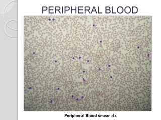
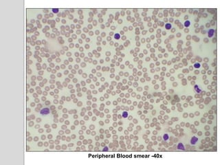
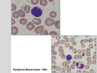
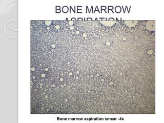
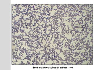
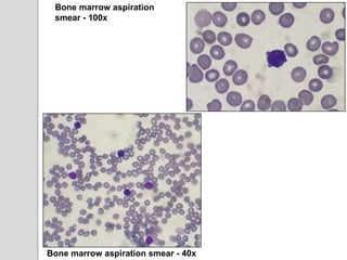
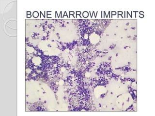
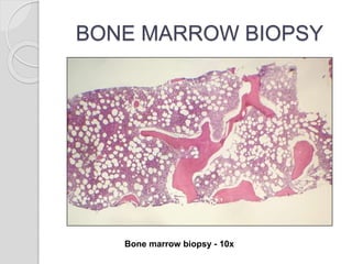
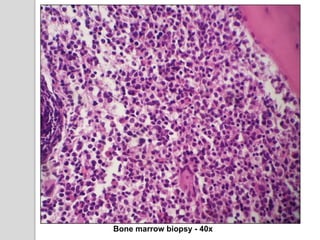
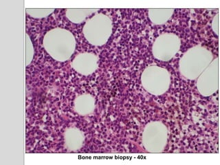
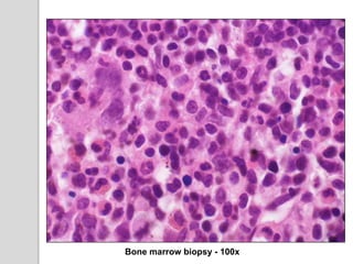
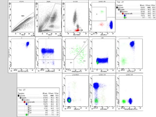
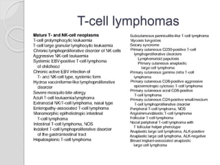
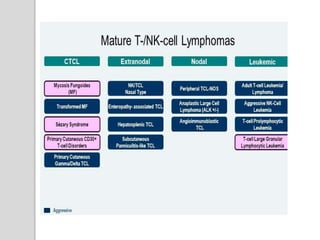
A rare cause of lymphocytosis
Dr. Soma Pradhan
Senior Resident
Department of Hematology
Sir Ganga Ram Hospital, New Delhi.
HISTORY
๏‚ง On 13th July 2019, a 42 yrs old male presented to
Clinical Hematology OPD with a complaints of pain
abdomen since last 15 days.
๏‚ง There was no history of fever and weight loss.
๏‚ง On examination, there is no pallor, lymphadenopathy
and spleen was not palpable.
๏‚— The patient was evaluated outside:
๏‚— CBC:
๏ƒ˜ Hb- 13.5 gm/dl
๏ƒ˜ TLC : 31,200/ยตl
๏ƒ˜ Platelet count :1,16,000/ยตl
๏‚— Peripheral smear examination:
๏ƒ˜ Marked lymphocytosis with 85% lymphocytes and
atypical lymphoid morphology.
INVESTIGATIONS
๏‚— CBC:
๏ƒ˜ Hb- 13.3 gm/dL
๏ƒ˜ RBC: 4.13 millions/ยตl
๏ƒ˜ PCV :38.9%
๏ƒ˜ MCV:94.2 fl
๏ƒ˜ MCH:32.2 pg
๏ƒ˜ RDW:12.5 %
๏ƒ˜ Platelets : 80,000/ยตl
๏ƒ˜ TLC : 31,660/ยตl
๏‚— DLC :
๏ƒ˜ Neutrophils- 11%, Lymphocytes-08%, Monocytes- 03%,
Eosinophils-01%, Abnormal lymphoid cells-77%
PERIPHERAL BLOOD
Peripheral Blood smear -4x
Peripheral Blood smear -40x
Peripheral Blood smear -100x
BONE MARROW
ASPIRATION
Bone marrow aspiration smear -4x
Bone marrow aspiration smear - 10x
Bone marrow aspiration
smear - 100x
Bone marrow aspiration smear - 40x
BONE MARROW IMPRINTS
BONE MARROW BIOPSY
Bone marrow biopsy - 10x
Bone marrow biopsy - 40x
Bone marrow biopsy - 100x
DIAGNOSIS
T- CELL PROLYMPHOCYTIC LEUKEMIA.
T-cell lymphomas
T-cell Prolymphocytic Leukemia:(T-PLL)
๏ฑ T-PLL accounts for 2% of mature lymphocytic leukemia
in adults.
๏ฑ Median Age : 61 yrs (30 -94 yrs)
๏ฑ Male >>> Female
๏ฑ Rare, aggressive T-cell neoplasm.
๏ฑ Patients with ataxia telangiectasia are at increased risk of
developing T-PLL
CLINICAL FEATURES
๏ƒ˜ Lymphocytosis with lymphocyte count >100 x 109/L
๏ƒ˜ Generalised lymphadenopathy
๏ƒ˜ Splenomegaly
๏ƒ˜ Skin infiltration โ€“ 20% of the cases
๏ƒ˜ Anemia
๏ƒ˜ Thrombocytopenia
DIFFERENTIATING FEATURES
Features T-PLL LGL leukemia Sezary
Syndrome
ATLL
Morphology prolymphocyte Large granular
lymphocyte
cerebriform Flower cell
Phenotype CD4+ CD8- CD8+ CD4- CD4+CD8- CD4+ CD8-
CD4+ CD8+ CD57+ CD3+ CD 25 +
CD7+ CD52+ CD16+/CD94+ CD7- CD26- CD7-
Histology
Spleen Red pulp Red pulp - -
Skin dermal - Dermal and
epidermal
Dermal and
epidermal
HTLV-1 Negative Negative Negative Positive
Clinical
course
Aggressive Indolent Chronic Aggressive-
Acute,
lymphatous
types
A B
C D
๏‚— A- T-PLL
๏‚— B- LGL Leukemia
๏‚— C- Sezary Syndrome
๏‚— D- ATLL
MOLECULAR GENETICS
๏‚ง T-cell receptor genes are rearranged and are identical, confirming a
clonal expansion of T-cells.
๏‚ง The most frequently observed group of cytogenetic abnormalities
involve chromosome 14.
๏‚ง inv(14) (q11;q32), t(14;14)(q11;q32)
๏‚ง The above translocations juxtaposes between TRA LOCUS with
TCL1A & TCL1B oncogenes at14q32.1
๏‚ง t(X;14)(q28;q11) :MTCP1 (mature T cell proliferation 1 gene),
which is located on the X chromosome.
๏‚ง Cytogenetic abnormalities involving chromosome 8 are the next
most frequently observed (idic(8p11), t(8;8) and trisomy 8q).
๏‚— Other recurrent abnormalities seen with conventional
techniques include :
๏ƒ˜ loss of 11q23 (ATM inactivation) together with additional
losses (22q, 13q, 6q, 9p, 12p,17p) and gains (22q and 6p).
๏ƒ˜ 12p13 deletion occurs in 50% of the cases.
๏ƒ˜ highly recurrent, largely exclusive, gain-of-function
mutations involving IL2RG, JAK1/3, and STAT5B,
which lead to constitutive STAT5 signaling.
DISEASE PROGRESSION &
PROGNOSIS
๏‚— T-PLL is aggressive and often resistant to therapy.
๏‚— Median survival is 1-2 years.
๏‚— The best responses have been reported with the monoclonal
antibody alemtuzumab (antiโ€•CD52).
๏‚— Autologous or allogeneic stem cell transplantation.
๏‚— High levels of expression of both TCL1 and AKT1 have been
identified as poor prognostic markers.
๏‚— STAT5B mutations have been documented to have a negative
prognostic impact.
T-PLL.pptx

T-PLL.pptx

  • 1.
    A rare causeof lymphocytosis Dr. Soma Pradhan Senior Resident Department of Hematology Sir Ganga Ram Hospital, New Delhi.
  • 2.
    HISTORY ๏‚ง On 13thJuly 2019, a 42 yrs old male presented to Clinical Hematology OPD with a complaints of pain abdomen since last 15 days. ๏‚ง There was no history of fever and weight loss. ๏‚ง On examination, there is no pallor, lymphadenopathy and spleen was not palpable.
  • 3.
    ๏‚— The patientwas evaluated outside: ๏‚— CBC: ๏ƒ˜ Hb- 13.5 gm/dl ๏ƒ˜ TLC : 31,200/ยตl ๏ƒ˜ Platelet count :1,16,000/ยตl ๏‚— Peripheral smear examination: ๏ƒ˜ Marked lymphocytosis with 85% lymphocytes and atypical lymphoid morphology.
  • 4.
    INVESTIGATIONS ๏‚— CBC: ๏ƒ˜ Hb-13.3 gm/dL ๏ƒ˜ RBC: 4.13 millions/ยตl ๏ƒ˜ PCV :38.9% ๏ƒ˜ MCV:94.2 fl ๏ƒ˜ MCH:32.2 pg ๏ƒ˜ RDW:12.5 % ๏ƒ˜ Platelets : 80,000/ยตl ๏ƒ˜ TLC : 31,660/ยตl ๏‚— DLC : ๏ƒ˜ Neutrophils- 11%, Lymphocytes-08%, Monocytes- 03%, Eosinophils-01%, Abnormal lymphoid cells-77%
  • 5.
  • 6.
  • 7.
  • 8.
  • 9.
  • 10.
    Bone marrow aspiration smear- 100x Bone marrow aspiration smear - 40x
  • 11.
  • 12.
    BONE MARROW BIOPSY Bonemarrow biopsy - 10x
  • 13.
  • 14.
  • 15.
  • 17.
  • 18.
  • 20.
    T-cell Prolymphocytic Leukemia:(T-PLL) ๏ฑT-PLL accounts for 2% of mature lymphocytic leukemia in adults. ๏ฑ Median Age : 61 yrs (30 -94 yrs) ๏ฑ Male >>> Female ๏ฑ Rare, aggressive T-cell neoplasm. ๏ฑ Patients with ataxia telangiectasia are at increased risk of developing T-PLL
  • 21.
    CLINICAL FEATURES ๏ƒ˜ Lymphocytosiswith lymphocyte count >100 x 109/L ๏ƒ˜ Generalised lymphadenopathy ๏ƒ˜ Splenomegaly ๏ƒ˜ Skin infiltration โ€“ 20% of the cases ๏ƒ˜ Anemia ๏ƒ˜ Thrombocytopenia
  • 22.
    DIFFERENTIATING FEATURES Features T-PLLLGL leukemia Sezary Syndrome ATLL Morphology prolymphocyte Large granular lymphocyte cerebriform Flower cell Phenotype CD4+ CD8- CD8+ CD4- CD4+CD8- CD4+ CD8- CD4+ CD8+ CD57+ CD3+ CD 25 + CD7+ CD52+ CD16+/CD94+ CD7- CD26- CD7- Histology Spleen Red pulp Red pulp - - Skin dermal - Dermal and epidermal Dermal and epidermal HTLV-1 Negative Negative Negative Positive Clinical course Aggressive Indolent Chronic Aggressive- Acute, lymphatous types
  • 23.
    A B C D ๏‚—A- T-PLL ๏‚— B- LGL Leukemia ๏‚— C- Sezary Syndrome ๏‚— D- ATLL
  • 24.
    MOLECULAR GENETICS ๏‚ง T-cellreceptor genes are rearranged and are identical, confirming a clonal expansion of T-cells. ๏‚ง The most frequently observed group of cytogenetic abnormalities involve chromosome 14. ๏‚ง inv(14) (q11;q32), t(14;14)(q11;q32) ๏‚ง The above translocations juxtaposes between TRA LOCUS with TCL1A & TCL1B oncogenes at14q32.1 ๏‚ง t(X;14)(q28;q11) :MTCP1 (mature T cell proliferation 1 gene), which is located on the X chromosome. ๏‚ง Cytogenetic abnormalities involving chromosome 8 are the next most frequently observed (idic(8p11), t(8;8) and trisomy 8q).
  • 26.
    ๏‚— Other recurrentabnormalities seen with conventional techniques include : ๏ƒ˜ loss of 11q23 (ATM inactivation) together with additional losses (22q, 13q, 6q, 9p, 12p,17p) and gains (22q and 6p). ๏ƒ˜ 12p13 deletion occurs in 50% of the cases. ๏ƒ˜ highly recurrent, largely exclusive, gain-of-function mutations involving IL2RG, JAK1/3, and STAT5B, which lead to constitutive STAT5 signaling.
  • 28.
    DISEASE PROGRESSION & PROGNOSIS ๏‚—T-PLL is aggressive and often resistant to therapy. ๏‚— Median survival is 1-2 years. ๏‚— The best responses have been reported with the monoclonal antibody alemtuzumab (antiโ€•CD52). ๏‚— Autologous or allogeneic stem cell transplantation. ๏‚— High levels of expression of both TCL1 and AKT1 have been identified as poor prognostic markers. ๏‚— STAT5B mutations have been documented to have a negative prognostic impact.

Editor's Notes

  • #22ย Lymphadenopathy, although present in a majority of patients, is rarely bulky. Anemia and thrombocytopenia are seen in up to one-half of patients.3 Erythematous or nodular skin rashes involving the trunk or limbs, peripheral edema, and pleuroperitoneal effusions may be seen in up to one-quarter of patients with T-PLL