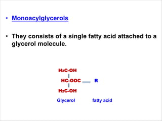
Lipids
Week 12
Dr. Sahena Ferdosh
What is lipid?
• Lipids are fatty acids & their derivatives, and
substances related biosynthetically or functionally to
these compounds.
• Fatty acids are merely carboxylic acids with long
hydrocarbon chains
• Esters ??
• Glycerol esters??
• Monoacylglycerols
• They consists of a single fatty acid attached to a
glycerol molecule.
H2C-OH
HC-OOC R
H2C-OH
Glycerol fatty acid
Diacylglycerol (DAG)
• These are also present in minor amounts.
H2C-OH
HC-OOC R1
H2C-OOC R2
Triacylglycerols (TAG)
• The majority of the glycerol esters exist in this form.
• Each OH group of the glycerol molecule is esterified with a
fatty acid molecule
H2C-OH H2C-OOC-R1
HC-OH + 3 Fatty acid (R1,2,3) HC-OOC-R2
H2C-OH H2C-OOC-R3
(Glycerol) (Triglyceride)
Basic structure of Triglyceride
Fatty acids
• Types of fatty acids
– Saturated & Unsaturated Fatty acids
– Cis: A cis configuration means that adjacent hydrogen atoms are
on the same side of the double bond.
– Trans: A trans configuration, by contrast, means that the next two
hydrogen atoms are bound to opposite sides of the double bond.
• Length of free fatty acid chains
– Short-chain fatty acids : aliphatic tails of fewer than
six carbons (i.e. butyric acid).
– Medium-chain fatty acids: aliphatic tails of 6–12 carbons
– Long-chain fatty acids: fatty acids with aliphatic tails 13 to
21 carbons.
– Very long chain fatty acids: fatty acids with aliphatic tails >
22 carbons.
Butyric (butanoic acid): CH3(CH2)2COOH C4:0
Caproic (hexanoic acid): CH3(CH2)4COOH C6:0
Caprylic (octanoic acid): CH3(CH2)6COOH C8:0
Capric (decanoic acid): CH3(CH2)8COOH C10:0
Lauric (dodecanoic acid): CH3(CH2)10COOH C12:0
Myristic (tetradecanoic
acid):
CH3(CH2)12COOH C14:0
Palmitic (hexadecanoic
acid):
CH3(CH2)14COOH C16:0
Stearic (octadecanoic acid): CH3(CH2)16COOH C18:0
Arachidic (eicosanoic acid): CH3(CH2)18COOH C20:0
Behenic (docosanoic acid): CH3(CH2)20COOH C22:0
Examples of Saturated Fatty Acids
Examples of unsaturated fatty acids:
Myristoleic
acid:
CH3(CH2)3CH=CH(CH2)7COOH C14:1
Palmitoleic
acid:
CH3(CH2)5CH=CH(CH2)7COOH C16:1
Oleic acid: CH3(CH2)7CH=CH(CH2)7COOH or cis-
Δ
9
C18:1
Linoleic acid: CH3(CH2)4CH=CHCH2CH=CH(CH2)7CO
OH
C18:2
Linolenic acid: CH3CH2CH=CHCH2CH=CHCH2CH=CH
(CH2)7COOH
C18:3
Arachidonic
acid:
CH3(CH2)4CH=CHCH2CH=CHCH2CH=
CHCH2CH=CH(CH2)3COOH
C20:4
Erucic acid: CH3(CH2)7CH=CH(CH2)11COOH C22:1
Docosahexaen
oic acid:
C22:6
Plant lipids
• Lipids are membrane constituents and function as carbon store
in seeds.
• Plant membrane lipids are mainly divided into glycerolipids,
sphingolipid and steroids.
• The polar glycerolipids are amphiphilic molecules. This property
enables them to form lipid bilayers.
• The polar glycerolipids are comprised mainly of fatty acids with
16 or 18 carbon atoms.
• The majority of these fatty acids are unsaturated and contain
one to three carbon-carbon double bonds.
• These double bonds are almost exclusively in the cis-
configuration and rarely in the trans-configuration.
• The fluidity of the membrane is governed by the proportion of
unsaturated fatty acids.
Membrane lipids
• In the phospholipids,
the head group
consists of a
phosphate residue
that is esterified with
a second alcoholic
compound such as
ethanolamine, choline,
serine, glycerol, or
inositol.
• In a green plant cell, about 70% to 80% of the total membrane
lipids are constituents of the thylakoid membranes.
• The main constituents of the chloroplast thylakoid and envelope
membranes are galactolipids.
• As a specialty of plants and cyanobacteria the galactolipids
MGDG and DGDG, and the sulfolipid (SL) are additionally
present in the membranes.
Membrane lipids
• Present in the plasma and ER membranes.
• This is a hydrocarbon chain containing double
bonds, an amino group in position 2, and two to
three hydroxyl groups in positions 1, 3, and 4.
eg: ceramide
Sphingolipid
• 17 carbon atoms in a 4-ring structure linked together from 3
cyclohexane rings & 1 cyclopentane ring and an eight-carbon
side chain on carbon 17.
• Example-cholesterol
Steroids
Triacylglycerols
• Triacylglycerols are primarily present in seeds but
also in some fruits such as olives or avocados.
• Triacylglycerols are deposited in oil bodies, also
termed oleosomes or lipid bodies.
• They are oil droplets, which are surrounded by a
lipid monolayer.
• Oil body proteins (oleosines, caloleosines,
steroleosines) are anchored to the lipid
monolayer and catalyze the mobilization of fatty
acids from the triacylglycerol store during seed
germination.
Fig. The incorporation of triacylglycerols in the ER membrane results
in the formation of oil bodies that are enclosed by a lipid monolayer.
The de novo synthesis of fatty acids
takes place in the plastids
• Acetyl CoA is a precursor for the synthesis of fatty acids.
• Sources of Acetyl CoA
– Plastids contain a pyruvate dehydrogenase complex, by
which pyruvate is oxidized to acetyl CoA, accompanied by
the reduction of NAD+.
– Chloroplasts contain a high activity of acetyl CoA synthetase,
which can convert acetate upon consumption of ATP to
acetyl CoA.
• In chloroplasts, photosynthesis provides the NADPH required for
the synthesis of fatty acids.
• In leucoplasts, it is provided by the oxidation of glucose 6-
phosphate via the oxidative pentose phosphate pathway.
Fig. Acetyl CoA synthesis
Plastids
Chloroplasts
Fatty acid synthesis
• Fatty acid synthesis starts with the carboxylation of acetyl CoA to
malonylCoA by acetyl CoA carboxylase, with the consumption of ATP.
• In a subsequent reaction, CoA is exchanged by acyl serine carrier
protein (ACP).
• Both ACP and CoA are covalently bound to a protein.
• The enzyme β-ketoacyl-ACP synthase III (KAS III) catalyzes the
condensation of acetyl CoA with malonyl-ACP.
• The reaction is irreversible due to the liberation of CO2.
• The acetoacetate thus formed remains bound as a thioester to ACP
and is reduced by NADPH to β-D-hydroxyacyl-ACP.
• Following the release of water, the carbon-carbon double bond
formed is reduced by NADPH to produce acyl ACP.
• The product is a fatty acid that has been elongated by two carbon
atoms
Step 1: Carboxilation
Fatty acid synthesis starts with the carboxylation of acetyl CoA
to malonyl CoA by acetyl CoA carboxylase, with the
consumption of ATP.
Acetyl-CoA
Step 2: Malonyl Transfer
Malonyl-CoA attaches to the acyl carrier protein
Step 3-Condensation
1. The enzyme β-ketoacyl-ACP synthase III catalyzes the
condensation of acetyl CoA with malonyl-ACP to produce β-
ketoacyl-ACP/acetoacetyl -ACP
2. The reaction is irreversible due to the liberation of CO2
Acetyl-CoA
Step 4: First reduction
The acetoacetate thus formed remains bound as a thioester to
ACP and is reduced by NADPH to β-D-hydroxyacyl-ACP.
Step 5: Dehydration
Carbon-carbon double bond is formed by releasing of water
Crotonyl-ACP
Step 6: 2nd reduction
Crotonyl--ACP is reduced by NADPH to produce acyl
ACP.
• Acyl ACP produced in the plastids has two
important functions:
– It acts as an acyl-donor for the synthesis of
plastid membrane lipids.
– Outside the plastids, acyl ACP is hydrolyzed by
acyl ACP thioesterases to release fatty acids
Acyl ACP
12P2-28
Chain Elongation (>16C)
• Acyl-ACP is a fatty acid that has been
elongated by two carbon atoms
• Two sites for chain lengthening:
1. Endoplasmic reticulum
–source of two carbons is malonyl-CoA
2. Mitochondrion
- source of two carbons is acetyl-CoA
Elongation of fatty acids in ER membrane
--Free palmitic acid
(16:0) synthesized in
cytoplasm is
elongated to stearic
acid (18:0) by the
addition 2 C at the
carboxyl terminal.
-2 carbons is added
essentially as in the
biosynthetic pathway.
ELONGATION IN ENDOPLASMIC
RETICULUM
Fig. A pool of acyl CoA with various chain lengths and desaturation is
present in the cytosol.
Glycerolipids synthesis in plastid
• G l y c e r o l 3 - p h o s p h a t e i s a p r e c u r s o r f o r
the synthesis of glycerolipids
• Glycerol 3-phosphate is synthesized by reduction of dihydroxyacetone
phosphate with NADH as reductant.
• Dihydroxyacetone phosphate reductases are present in the plastid
stroma as well as in the cytosol.
• In plastid lipid biosynthesis, the acyl residues are transferred directly
from acyl ACP to glycerol 3-phosphate.
• For the first acylation step, mostly an 18:1-, less frequently a 16:0-,
and more rarely an 18:0-acyl residue is esterified to carbon position 1
of glycerol 3-phosphate.
• The C-2-position, however, is always esterified with a 16:0-acyl
residue.
• Since this specificity is also observed in cyanobacteria, the glycerolipid
biosynthesis pathway of the plastids is called the prokaryotic pathway.
Fig. Membrane lipid synthesized in the plastids have
different fatty acids
Glycerolipids synthesis in plastid
• Acyl residues are transferred from acyl CoA.
• OH group in the C-1-position of glycerol 3-phosphate is
esterified with an 18:1-, 16:0-, or 18:0-acyl residue, and
position C-2 is always linked with a desaturated 18:n-acyl
residue.
Fig. Membrane lipid synthesized at the endoplasmic reticulum
Glycerolipid synthesis in ER; Eukaryotic pathway
Storage lipid mobilization
• At the beginning of germination, storage proteins are
degraded to amino acids, from which enzymes required
for the mobilization of the storage lipids.
• These enzymes include lipases, which catalyze the
hydrolysis of triacylglycerols to glycerol and fatty acids.
• The free fatty acids are first activated by reaction with
CoA to form thioesters and are then degraded to acetyl
CoA by β-oxidation.
• This process proceeds in specialized peroxisomes called
glyoxysomes.
β-oxidation of fatty acid
• β-oxidation represents the reversal of fatty acid synthesis.
• The fatty acids are first activated (CoA-thioesters) and then converted
to acetyl CoA and a fatty acid shortened by two carbon atoms.
• The sequence of reactions involve dehydrogenation via an FAD-
dependent oxidase, addition of water (hydroxylation), a second
dehydrogenation (by NAD+), and finally a thiolysis by CoA-SH.
• In acyl CoA dehydrogenation, hydrogen is transferred via an FAD
dependent oxidase to O2 to form H2O2. A catalase irreversibly
eliminates the toxic H2O2 at the site of its production by conversion to
water and oxygen.
• β-L Hydroxyacyl CoA is synthesized during the hydration of enoyl CoA.
• Hydrogen is transferred to NAD+ during the second dehydrogenation
step.
• In an irreversible reaction, CoA-SH-mediated thiolysis cleaves β-
ketoacyl CoA to synthesize one molecule each of acetyl CoA and of
acyl CoA shortened by two C atoms.
Fig. β-oxidation of
fatty acids in the
glyoxysomes
• 1. Acyl-CoA dehydrogenase
creates a double bond between
the alpha and beta carbons on
the fatty acid. The hydrogens are
added to FAD creating a molecule
of FADH2.
• A hydrogen is removed from the
alpha and beta carbon atoms.
Following the pink box, it will
become the acetyl CoA
• 2. Enoyl-CoA hydratase adds a
water to the double bond, putting
an OH onto the beta carbon and a
hydrogen onto the alpha carbon.
• Water is added to the molecule,
the pink box represents what will
become acetyl CoA
• 3.Beta-hydroxyacyl-CoA dehydrogenase
makes the carbon-OH bond a double
bond between the carbon and oxygen.
The lost hydrogen is added to NAD+
forming NADH.
• The hydrogen atoms are removed
forming a carbon oxygen double bond.
The pink box represents what will
become acetyl CoA
• 4. Acyl-CoA acetyltransferase cuts off
the first two carbon atoms, forming an
acetyl-CoA and a new fatty acid that is 2
carbons shorter than the previous fatty
acid.
• The acetyl CoA is removed from the
fatty acid, forming a shorter fatty acid
Comparison between fatty acid synthesis and fatty acid degradation
Lipoxygenase
• Lipoxygenase is involved in the synthesis of oxylipins,
which are defense and signal compounds.
• Oxylipins, which derive from the oxygenation of
unsaturated fatty acids, comprise a multiplicity of
various signal compounds in animals and plants.
• They are involved in defense reactions (e.g., as signal
components to regulate defense cascades), but also
as fungicides, bactericides, and insecticides, or as
volatile signals to attract predators, such as insects
that feed on herbivores.
• Moreover, they participate in wound healing,
regulate vegetative growth, and induce senescence

Topic 12.pdf

  • 1.
  • 2.
    What is lipid? •Lipids are fatty acids & their derivatives, and substances related biosynthetically or functionally to these compounds. • Fatty acids are merely carboxylic acids with long hydrocarbon chains • Esters ?? • Glycerol esters??
  • 3.
    • Monoacylglycerols • Theyconsists of a single fatty acid attached to a glycerol molecule. H2C-OH HC-OOC R H2C-OH Glycerol fatty acid
  • 4.
    Diacylglycerol (DAG) • Theseare also present in minor amounts. H2C-OH HC-OOC R1 H2C-OOC R2 Triacylglycerols (TAG) • The majority of the glycerol esters exist in this form. • Each OH group of the glycerol molecule is esterified with a fatty acid molecule
  • 5.
    H2C-OH H2C-OOC-R1 HC-OH +3 Fatty acid (R1,2,3) HC-OOC-R2 H2C-OH H2C-OOC-R3 (Glycerol) (Triglyceride) Basic structure of Triglyceride
  • 6.
    Fatty acids • Typesof fatty acids – Saturated & Unsaturated Fatty acids – Cis: A cis configuration means that adjacent hydrogen atoms are on the same side of the double bond. – Trans: A trans configuration, by contrast, means that the next two hydrogen atoms are bound to opposite sides of the double bond. • Length of free fatty acid chains – Short-chain fatty acids : aliphatic tails of fewer than six carbons (i.e. butyric acid). – Medium-chain fatty acids: aliphatic tails of 6–12 carbons – Long-chain fatty acids: fatty acids with aliphatic tails 13 to 21 carbons. – Very long chain fatty acids: fatty acids with aliphatic tails > 22 carbons.
  • 7.
    Butyric (butanoic acid):CH3(CH2)2COOH C4:0 Caproic (hexanoic acid): CH3(CH2)4COOH C6:0 Caprylic (octanoic acid): CH3(CH2)6COOH C8:0 Capric (decanoic acid): CH3(CH2)8COOH C10:0 Lauric (dodecanoic acid): CH3(CH2)10COOH C12:0 Myristic (tetradecanoic acid): CH3(CH2)12COOH C14:0 Palmitic (hexadecanoic acid): CH3(CH2)14COOH C16:0 Stearic (octadecanoic acid): CH3(CH2)16COOH C18:0 Arachidic (eicosanoic acid): CH3(CH2)18COOH C20:0 Behenic (docosanoic acid): CH3(CH2)20COOH C22:0 Examples of Saturated Fatty Acids
  • 8.
    Examples of unsaturatedfatty acids: Myristoleic acid: CH3(CH2)3CH=CH(CH2)7COOH C14:1 Palmitoleic acid: CH3(CH2)5CH=CH(CH2)7COOH C16:1 Oleic acid: CH3(CH2)7CH=CH(CH2)7COOH or cis- Δ 9 C18:1 Linoleic acid: CH3(CH2)4CH=CHCH2CH=CH(CH2)7CO OH C18:2 Linolenic acid: CH3CH2CH=CHCH2CH=CHCH2CH=CH (CH2)7COOH C18:3 Arachidonic acid: CH3(CH2)4CH=CHCH2CH=CHCH2CH= CHCH2CH=CH(CH2)3COOH C20:4 Erucic acid: CH3(CH2)7CH=CH(CH2)11COOH C22:1 Docosahexaen oic acid: C22:6
  • 9.
    Plant lipids • Lipidsare membrane constituents and function as carbon store in seeds. • Plant membrane lipids are mainly divided into glycerolipids, sphingolipid and steroids. • The polar glycerolipids are amphiphilic molecules. This property enables them to form lipid bilayers.
  • 10.
    • The polarglycerolipids are comprised mainly of fatty acids with 16 or 18 carbon atoms. • The majority of these fatty acids are unsaturated and contain one to three carbon-carbon double bonds. • These double bonds are almost exclusively in the cis- configuration and rarely in the trans-configuration. • The fluidity of the membrane is governed by the proportion of unsaturated fatty acids.
  • 11.
    Membrane lipids • Inthe phospholipids, the head group consists of a phosphate residue that is esterified with a second alcoholic compound such as ethanolamine, choline, serine, glycerol, or inositol.
  • 12.
    • In agreen plant cell, about 70% to 80% of the total membrane lipids are constituents of the thylakoid membranes. • The main constituents of the chloroplast thylakoid and envelope membranes are galactolipids. • As a specialty of plants and cyanobacteria the galactolipids MGDG and DGDG, and the sulfolipid (SL) are additionally present in the membranes. Membrane lipids
  • 13.
    • Present inthe plasma and ER membranes. • This is a hydrocarbon chain containing double bonds, an amino group in position 2, and two to three hydroxyl groups in positions 1, 3, and 4. eg: ceramide Sphingolipid
  • 14.
    • 17 carbonatoms in a 4-ring structure linked together from 3 cyclohexane rings & 1 cyclopentane ring and an eight-carbon side chain on carbon 17. • Example-cholesterol Steroids
  • 15.
    Triacylglycerols • Triacylglycerols areprimarily present in seeds but also in some fruits such as olives or avocados. • Triacylglycerols are deposited in oil bodies, also termed oleosomes or lipid bodies. • They are oil droplets, which are surrounded by a lipid monolayer. • Oil body proteins (oleosines, caloleosines, steroleosines) are anchored to the lipid monolayer and catalyze the mobilization of fatty acids from the triacylglycerol store during seed germination.
  • 16.
    Fig. The incorporationof triacylglycerols in the ER membrane results in the formation of oil bodies that are enclosed by a lipid monolayer.
  • 17.
    The de novosynthesis of fatty acids takes place in the plastids • Acetyl CoA is a precursor for the synthesis of fatty acids. • Sources of Acetyl CoA – Plastids contain a pyruvate dehydrogenase complex, by which pyruvate is oxidized to acetyl CoA, accompanied by the reduction of NAD+. – Chloroplasts contain a high activity of acetyl CoA synthetase, which can convert acetate upon consumption of ATP to acetyl CoA. • In chloroplasts, photosynthesis provides the NADPH required for the synthesis of fatty acids. • In leucoplasts, it is provided by the oxidation of glucose 6- phosphate via the oxidative pentose phosphate pathway.
  • 18.
    Fig. Acetyl CoAsynthesis Plastids Chloroplasts
  • 19.
    Fatty acid synthesis •Fatty acid synthesis starts with the carboxylation of acetyl CoA to malonylCoA by acetyl CoA carboxylase, with the consumption of ATP. • In a subsequent reaction, CoA is exchanged by acyl serine carrier protein (ACP). • Both ACP and CoA are covalently bound to a protein. • The enzyme β-ketoacyl-ACP synthase III (KAS III) catalyzes the condensation of acetyl CoA with malonyl-ACP. • The reaction is irreversible due to the liberation of CO2. • The acetoacetate thus formed remains bound as a thioester to ACP and is reduced by NADPH to β-D-hydroxyacyl-ACP. • Following the release of water, the carbon-carbon double bond formed is reduced by NADPH to produce acyl ACP. • The product is a fatty acid that has been elongated by two carbon atoms
  • 20.
    Step 1: Carboxilation Fattyacid synthesis starts with the carboxylation of acetyl CoA to malonyl CoA by acetyl CoA carboxylase, with the consumption of ATP. Acetyl-CoA
  • 21.
    Step 2: MalonylTransfer Malonyl-CoA attaches to the acyl carrier protein
  • 22.
    Step 3-Condensation 1. Theenzyme β-ketoacyl-ACP synthase III catalyzes the condensation of acetyl CoA with malonyl-ACP to produce β- ketoacyl-ACP/acetoacetyl -ACP 2. The reaction is irreversible due to the liberation of CO2 Acetyl-CoA
  • 23.
    Step 4: Firstreduction The acetoacetate thus formed remains bound as a thioester to ACP and is reduced by NADPH to β-D-hydroxyacyl-ACP.
  • 24.
    Step 5: Dehydration Carbon-carbondouble bond is formed by releasing of water Crotonyl-ACP
  • 25.
    Step 6: 2ndreduction Crotonyl--ACP is reduced by NADPH to produce acyl ACP.
  • 27.
    • Acyl ACPproduced in the plastids has two important functions: – It acts as an acyl-donor for the synthesis of plastid membrane lipids. – Outside the plastids, acyl ACP is hydrolyzed by acyl ACP thioesterases to release fatty acids Acyl ACP
  • 28.
    12P2-28 Chain Elongation (>16C) •Acyl-ACP is a fatty acid that has been elongated by two carbon atoms • Two sites for chain lengthening: 1. Endoplasmic reticulum –source of two carbons is malonyl-CoA 2. Mitochondrion - source of two carbons is acetyl-CoA
  • 29.
    Elongation of fattyacids in ER membrane --Free palmitic acid (16:0) synthesized in cytoplasm is elongated to stearic acid (18:0) by the addition 2 C at the carboxyl terminal. -2 carbons is added essentially as in the biosynthetic pathway.
  • 30.
  • 31.
    Fig. A poolof acyl CoA with various chain lengths and desaturation is present in the cytosol.
  • 32.
    Glycerolipids synthesis inplastid • G l y c e r o l 3 - p h o s p h a t e i s a p r e c u r s o r f o r the synthesis of glycerolipids • Glycerol 3-phosphate is synthesized by reduction of dihydroxyacetone phosphate with NADH as reductant. • Dihydroxyacetone phosphate reductases are present in the plastid stroma as well as in the cytosol. • In plastid lipid biosynthesis, the acyl residues are transferred directly from acyl ACP to glycerol 3-phosphate. • For the first acylation step, mostly an 18:1-, less frequently a 16:0-, and more rarely an 18:0-acyl residue is esterified to carbon position 1 of glycerol 3-phosphate. • The C-2-position, however, is always esterified with a 16:0-acyl residue. • Since this specificity is also observed in cyanobacteria, the glycerolipid biosynthesis pathway of the plastids is called the prokaryotic pathway.
  • 33.
    Fig. Membrane lipidsynthesized in the plastids have different fatty acids Glycerolipids synthesis in plastid
  • 34.
    • Acyl residuesare transferred from acyl CoA. • OH group in the C-1-position of glycerol 3-phosphate is esterified with an 18:1-, 16:0-, or 18:0-acyl residue, and position C-2 is always linked with a desaturated 18:n-acyl residue. Fig. Membrane lipid synthesized at the endoplasmic reticulum Glycerolipid synthesis in ER; Eukaryotic pathway
  • 35.
    Storage lipid mobilization •At the beginning of germination, storage proteins are degraded to amino acids, from which enzymes required for the mobilization of the storage lipids. • These enzymes include lipases, which catalyze the hydrolysis of triacylglycerols to glycerol and fatty acids. • The free fatty acids are first activated by reaction with CoA to form thioesters and are then degraded to acetyl CoA by β-oxidation. • This process proceeds in specialized peroxisomes called glyoxysomes.
  • 36.
    β-oxidation of fattyacid • β-oxidation represents the reversal of fatty acid synthesis. • The fatty acids are first activated (CoA-thioesters) and then converted to acetyl CoA and a fatty acid shortened by two carbon atoms. • The sequence of reactions involve dehydrogenation via an FAD- dependent oxidase, addition of water (hydroxylation), a second dehydrogenation (by NAD+), and finally a thiolysis by CoA-SH. • In acyl CoA dehydrogenation, hydrogen is transferred via an FAD dependent oxidase to O2 to form H2O2. A catalase irreversibly eliminates the toxic H2O2 at the site of its production by conversion to water and oxygen. • β-L Hydroxyacyl CoA is synthesized during the hydration of enoyl CoA. • Hydrogen is transferred to NAD+ during the second dehydrogenation step. • In an irreversible reaction, CoA-SH-mediated thiolysis cleaves β- ketoacyl CoA to synthesize one molecule each of acetyl CoA and of acyl CoA shortened by two C atoms.
  • 37.
    Fig. β-oxidation of fattyacids in the glyoxysomes
  • 38.
    • 1. Acyl-CoAdehydrogenase creates a double bond between the alpha and beta carbons on the fatty acid. The hydrogens are added to FAD creating a molecule of FADH2. • A hydrogen is removed from the alpha and beta carbon atoms. Following the pink box, it will become the acetyl CoA • 2. Enoyl-CoA hydratase adds a water to the double bond, putting an OH onto the beta carbon and a hydrogen onto the alpha carbon. • Water is added to the molecule, the pink box represents what will become acetyl CoA
  • 39.
    • 3.Beta-hydroxyacyl-CoA dehydrogenase makesthe carbon-OH bond a double bond between the carbon and oxygen. The lost hydrogen is added to NAD+ forming NADH. • The hydrogen atoms are removed forming a carbon oxygen double bond. The pink box represents what will become acetyl CoA • 4. Acyl-CoA acetyltransferase cuts off the first two carbon atoms, forming an acetyl-CoA and a new fatty acid that is 2 carbons shorter than the previous fatty acid. • The acetyl CoA is removed from the fatty acid, forming a shorter fatty acid
  • 40.
    Comparison between fattyacid synthesis and fatty acid degradation
  • 41.
    Lipoxygenase • Lipoxygenase isinvolved in the synthesis of oxylipins, which are defense and signal compounds. • Oxylipins, which derive from the oxygenation of unsaturated fatty acids, comprise a multiplicity of various signal compounds in animals and plants. • They are involved in defense reactions (e.g., as signal components to regulate defense cascades), but also as fungicides, bactericides, and insecticides, or as volatile signals to attract predators, such as insects that feed on herbivores. • Moreover, they participate in wound healing, regulate vegetative growth, and induce senescence