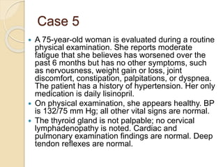
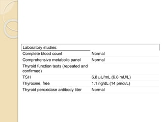
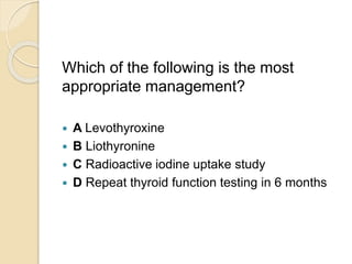
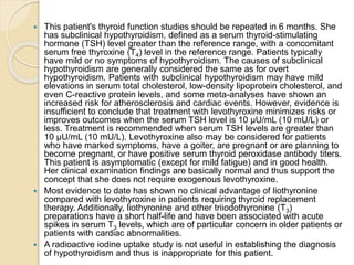
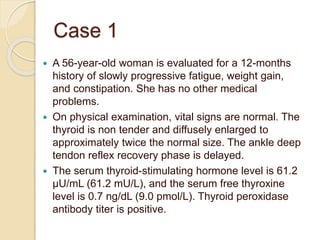
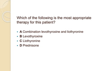
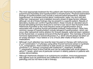
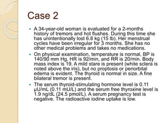
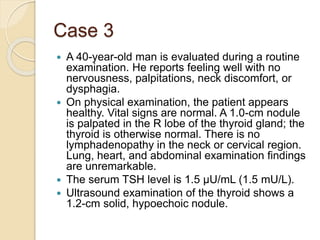
Thyroid Disorders
By Carla Sawan, MD.
Assistant Professor of Endocrinology,
University of Balamand, SGHUMC
Case 1
 A 56-year-old woman is evaluated for a 12-months
history of slowly progressive fatigue, weight gain,
and constipation. She has no other medical
problems.
 On physical examination, vital signs are normal. The
thyroid is non tender and diffusely enlarged to
approximately twice the normal size. The ankle deep
tendon reflex recovery phase is delayed.
 The serum thyroid-stimulating hormone level is 61.2
µU/mL (61.2 mU/L), and the serum free thyroxine
level is 0.7 ng/dL (9.0 pmol/L). Thyroid peroxidase
antibody titer is positive.
Which of the following is the most appropriate
therapy for this patient?
 A Combination levothyroxine and liothyronine
 B Levothyroxine
 C Liothyronine
 D Prednisone
 The most appropriate treatment for this patient with Hashimoto thyroiditis (chronic
autoimmune thyroiditis) and hypothyroidism is levothyroxine. Physical examination
findings of hypothyroidism may include a reduced basal temperature, diastolic
hypertension, an enlarged thyroid gland, bradycardia, pallor, dry and cold skin,
brittle hair, hoarseness, and a delayed recovery phase of deep tendon reflexes,
although not all of these findings may be present in a specific patient. Results of
laboratory studies can confirm hypothyroidism; these include an elevated serum
thyroid-stimulating hormone level and a normal or low serum free thyroxine level.
Hashimoto thyroiditis is the most frequent cause of hypothyroidism and is
associated with a positive thyroid peroxidase antibody measurement. Other less
common causes of hypothyroidism include iatrogenic hypothyroidism, which can
occur after radioactive iodine ablation for Graves disease, external-beam radiation
to the thyroid bed, or surgical removal of the thyroid gland. The mainstay of thyroid
hormone replacement is oral levothyroxine (T4), which should always be taken on
an empty stomach 1 hour before or 2 to 3 hours after intake of food or other
medications.
 Although much attention has recently been focused on therapy with liothyronine
(T3) or combination T3/T4therapy using either thyroid hormone extract or synthetic
T3/T4 combinations, most evidence to date shows no clinical advantage of
combined T3/T4 therapy compared with traditional T4 treatment. Available
T3 preparations have a short half-life and can be associated with acute spikes in
serum T3 levels, which are of particular concern in elderly patients or patients with
cardiac abnormalities.
 Although Hashimoto disease is an autoimmune condition, immunosuppression
with agents such as prednisone are ineffective in addressing the underlying
pathology and do not have a role in therapy.
Case 2
 A 34-year-old woman is evaluated for a 2-months
history of tremors and hot flushes. During this time she
has unintentionally lost 6.8 kg (15 lb). Her menstrual
cycles have been irregular for 3 months. She has no
other medical problems and takes no medications.
 On physical examination, temperature is normal, BP is
140/90 mm Hg, HR is 92/min, and RR is 20/min. Body
mass index is 19. A mild stare is present (white sclera is
noted above the iris), but no proptosis or periorbital
edema is evident. The thyroid is normal in size. A fine
bilateral tremor is present.
 The serum thyroid-stimulating hormone level is 0.11
µU/mL (0.11 mU/L) and the serum free thyroxine level is
1.9 ng/dL (24.5 pmol/L). A serum pregnancy test is
negative. The radioactive iodine uptake is low.
Which of the following is the most likely
diagnosis?
 A Graves disease
 B Lymphocytic thyroiditis
 C Toxic adenoma
 D Toxic multinodular goiter
 This patient most likely has silent lymphocytic thyroiditis (also known as chronic
thyroiditis). Thyroiditis involves transient destruction of thyroid tissue, which disrupts
follicles and causes the release of preformed thyroid hormone into the circulation.
Forms of destructive thyroiditis include subacute (de Quervain), silent, and
postpartum thyroiditis. Subacute thyroiditis most commonly occurs after a viral
infection and usually involves severe thyroid and neck pain; fever, fatigue, malaise,
anorexia, and myalgia are common. Silent thyroiditis is painless. Postpartum
thyroiditis is a subset of painless autoimmune thyroiditis and can occur up to 12
months after parturition. It affects 5% to 8% of pregnant women in the United States
and can recur with each pregnancy. Thyroiditis is associated with elevated serum
free thyroxine (T4) and triiodothyronine (T3) levels and a low serum thyroid-
stimulating hormone (TSH) level. The disorder usually follows a classic course of
approximately 6 weeks of thyrotoxicosis, a shorter period of euthyroidism, 4 to 6
weeks of hypothyroidism, and then restoration of euthyroidism. In patients with
subacute, silent, or postpartum thyroiditis or exposure to exogenous thyroid
hormones, the radioactive iodine uptake (RAIU) will be very low (<5% at 24 hours),
which indicates very little endogenous thyroid production.
 Graves disease is an autoimmune disorder that can affect the thyroid gland, ocular
muscles, orbital fat, and skin. Hyperthyroidism is by far the most common
manifestation. Examination of the thyroid may reveal the classic smooth, rubbery,
firm goiter, often associated with a bruit. Thyroid gland examination of patients with
toxic adenoma or toxic multinodular goiter usually reveals one or more palpable
nodule(s) or overall gland enlargement. Toxic multinodular goiter and toxic adenoma
result from an activating somatic mutation in the TSH receptor gene, which leads to
autonomy of function and secretion of excess T4 and T3 from the affected nodule(s).
Patients with Graves disease, toxic adenoma, or toxic multinodular goiter have an
elevated or high-normal RAIU, which indicates endogenous production of thyroid
hormones. A thyroid scan will show diffuse uptake of radioactive iodine in patients
with Graves disease or more focal uptake in those with toxic multinodular goiter or
toxic adenoma.
Case 3
 A 40-year-old man is evaluated during a routine
examination. He reports feeling well with no
nervousness, palpitations, neck discomfort, or
dysphagia.
 On physical examination, the patient appears
healthy. Vital signs are normal. A 1.0-cm nodule
is palpated in the R lobe of the thyroid gland; the
thyroid is otherwise normal. There is no
lymphadenopathy in the neck or cervical region.
Lung, heart, and abdominal examination findings
are unremarkable.
 The serum TSH level is 1.5 µU/mL (1.5 mU/L).
 Ultrasound examination of the thyroid shows a
1.2-cm solid, hypoechoic nodule.
Which of the following is the most
appropriate management?
 A Fine-needle aspiration biopsy
 B Radioactive iodine uptake scan
 C Repeat thyroid ultrasonography in 3 months
 D Right thyroid lobectomy
 This patient should have a fine-needle aspiration (FNA) biopsy of the thyroid nodule.
Most thyroid nodules are benign; only approximately 5% to 15% are malignant. FNA
biopsy is the most accurate method to determine whether a nodule is benign or
malignant. This is an outpatient procedure that allows cytologic categorization of the
cells within a nodule as benign or suspicious for malignancy, follicular neoplasm, or
papillary thyroid cancer. FNA biopsy is also the most sensitive and specific method to
help diagnose the cause of a thyroid nodule.
 Because thyroid nodules are extremely common (30% to 50% of healthy persons are
estimated to have a thyroid nodule on thyroid ultrasonography), guidelines have been
developed by the American Thyroid Association to maximize the effectiveness of thyroid
FNA biopsy in diagnosing malignancy in a cost-effective manner. These guidelines take
into account personal history and risk factors, family history, and ultrasound
characteristics of the nodule to help predict the likelihood of malignancy and the need for
thyroid FNA biopsy. FNA biopsy is recommended for any nodule greater than 1 cm in
diameter that is solid and hypoechoic on ultrasonography and for any nodule 2 cm or
greater that is mixed cystic-solid without worrisome sonographic characteristics. Biopsy
may be appropriate for smaller nodules (at least 5 mm in diameter) in patients with risk
factors, such as a history of radiation exposure, a family or personal history of thyroid
cancer, cervical lymphadenopathy, or suspicious ultrasound characteristics. FNA biopsy
is not routinely recommended for thyroid nodules less than 1 cm in diameter.
 A radioactive iodine uptake scan is appropriate for patients with a suppressed serum
thyroid-stimulating hormone level to evaluate for a possible toxic nodule (or toxic
multinodular goiter), which is not applicable in this patient.
 Because the size and characteristics of this patient's nodule meet the guideline criteria
for biopsy, repeat thyroid ultrasonography would not be appropriate. However,
ultrasound characteristics of thyroid nodules are very helpful in assessing malignant
potential, and repeat ultrasonography for follow-up of lesions without clear indications for
biopsy but with suspicious features may be helpful in selected patients.
 Thyroid lobectomy is generally reserved for treatment of cancerous thyroid nodules but
would be premature in this patient in whom a diagnosis of malignancy has not been
established.
Case 4
 A 27-year-old woman is evaluated during a routine
examination. She is in the 4th week of an uneventful
pregnancy. The patient has a 3-year history of
primary hypothyroidism due to Hashimoto
thyroiditis (chronic autoimmune thyroiditis) that is
treated with levothyroxine, 125 µg/d. She also
takes prenatal vitamins and iron sulfate.
 On physical examination, temperature is normal,
BP is 128/80 mm Hg, HR is 95/min, and RR is
18/min and regular. Body mass index is 25. The
thyroid gland is smooth and slightly enlarged
without a bruit or nodules. Lung, cardiac, skin, and
neurologic examination findings are normal.
 The serum TSH level is 4.2 µU/mL (4.2 mU/L) and
the serum free T4 level is 1.6 ng/dL (21 pmol/L).
Which of the following is the most
appropriate management?
 A Increase the levothyroxine dose by 10% now
 B Increase the levothyroxine dose by 30% now
 C Repeat thyroid function tests in 5 weeks
 D Repeat thyroid function tests in the second
trimester
 This patient's levothyroxine (T4) dose should be increased by 30%
now, and the thyroid function tests should be repeated in 2 to 4 weeks.
Pregnancy is known to increase levothyroxine requirements in most
patients receiving thyroid replacement therapy, and this expected
increase should be anticipated by increasing the levothyroxine dose.
This is typically increased in the first (and sometimes in the second)
trimester of pregnancy, with a possible total increase of 30% to 50%,
and an increase in levothyroxine dose in this range to maintain the
thyroid-stimulating hormone (TSH) level between approximately 0.1
and 2.5 µU/mL (0.1-2.5 mU/L) is associated with fewer maternal and
fetal complications. The fetus is largely dependent on transplacental
transfer of maternal thyroid hormones during the first 12 weeks of
gestation, and the presence of maternal subclinical or overt
hypothyroidism may be associated with subsequent fetal
neurocognitive impairment, increased risk of premature birth, low birth
weight, increased miscarriage rate, and even an increased risk of fetal
death. In pregnant women with hypothyroidism, thyroid function testing
should be frequent, preferably every 4 weeks, to protect the health of
mother and fetus and to avoid pregnancy complications.
 Continuing the current levothyroxine dose is inappropriate in this
patient because her TSH level is already too high (4.2 µU/mL [4.2
mU/L]). TSH levels generally should range from 0.1 to 2.5 µU/mL (0.1-
2.5 mU/L) in the first trimester, 0.2 to 3.0 µU/mL (0.2-3.0 mU/L) in the
second trimester, and 0.3 to 3.0 µU/mL (0.3-3.0 mU/L) in the third
trimester.
Case 5
 A 75-year-old woman is evaluated during a routine
physical examination. She reports moderate
fatigue that she believes has worsened over the
past 6 months but has no other symptoms, such
as nervousness, weight gain or loss, joint
discomfort, constipation, palpitations, or dyspnea.
The patient has a history of hypertension. Her only
medication is daily lisinopril.
 On physical examination, she appears healthy. BP
is 132/75 mm Hg; all other vital signs are normal.
 The thyroid gland is not palpable; no cervical
lymphadenopathy is noted. Cardiac and
pulmonary examination findings are normal. Deep
tendon reflexes are normal.
Laboratory studies:
Complete blood count Normal
Comprehensive metabolic panel Normal
Thyroid function tests (repeated and
confirmed)
TSH 6.8 µU/mL (6.8 mU/L)
Thyroxine, free 1.1 ng/dL (14 pmol/L)
Thyroid peroxidase antibody titer Normal
Which of the following is the most
appropriate management?
 A Levothyroxine
 B Liothyronine
 C Radioactive iodine uptake study
 D Repeat thyroid function testing in 6 months
 This patient's thyroid function studies should be repeated in 6 months. She
has subclinical hypothyroidism, defined as a serum thyroid-stimulating
hormone (TSH) level greater than the reference range, with a concomitant
serum free thyroxine (T4) level in the reference range. Patients typically
have mild or no symptoms of hypothyroidism. The causes of subclinical
hypothyroidism are generally considered the same as for overt
hypothyroidism. Patients with subclinical hypothyroidism may have mild
elevations in serum total cholesterol, low-density lipoprotein cholesterol, and
even C-reactive protein levels, and some meta-analyses have shown an
increased risk for atherosclerosis and cardiac events. However, evidence is
insufficient to conclude that treatment with levothyroxine minimizes risks or
improves outcomes when the serum TSH level is 10 µU/mL (10 mU/L) or
less. Treatment is recommended when serum TSH levels are greater than
10 µU/mL (10 mU/L). Levothyroxine also may be considered for patients
who have marked symptoms, have a goiter, are pregnant or are planning to
become pregnant, or have positive serum thyroid peroxidase antibody titers.
This patient is asymptomatic (except for mild fatigue) and in good health.
Her clinical examination findings are basically normal and thus support the
concept that she does not require exogenous levothyroxine.
 Most evidence to date has shown no clinical advantage of liothyronine
compared with levothyroxine in patients requiring thyroid replacement
therapy. Additionally, liothyronine and other triiodothyronine (T3)
preparations have a short half-life and have been associated with acute
spikes in serum T3 levels, which are of particular concern in older patients or
patients with cardiac abnormalities.
 A radioactive iodine uptake study is not useful in establishing the diagnosis
of hypothyroidism and thus is inappropriate for this patient.
Case 6
 An 18-year-old woman is evaluated for progressively
worsening tachycardia, nervousness, decreased exercise
tolerance, and weight loss over the past 6 months. Her
medical history is otherwise unremarkable, and she takes
no medications.
 On physical examination, temperature is normal, BP is
152/82 mm Hg, HR is 122/min, and RR is 16/min. Body
mass index is 19.
 There is no proptosis. Examination of the thyroid gland
shows symmetric, non-nodular enlargement to
approximately twice normal size. Cardiac examination
reveals regular tachycardia with a grade 2/6 systolic
ejection murmur at the right upper sternal border. The
remainder of the examination is unremarkable.
 The serum TSH level is less than 0.01 µU/mL (0.01 mU/L)
and the serum free thyroxine level is 5.5 ng/dL (71.0
pmol/L). A urine pregnancy test is negative.
Which of the following is the most
appropriate treatment for this patient?
 A Atenolol
 B Atenolol and methimazole
 C Methimazole
 D Radioactive iodine and methimazole
 The most appropriate medical regimen for this patient with Graves disease is atenolol
and methimazole. Graves disease can present with either subclinical or overt
thyrotoxicosis. Physical examination findings may include tachycardia; an elevated
systolic blood pressure with a widened pulse pressure; a palpable goiter, which is
classically smooth; a thyrotoxic stare due to lid retraction; proptosis; and, infrequently,
an infiltrative dermopathy. To control this patient's tachycardia, a β-blocker is
indicated. Although all β-blockers are effective, atenolol is frequently used because of
its β-1 selectivity and long half-life that allows once daily dosing in some patients.
Given the clinical and laboratory findings, this patient also has moderate
hyperthyroidism that can be treated with either methimazole or propylthiouracil.
Methimazole, which generally has fewer side effects and results in quicker
achievement of the euthyroid state than propylthiouracil, is preferred in most patients.
Because of a presumed immunomodulatory effect, antithyroidal drugs result in drug-
free remission rates of between 30% and 50% in patients with Graves disease who
are treated for 1 year.
 Atenolol alone would only address this patient's adrenergic symptoms and not reduce
her thyroid hormone levels, and methimazole alone would not immediately address
her tachycardia.
 Radioactive iodine therapy preceded or followed by adjunctive therapy with an
antithyroidal drug is occasionally used to treat Graves disease. An antithyroidal drug
is given in an attempt to decrease the risk of a transient worsening of the
thyrotoxicosis after thyroid ablation. Because antithyroidal drugs render the thyroid
radioresistant, they must be stopped for several days before and after giving the
radioactive iodine. Although an occasional patient becomes euthyroid after radioactive
iodine administration, the expected outcome is hypothyroidism, which typically occurs
within 2 to 3 months of therapy, at which time thyroid hormone replacement therapy is
begun. Although this therapy might be a reasonable long term management option in
this patient, it would not address her acute tachycardia and other hyperthyroid
symptoms, which are more effectively treated with β-blockade.

Thyroid disorders

  • 1.
    Thyroid Disorders By CarlaSawan, MD. Assistant Professor of Endocrinology, University of Balamand, SGHUMC
  • 2.
    Case 1  A56-year-old woman is evaluated for a 12-months history of slowly progressive fatigue, weight gain, and constipation. She has no other medical problems.  On physical examination, vital signs are normal. The thyroid is non tender and diffusely enlarged to approximately twice the normal size. The ankle deep tendon reflex recovery phase is delayed.  The serum thyroid-stimulating hormone level is 61.2 µU/mL (61.2 mU/L), and the serum free thyroxine level is 0.7 ng/dL (9.0 pmol/L). Thyroid peroxidase antibody titer is positive.
  • 3.
    Which of thefollowing is the most appropriate therapy for this patient?  A Combination levothyroxine and liothyronine  B Levothyroxine  C Liothyronine  D Prednisone
  • 4.
     The mostappropriate treatment for this patient with Hashimoto thyroiditis (chronic autoimmune thyroiditis) and hypothyroidism is levothyroxine. Physical examination findings of hypothyroidism may include a reduced basal temperature, diastolic hypertension, an enlarged thyroid gland, bradycardia, pallor, dry and cold skin, brittle hair, hoarseness, and a delayed recovery phase of deep tendon reflexes, although not all of these findings may be present in a specific patient. Results of laboratory studies can confirm hypothyroidism; these include an elevated serum thyroid-stimulating hormone level and a normal or low serum free thyroxine level. Hashimoto thyroiditis is the most frequent cause of hypothyroidism and is associated with a positive thyroid peroxidase antibody measurement. Other less common causes of hypothyroidism include iatrogenic hypothyroidism, which can occur after radioactive iodine ablation for Graves disease, external-beam radiation to the thyroid bed, or surgical removal of the thyroid gland. The mainstay of thyroid hormone replacement is oral levothyroxine (T4), which should always be taken on an empty stomach 1 hour before or 2 to 3 hours after intake of food or other medications.  Although much attention has recently been focused on therapy with liothyronine (T3) or combination T3/T4therapy using either thyroid hormone extract or synthetic T3/T4 combinations, most evidence to date shows no clinical advantage of combined T3/T4 therapy compared with traditional T4 treatment. Available T3 preparations have a short half-life and can be associated with acute spikes in serum T3 levels, which are of particular concern in elderly patients or patients with cardiac abnormalities.  Although Hashimoto disease is an autoimmune condition, immunosuppression with agents such as prednisone are ineffective in addressing the underlying pathology and do not have a role in therapy.
  • 5.
    Case 2  A34-year-old woman is evaluated for a 2-months history of tremors and hot flushes. During this time she has unintentionally lost 6.8 kg (15 lb). Her menstrual cycles have been irregular for 3 months. She has no other medical problems and takes no medications.  On physical examination, temperature is normal, BP is 140/90 mm Hg, HR is 92/min, and RR is 20/min. Body mass index is 19. A mild stare is present (white sclera is noted above the iris), but no proptosis or periorbital edema is evident. The thyroid is normal in size. A fine bilateral tremor is present.  The serum thyroid-stimulating hormone level is 0.11 µU/mL (0.11 mU/L) and the serum free thyroxine level is 1.9 ng/dL (24.5 pmol/L). A serum pregnancy test is negative. The radioactive iodine uptake is low.
  • 6.
    Which of thefollowing is the most likely diagnosis?  A Graves disease  B Lymphocytic thyroiditis  C Toxic adenoma  D Toxic multinodular goiter
  • 7.
     This patientmost likely has silent lymphocytic thyroiditis (also known as chronic thyroiditis). Thyroiditis involves transient destruction of thyroid tissue, which disrupts follicles and causes the release of preformed thyroid hormone into the circulation. Forms of destructive thyroiditis include subacute (de Quervain), silent, and postpartum thyroiditis. Subacute thyroiditis most commonly occurs after a viral infection and usually involves severe thyroid and neck pain; fever, fatigue, malaise, anorexia, and myalgia are common. Silent thyroiditis is painless. Postpartum thyroiditis is a subset of painless autoimmune thyroiditis and can occur up to 12 months after parturition. It affects 5% to 8% of pregnant women in the United States and can recur with each pregnancy. Thyroiditis is associated with elevated serum free thyroxine (T4) and triiodothyronine (T3) levels and a low serum thyroid- stimulating hormone (TSH) level. The disorder usually follows a classic course of approximately 6 weeks of thyrotoxicosis, a shorter period of euthyroidism, 4 to 6 weeks of hypothyroidism, and then restoration of euthyroidism. In patients with subacute, silent, or postpartum thyroiditis or exposure to exogenous thyroid hormones, the radioactive iodine uptake (RAIU) will be very low (<5% at 24 hours), which indicates very little endogenous thyroid production.  Graves disease is an autoimmune disorder that can affect the thyroid gland, ocular muscles, orbital fat, and skin. Hyperthyroidism is by far the most common manifestation. Examination of the thyroid may reveal the classic smooth, rubbery, firm goiter, often associated with a bruit. Thyroid gland examination of patients with toxic adenoma or toxic multinodular goiter usually reveals one or more palpable nodule(s) or overall gland enlargement. Toxic multinodular goiter and toxic adenoma result from an activating somatic mutation in the TSH receptor gene, which leads to autonomy of function and secretion of excess T4 and T3 from the affected nodule(s). Patients with Graves disease, toxic adenoma, or toxic multinodular goiter have an elevated or high-normal RAIU, which indicates endogenous production of thyroid hormones. A thyroid scan will show diffuse uptake of radioactive iodine in patients with Graves disease or more focal uptake in those with toxic multinodular goiter or toxic adenoma.
  • 8.
    Case 3  A40-year-old man is evaluated during a routine examination. He reports feeling well with no nervousness, palpitations, neck discomfort, or dysphagia.  On physical examination, the patient appears healthy. Vital signs are normal. A 1.0-cm nodule is palpated in the R lobe of the thyroid gland; the thyroid is otherwise normal. There is no lymphadenopathy in the neck or cervical region. Lung, heart, and abdominal examination findings are unremarkable.  The serum TSH level is 1.5 µU/mL (1.5 mU/L).  Ultrasound examination of the thyroid shows a 1.2-cm solid, hypoechoic nodule.
  • 9.
    Which of thefollowing is the most appropriate management?  A Fine-needle aspiration biopsy  B Radioactive iodine uptake scan  C Repeat thyroid ultrasonography in 3 months  D Right thyroid lobectomy
  • 10.
     This patientshould have a fine-needle aspiration (FNA) biopsy of the thyroid nodule. Most thyroid nodules are benign; only approximately 5% to 15% are malignant. FNA biopsy is the most accurate method to determine whether a nodule is benign or malignant. This is an outpatient procedure that allows cytologic categorization of the cells within a nodule as benign or suspicious for malignancy, follicular neoplasm, or papillary thyroid cancer. FNA biopsy is also the most sensitive and specific method to help diagnose the cause of a thyroid nodule.  Because thyroid nodules are extremely common (30% to 50% of healthy persons are estimated to have a thyroid nodule on thyroid ultrasonography), guidelines have been developed by the American Thyroid Association to maximize the effectiveness of thyroid FNA biopsy in diagnosing malignancy in a cost-effective manner. These guidelines take into account personal history and risk factors, family history, and ultrasound characteristics of the nodule to help predict the likelihood of malignancy and the need for thyroid FNA biopsy. FNA biopsy is recommended for any nodule greater than 1 cm in diameter that is solid and hypoechoic on ultrasonography and for any nodule 2 cm or greater that is mixed cystic-solid without worrisome sonographic characteristics. Biopsy may be appropriate for smaller nodules (at least 5 mm in diameter) in patients with risk factors, such as a history of radiation exposure, a family or personal history of thyroid cancer, cervical lymphadenopathy, or suspicious ultrasound characteristics. FNA biopsy is not routinely recommended for thyroid nodules less than 1 cm in diameter.  A radioactive iodine uptake scan is appropriate for patients with a suppressed serum thyroid-stimulating hormone level to evaluate for a possible toxic nodule (or toxic multinodular goiter), which is not applicable in this patient.  Because the size and characteristics of this patient's nodule meet the guideline criteria for biopsy, repeat thyroid ultrasonography would not be appropriate. However, ultrasound characteristics of thyroid nodules are very helpful in assessing malignant potential, and repeat ultrasonography for follow-up of lesions without clear indications for biopsy but with suspicious features may be helpful in selected patients.  Thyroid lobectomy is generally reserved for treatment of cancerous thyroid nodules but would be premature in this patient in whom a diagnosis of malignancy has not been established.
  • 11.
    Case 4  A27-year-old woman is evaluated during a routine examination. She is in the 4th week of an uneventful pregnancy. The patient has a 3-year history of primary hypothyroidism due to Hashimoto thyroiditis (chronic autoimmune thyroiditis) that is treated with levothyroxine, 125 µg/d. She also takes prenatal vitamins and iron sulfate.  On physical examination, temperature is normal, BP is 128/80 mm Hg, HR is 95/min, and RR is 18/min and regular. Body mass index is 25. The thyroid gland is smooth and slightly enlarged without a bruit or nodules. Lung, cardiac, skin, and neurologic examination findings are normal.  The serum TSH level is 4.2 µU/mL (4.2 mU/L) and the serum free T4 level is 1.6 ng/dL (21 pmol/L).
  • 12.
    Which of thefollowing is the most appropriate management?  A Increase the levothyroxine dose by 10% now  B Increase the levothyroxine dose by 30% now  C Repeat thyroid function tests in 5 weeks  D Repeat thyroid function tests in the second trimester
  • 13.
     This patient'slevothyroxine (T4) dose should be increased by 30% now, and the thyroid function tests should be repeated in 2 to 4 weeks. Pregnancy is known to increase levothyroxine requirements in most patients receiving thyroid replacement therapy, and this expected increase should be anticipated by increasing the levothyroxine dose. This is typically increased in the first (and sometimes in the second) trimester of pregnancy, with a possible total increase of 30% to 50%, and an increase in levothyroxine dose in this range to maintain the thyroid-stimulating hormone (TSH) level between approximately 0.1 and 2.5 µU/mL (0.1-2.5 mU/L) is associated with fewer maternal and fetal complications. The fetus is largely dependent on transplacental transfer of maternal thyroid hormones during the first 12 weeks of gestation, and the presence of maternal subclinical or overt hypothyroidism may be associated with subsequent fetal neurocognitive impairment, increased risk of premature birth, low birth weight, increased miscarriage rate, and even an increased risk of fetal death. In pregnant women with hypothyroidism, thyroid function testing should be frequent, preferably every 4 weeks, to protect the health of mother and fetus and to avoid pregnancy complications.  Continuing the current levothyroxine dose is inappropriate in this patient because her TSH level is already too high (4.2 µU/mL [4.2 mU/L]). TSH levels generally should range from 0.1 to 2.5 µU/mL (0.1- 2.5 mU/L) in the first trimester, 0.2 to 3.0 µU/mL (0.2-3.0 mU/L) in the second trimester, and 0.3 to 3.0 µU/mL (0.3-3.0 mU/L) in the third trimester.
  • 14.
    Case 5  A75-year-old woman is evaluated during a routine physical examination. She reports moderate fatigue that she believes has worsened over the past 6 months but has no other symptoms, such as nervousness, weight gain or loss, joint discomfort, constipation, palpitations, or dyspnea. The patient has a history of hypertension. Her only medication is daily lisinopril.  On physical examination, she appears healthy. BP is 132/75 mm Hg; all other vital signs are normal.  The thyroid gland is not palpable; no cervical lymphadenopathy is noted. Cardiac and pulmonary examination findings are normal. Deep tendon reflexes are normal.
  • 15.
    Laboratory studies: Complete bloodcount Normal Comprehensive metabolic panel Normal Thyroid function tests (repeated and confirmed) TSH 6.8 µU/mL (6.8 mU/L) Thyroxine, free 1.1 ng/dL (14 pmol/L) Thyroid peroxidase antibody titer Normal
  • 16.
    Which of thefollowing is the most appropriate management?  A Levothyroxine  B Liothyronine  C Radioactive iodine uptake study  D Repeat thyroid function testing in 6 months
  • 17.
     This patient'sthyroid function studies should be repeated in 6 months. She has subclinical hypothyroidism, defined as a serum thyroid-stimulating hormone (TSH) level greater than the reference range, with a concomitant serum free thyroxine (T4) level in the reference range. Patients typically have mild or no symptoms of hypothyroidism. The causes of subclinical hypothyroidism are generally considered the same as for overt hypothyroidism. Patients with subclinical hypothyroidism may have mild elevations in serum total cholesterol, low-density lipoprotein cholesterol, and even C-reactive protein levels, and some meta-analyses have shown an increased risk for atherosclerosis and cardiac events. However, evidence is insufficient to conclude that treatment with levothyroxine minimizes risks or improves outcomes when the serum TSH level is 10 µU/mL (10 mU/L) or less. Treatment is recommended when serum TSH levels are greater than 10 µU/mL (10 mU/L). Levothyroxine also may be considered for patients who have marked symptoms, have a goiter, are pregnant or are planning to become pregnant, or have positive serum thyroid peroxidase antibody titers. This patient is asymptomatic (except for mild fatigue) and in good health. Her clinical examination findings are basically normal and thus support the concept that she does not require exogenous levothyroxine.  Most evidence to date has shown no clinical advantage of liothyronine compared with levothyroxine in patients requiring thyroid replacement therapy. Additionally, liothyronine and other triiodothyronine (T3) preparations have a short half-life and have been associated with acute spikes in serum T3 levels, which are of particular concern in older patients or patients with cardiac abnormalities.  A radioactive iodine uptake study is not useful in establishing the diagnosis of hypothyroidism and thus is inappropriate for this patient.
  • 18.
    Case 6  An18-year-old woman is evaluated for progressively worsening tachycardia, nervousness, decreased exercise tolerance, and weight loss over the past 6 months. Her medical history is otherwise unremarkable, and she takes no medications.  On physical examination, temperature is normal, BP is 152/82 mm Hg, HR is 122/min, and RR is 16/min. Body mass index is 19.  There is no proptosis. Examination of the thyroid gland shows symmetric, non-nodular enlargement to approximately twice normal size. Cardiac examination reveals regular tachycardia with a grade 2/6 systolic ejection murmur at the right upper sternal border. The remainder of the examination is unremarkable.  The serum TSH level is less than 0.01 µU/mL (0.01 mU/L) and the serum free thyroxine level is 5.5 ng/dL (71.0 pmol/L). A urine pregnancy test is negative.
  • 19.
    Which of thefollowing is the most appropriate treatment for this patient?  A Atenolol  B Atenolol and methimazole  C Methimazole  D Radioactive iodine and methimazole
  • 20.
     The mostappropriate medical regimen for this patient with Graves disease is atenolol and methimazole. Graves disease can present with either subclinical or overt thyrotoxicosis. Physical examination findings may include tachycardia; an elevated systolic blood pressure with a widened pulse pressure; a palpable goiter, which is classically smooth; a thyrotoxic stare due to lid retraction; proptosis; and, infrequently, an infiltrative dermopathy. To control this patient's tachycardia, a β-blocker is indicated. Although all β-blockers are effective, atenolol is frequently used because of its β-1 selectivity and long half-life that allows once daily dosing in some patients. Given the clinical and laboratory findings, this patient also has moderate hyperthyroidism that can be treated with either methimazole or propylthiouracil. Methimazole, which generally has fewer side effects and results in quicker achievement of the euthyroid state than propylthiouracil, is preferred in most patients. Because of a presumed immunomodulatory effect, antithyroidal drugs result in drug- free remission rates of between 30% and 50% in patients with Graves disease who are treated for 1 year.  Atenolol alone would only address this patient's adrenergic symptoms and not reduce her thyroid hormone levels, and methimazole alone would not immediately address her tachycardia.  Radioactive iodine therapy preceded or followed by adjunctive therapy with an antithyroidal drug is occasionally used to treat Graves disease. An antithyroidal drug is given in an attempt to decrease the risk of a transient worsening of the thyrotoxicosis after thyroid ablation. Because antithyroidal drugs render the thyroid radioresistant, they must be stopped for several days before and after giving the radioactive iodine. Although an occasional patient becomes euthyroid after radioactive iodine administration, the expected outcome is hypothyroidism, which typically occurs within 2 to 3 months of therapy, at which time thyroid hormone replacement therapy is begun. Although this therapy might be a reasonable long term management option in this patient, it would not address her acute tachycardia and other hyperthyroid symptoms, which are more effectively treated with β-blockade.