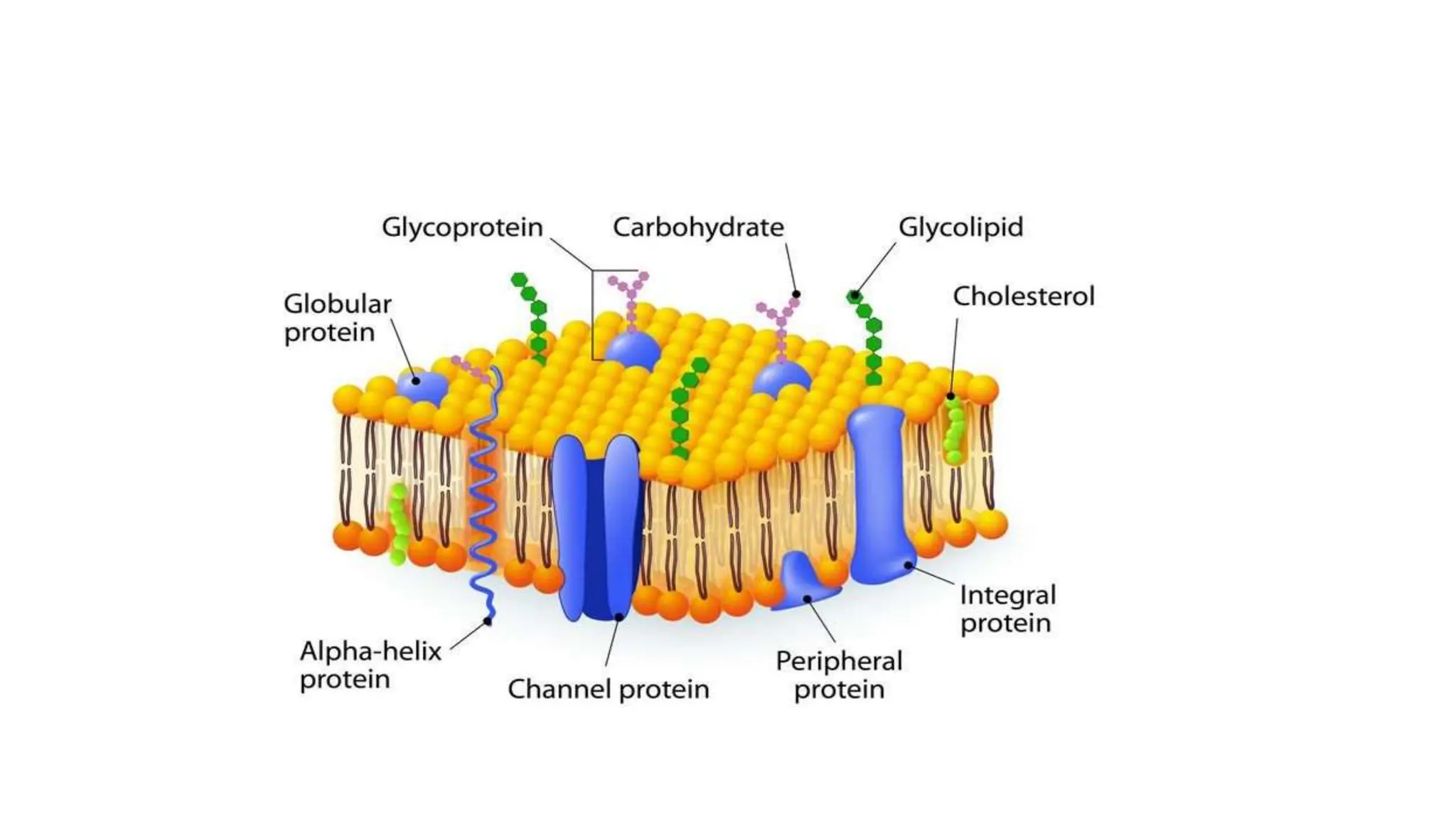
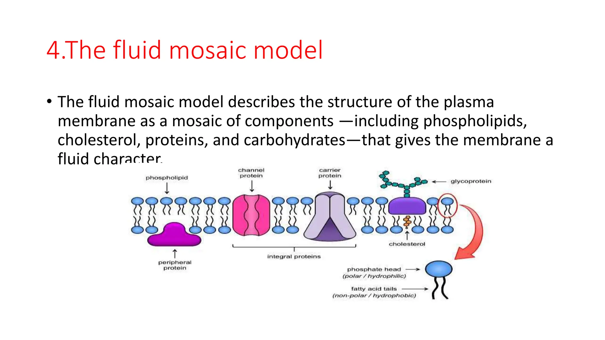
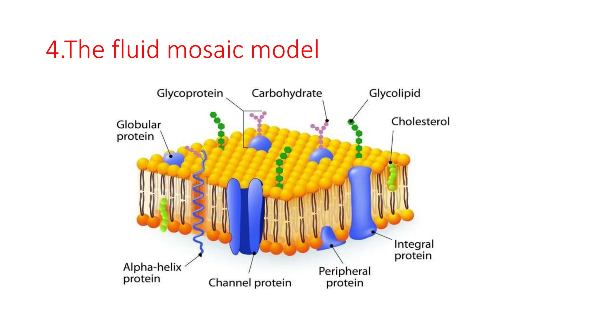
Patheri kuniyil kaleena
Plasma Membrane :
Structure and Functions-Part
I
Plasma membrane structure
• The plasma membrane, which is also called the cell membrane, has
many functions, but the most basic one is to define the borders of the
cell and keep the cell functional. The plasma membrane is selectively
permeable.
• This means that the membrane allows some materials to freely enter
or leave the cell, while other materials cannot move freely, but
require the use of a specialized structure, and occasionally, even
energy investment for crossing.
• The plasma membrane must be very flexible to allow certain cells,
such as red blood cells and white blood cells, to change shape as they
pass through narrow capillaries.
• In addition, the surface of the plasma membrane carries markers that
allow cells to recognize one another, which is vital for tissue and
organ formation during early development.
Various models –Structure of Plasma
membrane
• Dicovered by Karl Nageli and Crammer in 1885
• Term proposed by Karl Wilhelm Nageli
• 1. Lipid and Lipid Bilayer Model (1925-26)
• 2. Dannelli Model. (1935)-First widely accepted model
• Hugh Daaavson aand James Danielli -1935
• 3. Unit Membrane Model (Protein-Lipid Bilayer-Protein) (1953)
• 4. Fluid Mosaic Model (1972)
.1. Lipid and Lipid Bilayer Model:
In 1902, Overton observed that substances soluble in lipid could
selectively pass through the membranes. On this basis he stated that
plasma membrane is composed of
a thin layer of lipid Layer
Gorter and Grendel (1925)’ were
the first to suggest the
bimolecualar lipid model of
plasma membrane
the membrane consisted of
double layers of lipid molecules,
the polar hydrophilic groups of
the molecules being situated on
the outside and hydrophobic
ends standing at right angles to
the surface are oriented inwardly.
models of Gorter and Grendel could not explain the proper structure
of plasma
membrane but they put the foundation of future models of
membrane structure
2.Dannelli Model (Sandwich Models):
 James Danielli and Hugh Davson in 1935 have suggested bimolecular
leaflet model of cell membrane.
 Danielli and Davson model was the first highly accepted model of
plasma membrane
 According to bimolecular model of Danielli and Davson, plasma
membrane consists of two layers of phospholipid molecules (a
bimolecular leaflet) in which phospholipid molecules are arranged in
such a way that hydrophilic heads of the phospholipid molecules face
outside and hydrophobic non-polar lipid chains are associated in the
inner region of leaflet.
The hypothesis also suggested that the polar ends of lipid molecules are
associated with monomolecular layer of globular proteins. The plasma
The membrane would thus
consist of a double layer of
phospholipid molecules
sandwiched between two
essentially continuous layers of
protein.
The model was also described as
a ‘lipo-protein sandwich’, as the
lipid layer was
sandwiched between two protein
layers.
The Davson–Danielli model
predominated until Singer and
Nicolson advanced the fluid
mosaic model in 1972.
The fluid mosaic model expanded on the Davson–
Danielli model by including transmembrane
proteins and eliminated the previously-proposed
flanking protein layers that were not well-
supported by experimental evidence.
3.Unit Membrane Model
• Was given by in the late 1950s proposed the unit membrane
hypothesis according
• to which all biological membranes show generalized unit membrane
construction.
• The unit membrane model visualizes cell membrane as a trilaminar
and indicates structure consisting of two dark osmiophilic layers
separated by a light osmiophilic layer.
Unit Membrane Model…..
The physical appearance of
this trilaminar model has
led to the term unit
membrane
The unit membrane
concept implies a
trilaminar appearance with
a bimolecular lipid layer
between two protein layers.
Each dense osmiophilic
band is made up of protein
(20 Å) and the polar groups
of phospholipids (5 A) and
is thus 25 Å thick.
4.The fluid mosaic model
• The fluid mosaic model was first proposed by S.J. Singer and Garth L.
Nicolson in 1972 to explain the structure of the plasma membrane.
The model has evolved somewhat over time, but it still best accounts
for the structure and functions of the plasma membrane as we now
understand them.
4.The fluid mosaic model
• The fluid mosaic model describes the structure of the plasma
membrane as a mosaic of components —including phospholipids,
cholesterol, proteins, and carbohydrates—that gives the membrane a
fluid character.
The fluid mosaic model……
• structure of the plasma membrane as a mosaic of components —
including
• phospholipids, cholesterol, proteins, and carbohydrates—that gives
the membrane a fluid character. Plasma membranes range from 5 to
10 nm in thickness.
• The proportions of proteins, lipids, and carbohydrates in the plasma
membrane vary with cell type.
• example, myelin contains 18% protein and 76% lipid. The
mitochondrial inner membrane contains 76% protein and 24% lipid.
4.The fluid mosaic model
 The lipids include phospholipids and cholesterol.
Proteins either float in the bilayer or are attached to
one side or the other of it.
 Carbohydrate chains are attached to the proteins and
lipids on the outside surface of the membrane.
 The proportions of proteins, lipids, and carbohydrates
in the plasma membrane vary with cell type, but for a
typical human cell, protein accounts for about 50
percent of the composition by mass, lipids account for
about 40 percent of the composition by mass, with
the remaining 10 percent of the composition by mass
being carbohydrates.
 example, myelin contains 18% protein and 76% lipid.
The mitochondrial inner membrane contains 76%
protein and 24% lipid.

the Plasma Membrane models and accepted fluid mosaic model.pptx

  • 1.
    Patheri kuniyil kaleena PlasmaMembrane : Structure and Functions-Part I
  • 3.
    Plasma membrane structure •The plasma membrane, which is also called the cell membrane, has many functions, but the most basic one is to define the borders of the cell and keep the cell functional. The plasma membrane is selectively permeable. • This means that the membrane allows some materials to freely enter or leave the cell, while other materials cannot move freely, but require the use of a specialized structure, and occasionally, even energy investment for crossing.
  • 4.
    • The plasmamembrane must be very flexible to allow certain cells, such as red blood cells and white blood cells, to change shape as they pass through narrow capillaries. • In addition, the surface of the plasma membrane carries markers that allow cells to recognize one another, which is vital for tissue and organ formation during early development.
  • 5.
    Various models –Structureof Plasma membrane • Dicovered by Karl Nageli and Crammer in 1885 • Term proposed by Karl Wilhelm Nageli • 1. Lipid and Lipid Bilayer Model (1925-26) • 2. Dannelli Model. (1935)-First widely accepted model • Hugh Daaavson aand James Danielli -1935 • 3. Unit Membrane Model (Protein-Lipid Bilayer-Protein) (1953) • 4. Fluid Mosaic Model (1972)
  • 6.
    .1. Lipid andLipid Bilayer Model: In 1902, Overton observed that substances soluble in lipid could selectively pass through the membranes. On this basis he stated that plasma membrane is composed of a thin layer of lipid Layer
  • 7.
    Gorter and Grendel(1925)’ were the first to suggest the bimolecualar lipid model of plasma membrane the membrane consisted of double layers of lipid molecules, the polar hydrophilic groups of the molecules being situated on the outside and hydrophobic ends standing at right angles to the surface are oriented inwardly. models of Gorter and Grendel could not explain the proper structure of plasma membrane but they put the foundation of future models of membrane structure
  • 8.
    2.Dannelli Model (SandwichModels):  James Danielli and Hugh Davson in 1935 have suggested bimolecular leaflet model of cell membrane.  Danielli and Davson model was the first highly accepted model of plasma membrane  According to bimolecular model of Danielli and Davson, plasma membrane consists of two layers of phospholipid molecules (a bimolecular leaflet) in which phospholipid molecules are arranged in such a way that hydrophilic heads of the phospholipid molecules face outside and hydrophobic non-polar lipid chains are associated in the inner region of leaflet. The hypothesis also suggested that the polar ends of lipid molecules are associated with monomolecular layer of globular proteins. The plasma
  • 9.
    The membrane wouldthus consist of a double layer of phospholipid molecules sandwiched between two essentially continuous layers of protein. The model was also described as a ‘lipo-protein sandwich’, as the lipid layer was sandwiched between two protein layers. The Davson–Danielli model predominated until Singer and Nicolson advanced the fluid mosaic model in 1972. The fluid mosaic model expanded on the Davson– Danielli model by including transmembrane proteins and eliminated the previously-proposed flanking protein layers that were not well- supported by experimental evidence.
  • 10.
    3.Unit Membrane Model •Was given by in the late 1950s proposed the unit membrane hypothesis according • to which all biological membranes show generalized unit membrane construction. • The unit membrane model visualizes cell membrane as a trilaminar and indicates structure consisting of two dark osmiophilic layers separated by a light osmiophilic layer.
  • 11.
    Unit Membrane Model….. Thephysical appearance of this trilaminar model has led to the term unit membrane The unit membrane concept implies a trilaminar appearance with a bimolecular lipid layer between two protein layers. Each dense osmiophilic band is made up of protein (20 Å) and the polar groups of phospholipids (5 A) and is thus 25 Å thick.
  • 12.
    4.The fluid mosaicmodel • The fluid mosaic model was first proposed by S.J. Singer and Garth L. Nicolson in 1972 to explain the structure of the plasma membrane. The model has evolved somewhat over time, but it still best accounts for the structure and functions of the plasma membrane as we now understand them.
  • 13.
    4.The fluid mosaicmodel • The fluid mosaic model describes the structure of the plasma membrane as a mosaic of components —including phospholipids, cholesterol, proteins, and carbohydrates—that gives the membrane a fluid character.
  • 14.
    The fluid mosaicmodel…… • structure of the plasma membrane as a mosaic of components — including • phospholipids, cholesterol, proteins, and carbohydrates—that gives the membrane a fluid character. Plasma membranes range from 5 to 10 nm in thickness. • The proportions of proteins, lipids, and carbohydrates in the plasma membrane vary with cell type. • example, myelin contains 18% protein and 76% lipid. The mitochondrial inner membrane contains 76% protein and 24% lipid.
  • 15.
  • 16.
     The lipidsinclude phospholipids and cholesterol. Proteins either float in the bilayer or are attached to one side or the other of it.  Carbohydrate chains are attached to the proteins and lipids on the outside surface of the membrane.  The proportions of proteins, lipids, and carbohydrates in the plasma membrane vary with cell type, but for a typical human cell, protein accounts for about 50 percent of the composition by mass, lipids account for about 40 percent of the composition by mass, with the remaining 10 percent of the composition by mass being carbohydrates.  example, myelin contains 18% protein and 76% lipid. The mitochondrial inner membrane contains 76% protein and 24% lipid.