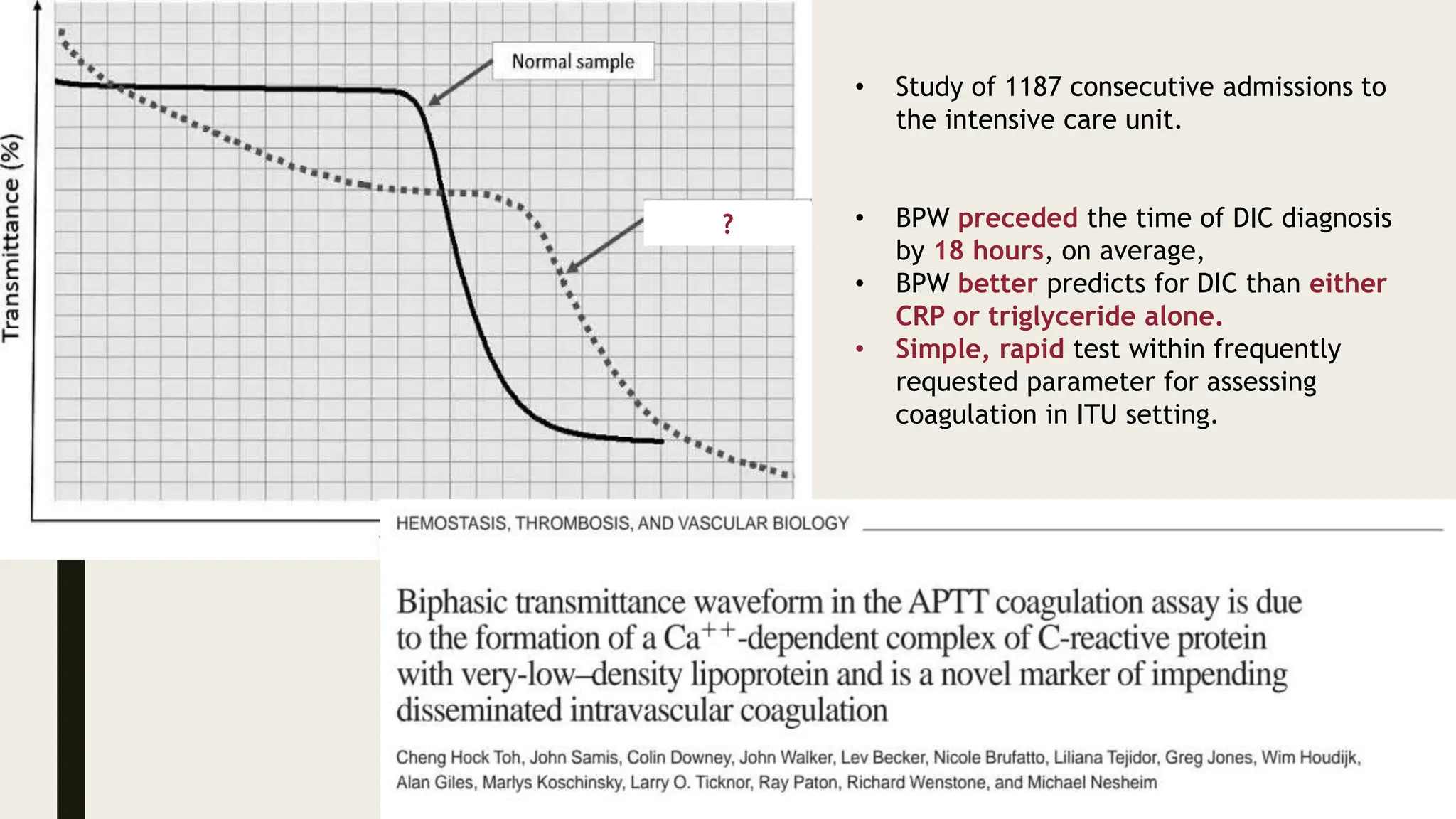
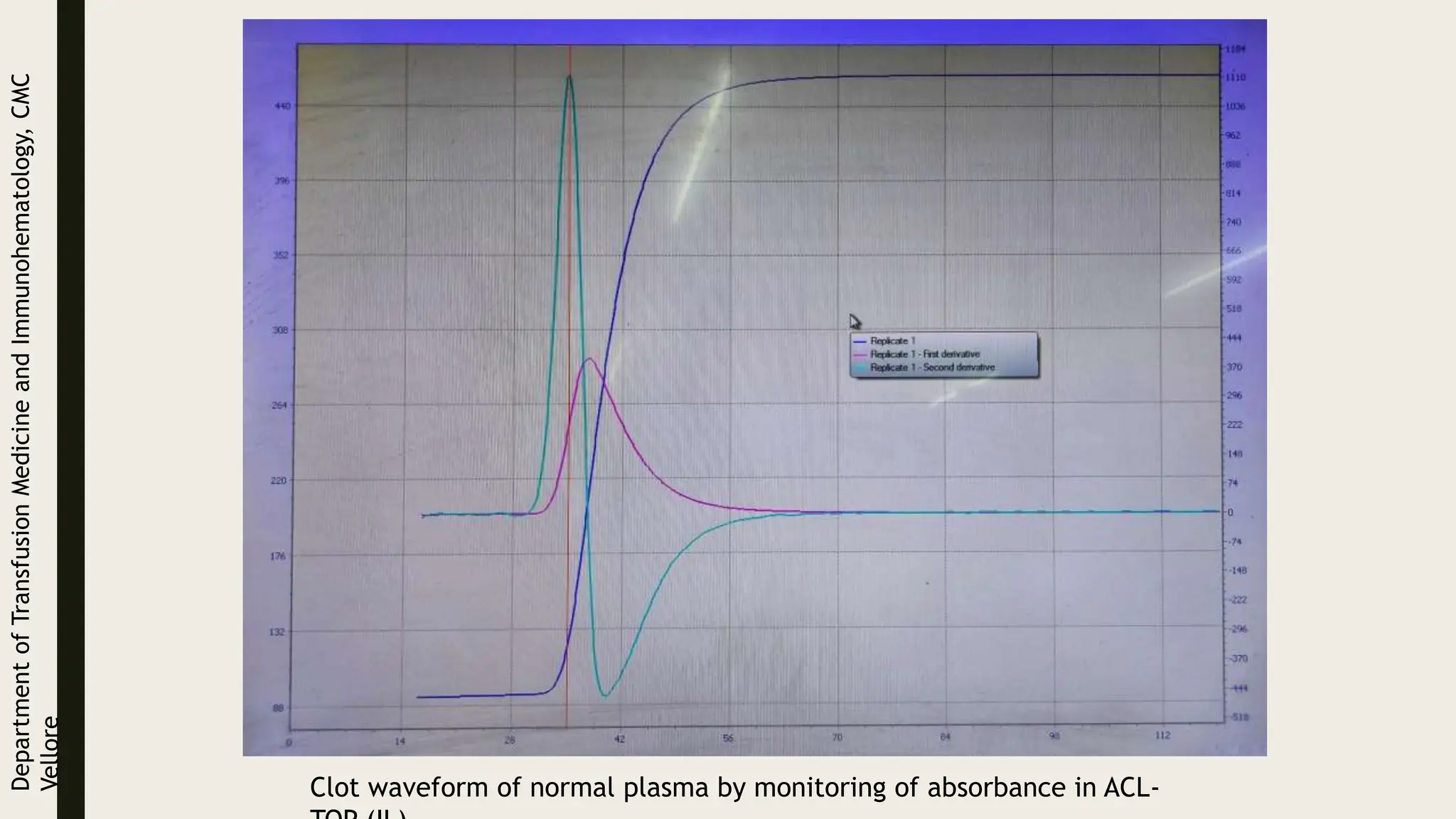
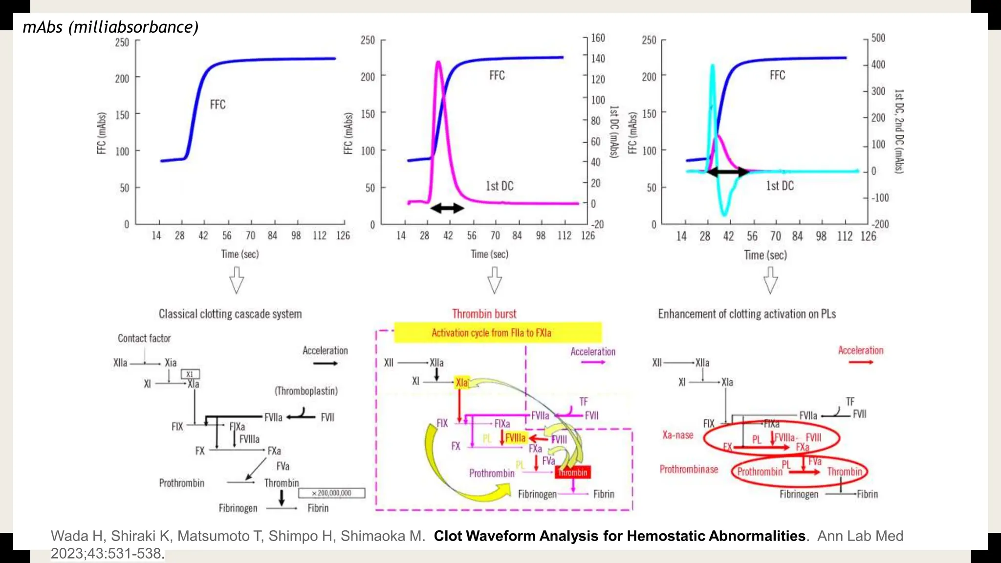
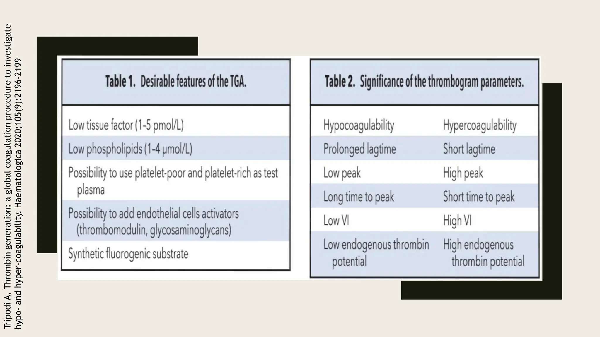
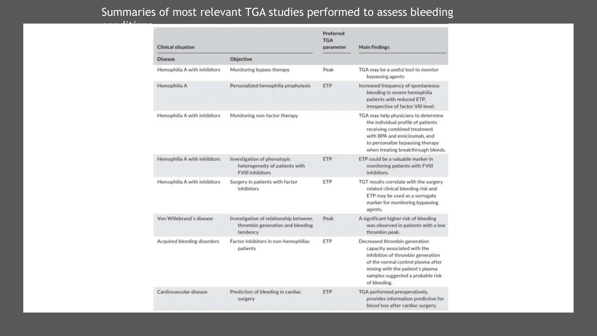
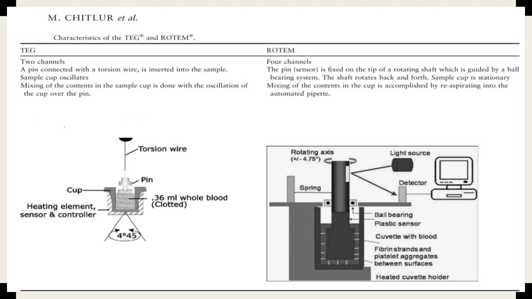
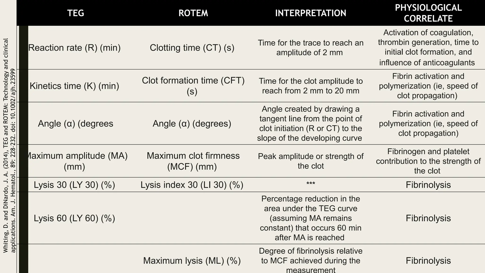
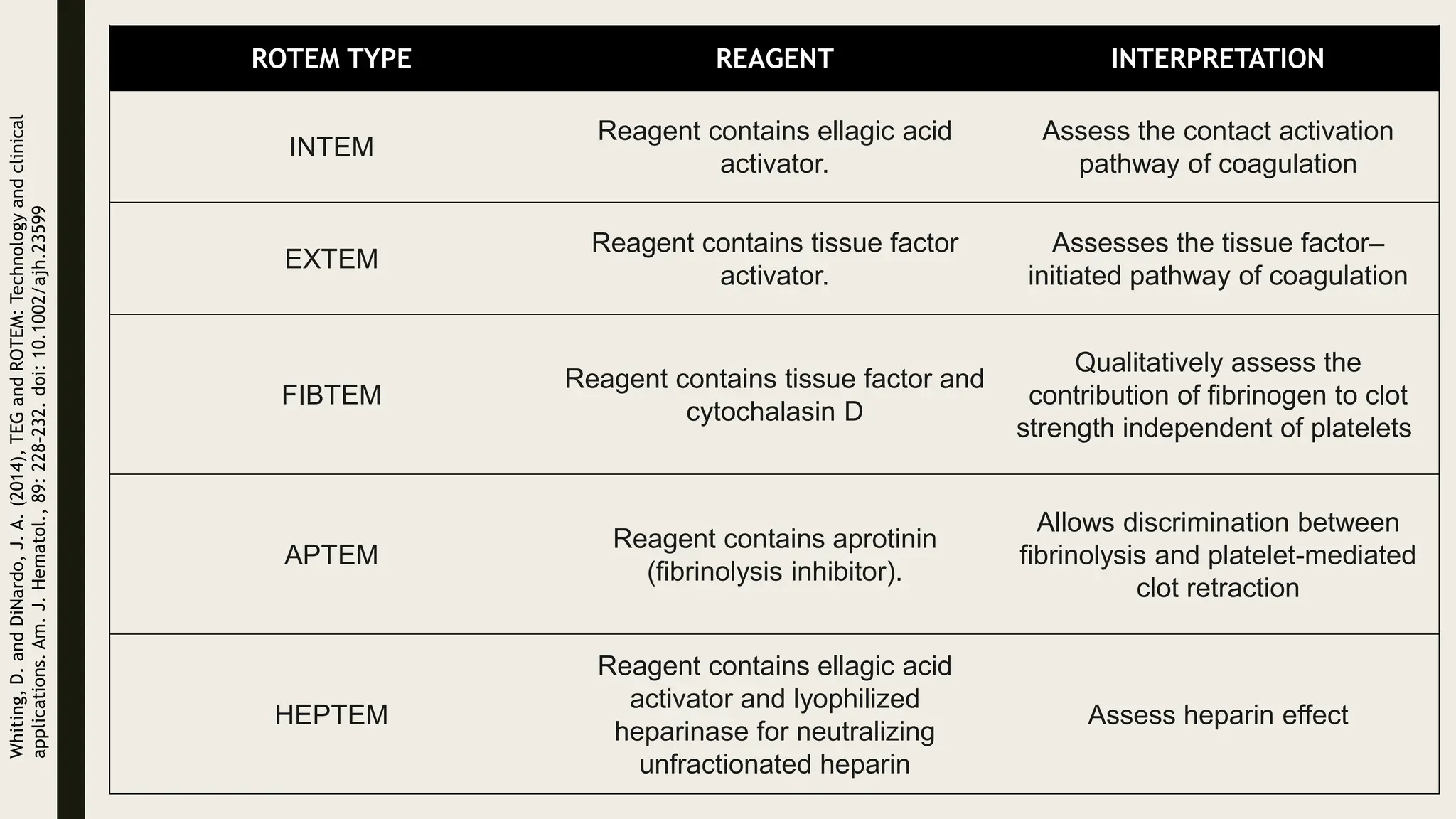
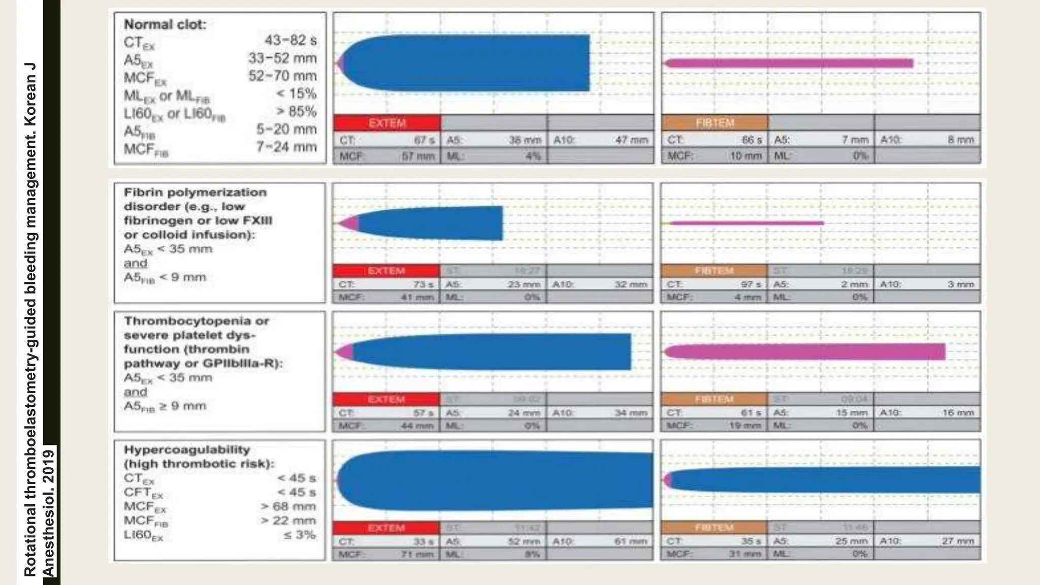
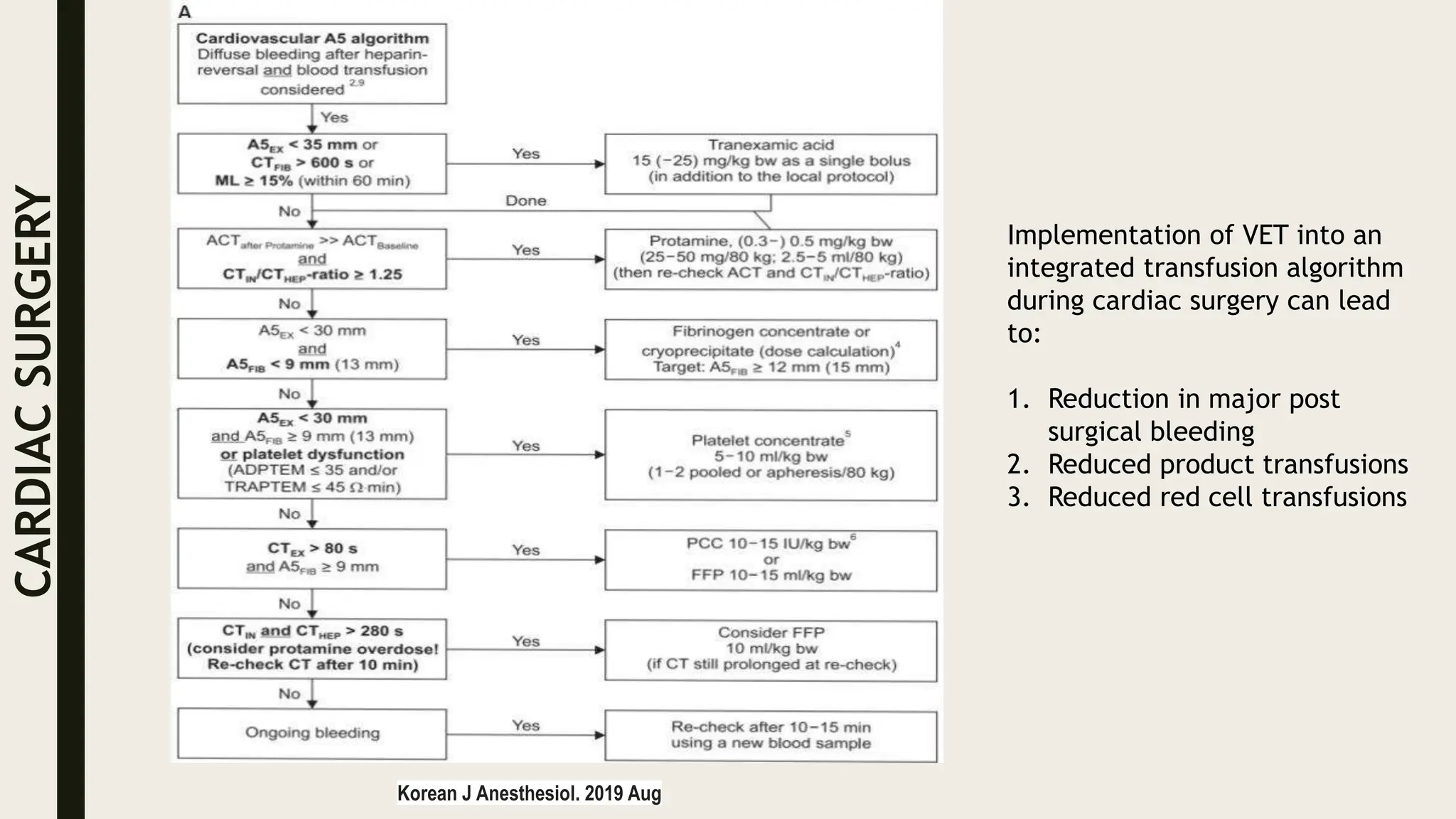
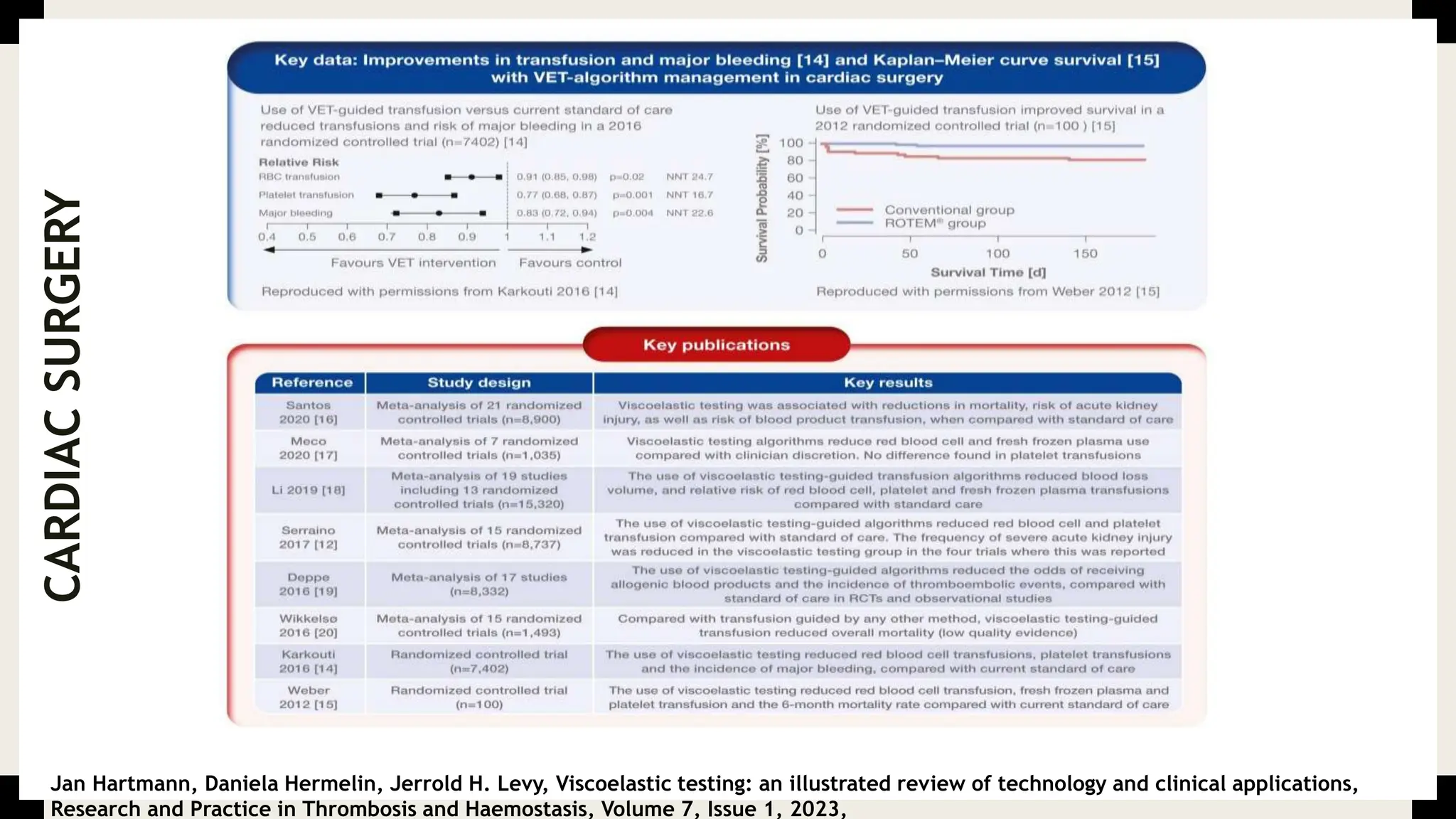
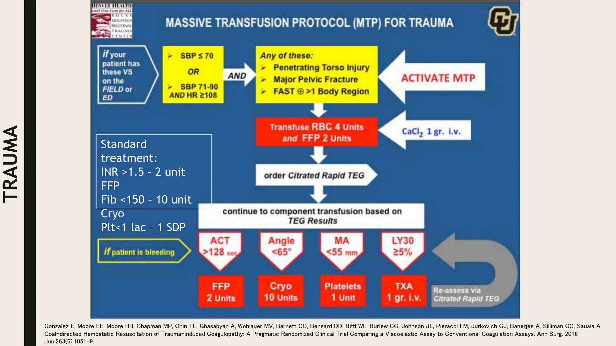
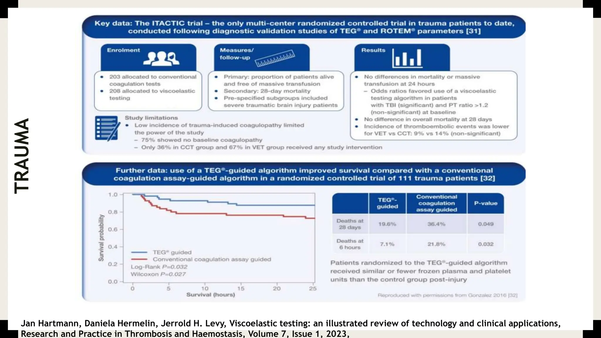
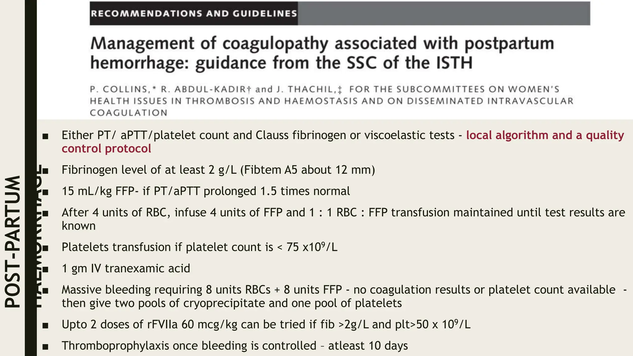
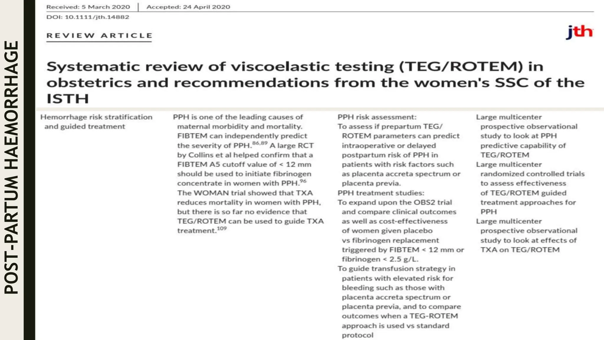
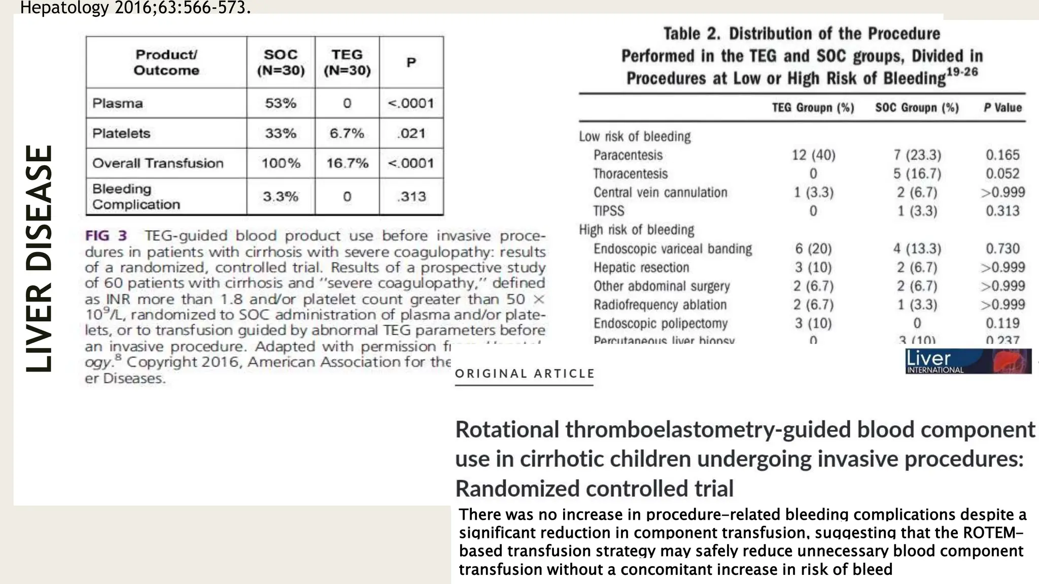
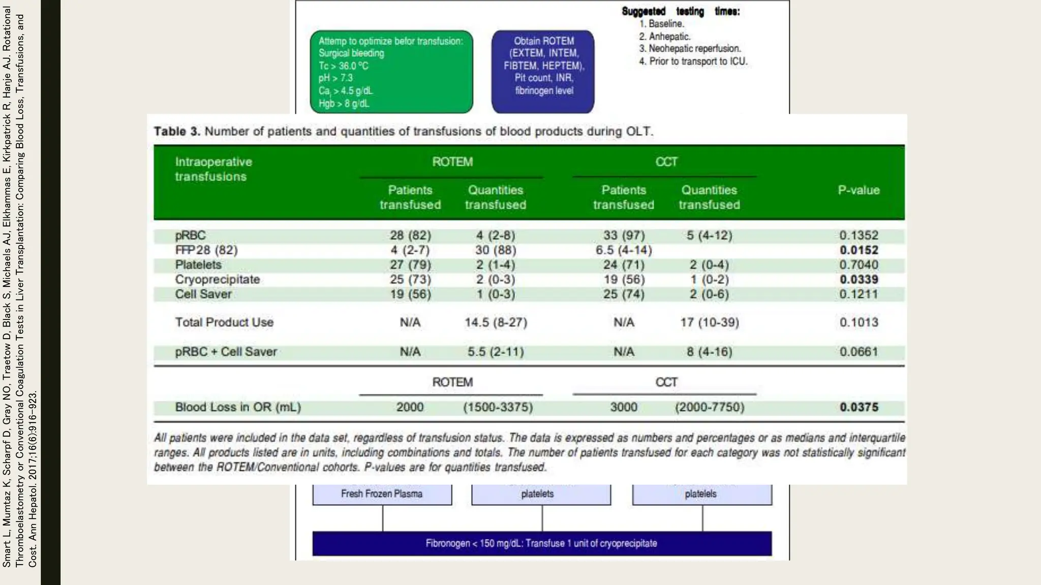
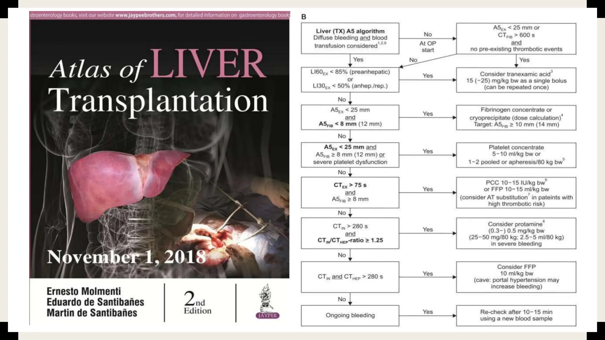
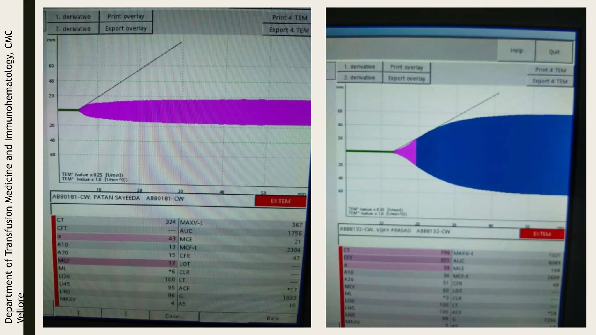
TESTS OF GLOBAL HAEMOSTASIS
Meenakshi Bhatia
28/06/2024
CASCADE MODEL OF COAGULATION CELL BASED MODEL OF COAGULATION
Hoffman,
Maureane
&
Monroe,
Dougald.
(2001).
A
Cell-based
Model
of
Hemostasis.
Thrombosis
and
haemostasis.
85.
958-65.
10.1055/s-0037-1615947.
■ Thrombin, terminal enzyme in the ?cascade (Janus-faced
protein): adopts opposing procoagulant and anticoagulant
(thrombomodulin on endothelial cells) functions.
■ PT/APTT normal in protein C, S deficiency: not
shortened.
■ Plasma tends to clot after 5% of the thrombin has been
formed: 95% thrombin formation not accounted for.
PROCOAGULANT
DRIVERS
ANTICOAGULANT
DRIVERS
Elbaz C, Sholzberg M. An illustrated review of bleeding assessment tools and common coagulation tests. Research and Practice in Thrombosis and Haemostasis. 2020 Jul;4(5):761-773. DOI: 10.1002/rth2.12339. PMID: 32685885;
PMCID: PMC7354401.
Clot Waveform Analysis
APTT
APTT Clot Waveform Analysis
Measured by photo-optical coagulometer which
monitors changes in light transmittance over
entire course of clot formation being reflected
as a waveform, which is processed
mathematically to give various parameters like:
■ First Derivative: (dT/dt): Rate of change in
light Transmittance –velocity of clotting process
– Min1: the minimum value of the first
derivative of the aPTT waveform
■ Second Derivative (d2T/dt2): Magnitude of
change in light Transmittance with respect to
time – coagulation acceleration
– Min2: minimum value of the second
derivative of the aPTT waveform.
– Max2: maximum value of the second
derivative of the aPTT waveform.
Nair
SC,
Dargaud
Y,
Chitlur
M,
Srivastava
A.
Haemophilia.
2010
Jul;16
Suppl
5:85-92.
dT/dt
d2T/dt2
Max2
Min1
Min2
?
• Study of 1187 consecutive admissions to
the intensive care unit.
• BPW preceded the time of DIC diagnosis
by 18 hours, on average,
• BPW better predicts for DIC than either
CRP or triglyceride alone.
• Simple, rapid test within frequently
requested parameter for assessing
coagulation in ITU setting.
Clot waveform of normal plasma by monitoring of absorbance in ACL-
Department
of
Transfusion
Medicine
and
Immunohematology,
CMC
Vellore
Wada H, Shiraki K, Matsumoto T, Shimpo H, Shimaoka M. Clot Waveform Analysis for Hemostatic Abnormalities. Ann Lab Med
2023;43:531-538.
mAbs (milliabsorbance)
RECOMMENDATIONS
TEST PLASMA To be prepared from fresh citrated whole
blood
REFERENCE PLASMA Pooled plasma from normal
individuals/commercial normal plasma
APTT REAGENTS (COLOR) Colourless APTT reagents without opacity
(sensitive to changes in transmittance/
absorbance)
APTT REAGENTS (TRANSMITTANCE)
{MDA-II/CS series}
Thrombocheck APTT-SLA/0.02M CaCl2
APTT REAGENTS (ABSORBANCE)
{ACL-Top series}
HemosIL SynthASiL
• Other instrument/reagent combinations need to be tested prior to use.
• APTT reagents for APLA antibodies not recommended (low sensitivity for assessing low levels of clotting
function)
RECOMMENDED
CLINICAL
APPLICATIONS Clotting function of various
bleeding disorders
Dose-dependent waveform changes in
plasma containing various conc. Of factor
VIII
Waveform changes in haemophilia A with
various levels of Factor VIII
CWA parameters and clinical severity of
severe haemophilia A
‘CWA is a reliable test in patients with both hypercoagulable and
hypocoagulable states.’
‘Routine use of clot waveform analysis may be promoted even in
laboratories not dedicated to haemostasis and thrombosis.’
HYPOCOAGUABLE STATES
HEMOPHILIA
Highly significant correlation between CWA and
thrombin generation throughout a wide range of
concentrations (including lower levels) of both FVIII
and FIX.
Useful for obtaining information on the possible
heterogeneity of clinical phenotypes among patients
with severe or moderate hemophilia.
EMICIZUMAB
Modified CWA with a PT/aPTT mixed reagent (TF and
ellagic acid) used to create an adjusted Min1
(first derivative).
addition of emicizumab to hemophiliac plasma with/without
inhibitors led to concentration-dependent increases in adjusted
Min1
Modified CWA can provide a useful tool for assessing
global coagulation activity in patients with
hemophilia treated with emicizumab
(Adjusted CWA to remove impact of fibrinogen Fig. AdMin1 in FVIII incubated with emicizumab
HYPERCOAGUABLE STATES
VENOUS THROMBOEMBOLISM
CWA parameters are significantly higher in patients
with VTE compared to controls.
CANCER
Elevated peak height of CWA-sTF/FIXa may be
considered as a risk factor for thrombosis in patients
with cancer.
VTE PATIENTS vs CONTROLS
Mean APTT similar
Min1, p<0.001
Min2, p=0.001
Max2, p=0.002
Deltachange, p<0.001
Clot waveform analysis data from patients
(N = 45) with objectively proven acute VTE
who had an aPTT performed prior to
initiation of anticoagulation were compared
with controls (N = 111). The CWA
parameters measured were min1, min2,
max2 and delta change.
Kobayashi
M,
Wada
H,
Fukui
S,
et
al.
Clot
waveform
analysis
showing
a
hypercoagulable
state
in
patients
with
malignant
neoplasms.
J
Clin
Med.
2021
Thrombin generation assays for global evaluation of the hemostatic system: perspectives and limitations, Revista Brasileira de Hematologia e
Hemoterapia, Volume 39, Issue 3, 2017,
THROMBIN GENERATION TEST
THROMBINOSCOPE BV SOFTWARE (RFU > nM)
Peak Height (PH)
Endogenous Thrombin
Potential (ETP): Area Under
The Curve
Lag Time
Time To Peak (TTP)
Start Tail
Tripodi
A.
Thrombin
generation:
a
global
coagulation
procedure
to
investigate
hypo-
and
hyper-coagulability.
Haematologica
2020;105(9):2196-2199
Tripodi
A.
Thrombin
generation:
a
global
coagulation
procedure
to
investigate
hypo-
and
hyper-coagulability.
Haematologica
2020;105(9):2196-2199
1. Emphasis has been placed on more reliable, more standardized and more user-friendly assays that have
now reached a high level of maturity.
2. Pre-analytical conditions have been extensively studied leading to precise yet manageable
recommendations.
3. Researchers have looked at the TGA potential in various clinical and industrial settings and demonstrated
the contributions of TGA in patient's diagnosis, prognosis evaluation and treatment monitoring.
4. Altogether, this has paved the way for an imminent transition of TGA from research to routine use.
ESTABILISHING STANDARDIZED TGA
■ A specific analyzer optimized for TGA including tightly controlled temperature
and light intensity
■ Validated pipetting schemes for the respective reagent including optimized
measurement times
■ Well controlled substrate including traceable thrombin and fluorescence
standards
■ Availability of specific QC controls covering the analytical measuring range
■ Use of standardized reagents or triggers
RECOMMENDATIONS
SUBSTRATE (FLUOROGENIC)
Z-Gly-Gly-Arg-AMC (ZGGR-AMC)
(7-Amino-4-methylcoumarin: fluorophore that generates
390 nm excitation/460 nm emission)
SUBSTRATE (CHROMOGENIC)
H-β-Ala-Gly-Arg-pNA
(absorption at 405 nm, requires defibrination, no longer
commercially available)
TRIGGER/REAGANT
• For bleeding tendencies testing on PPP
low TF concentrations 0.4 to 2 pM + phospholipid 0.4 to 4
µM
• Trigger reagents for thrombophilia testing
TF between 5 and 40 pM + phospholipid 0.4 to 4 µM
• To overcome anticoagulation
20 and 50 pM TF + 4µM phospholipids
CALIBRATION
Thrombin calibrator with known thrombin activity run
Summaries of most relevant TGA studies performed to assess bleeding
conditions.
Summaries of most relevant TGA studies for thrombotic
conditions.
Viscoelastic tests of coagulation
■ TEG
■ ROTEM
Setting
1. Cardiac surgery
2. Trauma
3. Post partum hemorrhage
4. Liver transplantation
TEG ROTEM INTERPRETATION
PHYSIOLOGICAL
CORRELATE
Reaction rate (R) (min) Clotting time (CT) (s)
Time for the trace to reach an
amplitude of 2 mm
Activation of coagulation,
thrombin generation, time to
initial clot formation, and
influence of anticoagulants
Kinetics time (K) (min)
Clot formation time (CFT)
(s)
Time for the clot amplitude to
reach from 2 mm to 20 mm
Fibrin activation and
polymerization (ie, speed of
clot propagation)
Angle (α) (degrees Angle (α) (degrees)
Angle created by drawing a
tangent line from the point of
clot initiation (R or CT) to the
slope of the developing curve
Fibrin activation and
polymerization (ie, speed of
clot propagation)
Maximum amplitude (MA)
(mm)
Maximum clot firmness
(MCF) (mm)
Peak amplitude or strength of
the clot
Fibrinogen and platelet
contribution to the strength of
the clot
Lysis 30 (LY 30) (%) Lysis index 30 (LI 30) (%) *** Fibrinolysis
Lysis 60 (LY 60) (%)
Percentage reduction in the
area under the TEG curve
(assuming MA remains
constant) that occurs 60 min
after MA is reached
Fibrinolysis
Maximum lysis (ML) (%)
Degree of fibrinolysis relative
to MCF achieved during the
measurement
Fibrinolysis
Whiting,
D.
and
DiNardo,
J.
A.
(2014),
TEG
and
ROTEM:
Technology
and
clinical
applications.
Am.
J.
Hematol.,
89:
228–232.
doi:
10.1002/ajh.23599
TEG v/s standard tests of coagulation
Whiting,
D.
and
DiNardo,
J.
A.
(2014),
TEG
and
ROTEM:
Technology
and
clinical
applications.
Am.
J.
Hematol.,
89:
228–232.
doi:
10.1002/ajh.23599
Müller,
M.C.,
Meijers,
J.C.,
Vroom,
M.B.
et
al.
Utility
of
thromboelastography
and/or
thromboelastometry
in
adults
with
sepsis:
a
systematic
review.
Crit
Care
18,
R30
(2014).
https://doi.org/10.1186/cc13721
Jan Hartmann, Daniela Hermelin, Jerrold H. Levy, Viscoelastic testing: an illustrated review of technology and clinical applications,
Research and Practice in Thrombosis and Haemostasis, Volume 7, Issue 1, 2023,
PARAMETER AB35946 NORMAL RANGE
CT 363 secs 324-565 secs
CFT 323 sec 112-224 secs
Alpha-angle 41 50-68 degrees
MCF 41mm 55-66 mm
ML 8% 0-15%
LAB PARAMETERS AB35946
PT 19.8 secs
INR 1.51
PT correction studies 16 secs
APTT 43.8 secs
APTT correction studies 31.3 secs
ROTEM TYPE REAGENT INTERPRETATION
INTEM
Reagent contains ellagic acid
activator.
Assess the contact activation
pathway of coagulation
EXTEM
Reagent contains tissue factor
activator.
Assesses the tissue factor–
initiated pathway of coagulation
FIBTEM
Reagent contains tissue factor and
cytochalasin D
Qualitatively assess the
contribution of fibrinogen to clot
strength independent of platelets
APTEM
Reagent contains aprotinin
(fibrinolysis inhibitor).
Allows discrimination between
fibrinolysis and platelet-mediated
clot retraction
HEPTEM
Reagent contains ellagic acid
activator and lyophilized
heparinase for neutralizing
unfractionated heparin
Assess heparin effect
Whiting,
D.
and
DiNardo,
J.
A.
(2014),
TEG
and
ROTEM:
Technology
and
clinical
applications.
Am.
J.
Hematol.,
89:
228–232.
doi:
10.1002/ajh.23599
Rotational
thromboelastometry-guided
bleeding
management.
Korean
J
Anesthesiol.
2019
CARDIAC SURGERY
www.nice.org.uk/guidance/dg13 published 20th August 2014
CARDIAC
SURGERY
CARDIAC
SURGERY
Wikkelsø A, Wetterslev J, Møller AM, Afshari A. Thromboelastography (TEG) or thromboelastometry (ROTEM) to monitor haemostatic treatment versus usual care in adults or children with bleeding. Cochrane Database Syst
Rev. 2016
CARDIAC
SURGERY
Korean J Anesthesiol. 2019 Aug
Implementation of VET into an
integrated transfusion algorithm
during cardiac surgery can lead
to:
1. Reduction in major post
surgical bleeding
2. Reduced product transfusions
3. Reduced red cell transfusions
CARDIAC
SURGERY
Jan Hartmann, Daniela Hermelin, Jerrold H. Levy, Viscoelastic testing: an illustrated review of technology and clinical applications,
Research and Practice in Thrombosis and Haemostasis, Volume 7, Issue 1, 2023,
Standard
treatment:
INR >1.5 – 2 unit
FFP
Fib <150 – 10 unit
Cryo
Plt<1 lac – 1 SDP
Gonzalez E, Moore EE, Moore HB, Chapman MP, Chin TL, Ghasabyan A, Wohlauer MV, Barnett CC, Bensard DD, Biffl WL, Burlew CC, Johnson JL, Pieracci FM, Jurkovich GJ, Banerjee A, Silliman CC, Sauaia A.
Goal-directed Hemostatic Resuscitation of Trauma-induced Coagulopathy: A Pragmatic Randomized Clinical Trial Comparing a Viscoelastic Assay to Conventional Coagulation Assays. Ann Surg. 2016
Jun;263(6):1051-9.
TRAUMA
Cochrane Database Syst Rev. 2015 Feb 16;(2):CD010438.
TRAUMA
Viscoelastic haemostatic assay augmented protocols for major trauma haemorrhage (ITACTIC): a randomized, controlled trial.
Intensive Care Med. 2021
TRAUMA
Jan Hartmann, Daniela Hermelin, Jerrold H. Levy, Viscoelastic testing: an illustrated review of technology and clinical applications,
Research and Practice in Thrombosis and Haemostasis, Volume 7, Issue 1, 2023,
TRAUMA
Charbit B, Mandelbrot L, Samain E, Baron G, Haddaoui B, Keita H, Sibony O, Mahieu-Caputo D, Hurtaud-Roux
MF, Huisse MG, Denninger MH, de Prost D, for the PPH Study Group. The decrease of fibrinogen is an early
predictor of the severity of postpartum hemorrhage. J Thromb Haemost 2007; 5: 266–73.
POST-PARTUM
HAEMORRHAGE
■ Either PT/ aPTT/platelet count and Clauss fibrinogen or viscoelastic tests - local algorithm and a quality
control protocol
■ Fibrinogen level of at least 2 g/L (Fibtem A5 about 12 mm)
■ 15 mL/kg FFP- if PT/aPTT prolonged 1.5 times normal
■ After 4 units of RBC, infuse 4 units of FFP and 1 : 1 RBC : FFP transfusion maintained until test results are
known
■ Platelets transfusion if platelet count is < 75 x109/L
■ 1 gm IV tranexamic acid
■ Massive bleeding requiring 8 units RBCs + 8 units FFP - no coagulation results or platelet count available -
then give two pools of cryoprecipitate and one pool of platelets
■ Upto 2 doses of rFVIIa 60 mcg/kg can be tried if fib >2g/L and plt>50 x 109/L
■ Thromboprophylaxis once bleeding is controlled – atleast 10 days
POST-PARTUM
HAEMORRHAGE
■ FIBTEM A5 <5 mm or A10 <6 mm - 5 to 15 units of cryoprecipitate - Goal - A10 of 8 mm if
bleeding controlled and 10 mm for patients with ongoing hemorrhage
■ Repeat test at the end of the cryoprecipitate transfusion and every 20 to 60 min thereafter till
bleeding stops
■ If CFT and alpha angle demonstrated hypocoagulation accompanied by a decrease in EXTEM
MCF to <45 mm - one bag (5 units) of platelet concentrate, followed by repeat testing.
■ If isolated CT prolongation was noticed in EXTEM and INTEM assays - 2 units of FFP, followed by
repeat testing.
■ Packed red blood cell transfusions were provided as needed in all the cases to maintain the
hematocrit above 21%
Snegovskikh
D,
Souza
D,
Walton
Z,
Dai
F,
Rachler
R,
Garay
A,
Snegovskikh
VV,
Braveman
FR,
Norwitz
ER.
Point-of-
care
viscoelastic
testing
improves
the
outcome
of
pregnancies
complicated
by
severe
postpartum
hemorrhage.
J
Clin
Anesth.
2018
Feb;44:50-56.
POST-PARTUM
HAEMORRHAGE
Lisman T, Hernandez-Gea V, Magnusson M, Roberts L, Stanworth S, Thachil J, Tripodi A. The
concept of rebalanced hemostasis in patients with liver disease: Communication from the
ISTH SSC working group on hemostatic management of patients with liver disease. J Thromb
Haemost. 2021 Apr
Patients with liver disease are in haemostatic balance due to the
simultaneous decline of pro‐ and anticoagulant drivers. The value
of the PT and APTT in patients with liver disease is limited and
may also provide misleading information
Prophylactic correction of abnormal laboratory tests of hemostasis
with blood products or pharmacological prohemostatic agents
aiming to prevent spontaneous or procedure‐related bleeding is
not indicated.
REBALANCE HEMOSTASIS
IN PATIENTS WITH LIVER DISEASE
Hepatology 2016;63:566-573.
LIVER
DISEASE
There was no increase in procedure-related bleeding complications despite a
significant reduction in component transfusion, suggesting that the ROTEM-
based transfusion strategy may safely reduce unnecessary blood component
transfusion without a concomitant increase in risk of bleed
Global assays such as ROTEM/TEG may be an attractive means to
reassure the proceduralist that haemostasis is ‘normal’ as these
assays are frequently normal despite prolonged coagulation times.
‘ We recommend ROTEM/TEG over measuring INR,
platelet, fibrinogen in critically ill patients with
ALF/ACLF undergoing procedures strongly'
Smart
L,
Mumtaz
K,
Scharpf
D,
Gray
NO,
Traetow
D,
Black
S,
Michaels
AJ,
Elkhammas
E,
Kirkpatrick
R,
Hanje
AJ.
Rotational
Thromboelastometry
or
Conventional
Coagulation
Tests
in
Liver
Transplantation:
Comparing
Blood
Loss,
Transfusions,
and
Cost.
Ann
Hepatol.
2017;16(6):916-923.
Department
of
Transfusion
Medicine
and
Immunohematology,
CMC
Vellore
THANK YOU

Tests of Global Hemostasis Hematological

  • 1.
    TESTS OF GLOBALHAEMOSTASIS Meenakshi Bhatia 28/06/2024
  • 2.
    CASCADE MODEL OFCOAGULATION CELL BASED MODEL OF COAGULATION Hoffman, Maureane & Monroe, Dougald. (2001). A Cell-based Model of Hemostasis. Thrombosis and haemostasis. 85. 958-65. 10.1055/s-0037-1615947.
  • 3.
    ■ Thrombin, terminalenzyme in the ?cascade (Janus-faced protein): adopts opposing procoagulant and anticoagulant (thrombomodulin on endothelial cells) functions. ■ PT/APTT normal in protein C, S deficiency: not shortened. ■ Plasma tends to clot after 5% of the thrombin has been formed: 95% thrombin formation not accounted for. PROCOAGULANT DRIVERS ANTICOAGULANT DRIVERS
  • 4.
    Elbaz C, SholzbergM. An illustrated review of bleeding assessment tools and common coagulation tests. Research and Practice in Thrombosis and Haemostasis. 2020 Jul;4(5):761-773. DOI: 10.1002/rth2.12339. PMID: 32685885; PMCID: PMC7354401.
  • 5.
  • 6.
    APTT Clot WaveformAnalysis Measured by photo-optical coagulometer which monitors changes in light transmittance over entire course of clot formation being reflected as a waveform, which is processed mathematically to give various parameters like: ■ First Derivative: (dT/dt): Rate of change in light Transmittance –velocity of clotting process – Min1: the minimum value of the first derivative of the aPTT waveform ■ Second Derivative (d2T/dt2): Magnitude of change in light Transmittance with respect to time – coagulation acceleration – Min2: minimum value of the second derivative of the aPTT waveform. – Max2: maximum value of the second derivative of the aPTT waveform. Nair SC, Dargaud Y, Chitlur M, Srivastava A. Haemophilia. 2010 Jul;16 Suppl 5:85-92. dT/dt d2T/dt2 Max2 Min1 Min2
  • 7.
    ? • Study of1187 consecutive admissions to the intensive care unit. • BPW preceded the time of DIC diagnosis by 18 hours, on average, • BPW better predicts for DIC than either CRP or triglyceride alone. • Simple, rapid test within frequently requested parameter for assessing coagulation in ITU setting.
  • 8.
    Clot waveform ofnormal plasma by monitoring of absorbance in ACL- Department of Transfusion Medicine and Immunohematology, CMC Vellore
  • 9.
    Wada H, ShirakiK, Matsumoto T, Shimpo H, Shimaoka M. Clot Waveform Analysis for Hemostatic Abnormalities. Ann Lab Med 2023;43:531-538. mAbs (milliabsorbance)
  • 11.
    RECOMMENDATIONS TEST PLASMA Tobe prepared from fresh citrated whole blood REFERENCE PLASMA Pooled plasma from normal individuals/commercial normal plasma APTT REAGENTS (COLOR) Colourless APTT reagents without opacity (sensitive to changes in transmittance/ absorbance) APTT REAGENTS (TRANSMITTANCE) {MDA-II/CS series} Thrombocheck APTT-SLA/0.02M CaCl2 APTT REAGENTS (ABSORBANCE) {ACL-Top series} HemosIL SynthASiL • Other instrument/reagent combinations need to be tested prior to use. • APTT reagents for APLA antibodies not recommended (low sensitivity for assessing low levels of clotting function)
  • 12.
    RECOMMENDED CLINICAL APPLICATIONS Clotting functionof various bleeding disorders Dose-dependent waveform changes in plasma containing various conc. Of factor VIII Waveform changes in haemophilia A with various levels of Factor VIII CWA parameters and clinical severity of severe haemophilia A
  • 13.
    ‘CWA is areliable test in patients with both hypercoagulable and hypocoagulable states.’ ‘Routine use of clot waveform analysis may be promoted even in laboratories not dedicated to haemostasis and thrombosis.’
  • 14.
    HYPOCOAGUABLE STATES HEMOPHILIA Highly significantcorrelation between CWA and thrombin generation throughout a wide range of concentrations (including lower levels) of both FVIII and FIX. Useful for obtaining information on the possible heterogeneity of clinical phenotypes among patients with severe or moderate hemophilia. EMICIZUMAB Modified CWA with a PT/aPTT mixed reagent (TF and ellagic acid) used to create an adjusted Min1 (first derivative). addition of emicizumab to hemophiliac plasma with/without inhibitors led to concentration-dependent increases in adjusted Min1 Modified CWA can provide a useful tool for assessing global coagulation activity in patients with hemophilia treated with emicizumab
  • 15.
    (Adjusted CWA toremove impact of fibrinogen Fig. AdMin1 in FVIII incubated with emicizumab
  • 16.
    HYPERCOAGUABLE STATES VENOUS THROMBOEMBOLISM CWAparameters are significantly higher in patients with VTE compared to controls. CANCER Elevated peak height of CWA-sTF/FIXa may be considered as a risk factor for thrombosis in patients with cancer.
  • 17.
    VTE PATIENTS vsCONTROLS Mean APTT similar Min1, p<0.001 Min2, p=0.001 Max2, p=0.002 Deltachange, p<0.001 Clot waveform analysis data from patients (N = 45) with objectively proven acute VTE who had an aPTT performed prior to initiation of anticoagulation were compared with controls (N = 111). The CWA parameters measured were min1, min2, max2 and delta change.
  • 18.
  • 19.
    Thrombin generation assaysfor global evaluation of the hemostatic system: perspectives and limitations, Revista Brasileira de Hematologia e Hemoterapia, Volume 39, Issue 3, 2017, THROMBIN GENERATION TEST THROMBINOSCOPE BV SOFTWARE (RFU > nM)
  • 20.
    Peak Height (PH) EndogenousThrombin Potential (ETP): Area Under The Curve Lag Time Time To Peak (TTP) Start Tail Tripodi A. Thrombin generation: a global coagulation procedure to investigate hypo- and hyper-coagulability. Haematologica 2020;105(9):2196-2199
  • 21.
  • 22.
    1. Emphasis hasbeen placed on more reliable, more standardized and more user-friendly assays that have now reached a high level of maturity. 2. Pre-analytical conditions have been extensively studied leading to precise yet manageable recommendations. 3. Researchers have looked at the TGA potential in various clinical and industrial settings and demonstrated the contributions of TGA in patient's diagnosis, prognosis evaluation and treatment monitoring. 4. Altogether, this has paved the way for an imminent transition of TGA from research to routine use.
  • 23.
    ESTABILISHING STANDARDIZED TGA ■A specific analyzer optimized for TGA including tightly controlled temperature and light intensity ■ Validated pipetting schemes for the respective reagent including optimized measurement times ■ Well controlled substrate including traceable thrombin and fluorescence standards ■ Availability of specific QC controls covering the analytical measuring range ■ Use of standardized reagents or triggers
  • 24.
    RECOMMENDATIONS SUBSTRATE (FLUOROGENIC) Z-Gly-Gly-Arg-AMC (ZGGR-AMC) (7-Amino-4-methylcoumarin:fluorophore that generates 390 nm excitation/460 nm emission) SUBSTRATE (CHROMOGENIC) H-β-Ala-Gly-Arg-pNA (absorption at 405 nm, requires defibrination, no longer commercially available) TRIGGER/REAGANT • For bleeding tendencies testing on PPP low TF concentrations 0.4 to 2 pM + phospholipid 0.4 to 4 µM • Trigger reagents for thrombophilia testing TF between 5 and 40 pM + phospholipid 0.4 to 4 µM • To overcome anticoagulation 20 and 50 pM TF + 4µM phospholipids CALIBRATION Thrombin calibrator with known thrombin activity run
  • 26.
    Summaries of mostrelevant TGA studies performed to assess bleeding conditions.
  • 27.
    Summaries of mostrelevant TGA studies for thrombotic conditions.
  • 28.
    Viscoelastic tests ofcoagulation ■ TEG ■ ROTEM Setting 1. Cardiac surgery 2. Trauma 3. Post partum hemorrhage 4. Liver transplantation
  • 30.
    TEG ROTEM INTERPRETATION PHYSIOLOGICAL CORRELATE Reactionrate (R) (min) Clotting time (CT) (s) Time for the trace to reach an amplitude of 2 mm Activation of coagulation, thrombin generation, time to initial clot formation, and influence of anticoagulants Kinetics time (K) (min) Clot formation time (CFT) (s) Time for the clot amplitude to reach from 2 mm to 20 mm Fibrin activation and polymerization (ie, speed of clot propagation) Angle (α) (degrees Angle (α) (degrees) Angle created by drawing a tangent line from the point of clot initiation (R or CT) to the slope of the developing curve Fibrin activation and polymerization (ie, speed of clot propagation) Maximum amplitude (MA) (mm) Maximum clot firmness (MCF) (mm) Peak amplitude or strength of the clot Fibrinogen and platelet contribution to the strength of the clot Lysis 30 (LY 30) (%) Lysis index 30 (LI 30) (%) *** Fibrinolysis Lysis 60 (LY 60) (%) Percentage reduction in the area under the TEG curve (assuming MA remains constant) that occurs 60 min after MA is reached Fibrinolysis Maximum lysis (ML) (%) Degree of fibrinolysis relative to MCF achieved during the measurement Fibrinolysis Whiting, D. and DiNardo, J. A. (2014), TEG and ROTEM: Technology and clinical applications. Am. J. Hematol., 89: 228–232. doi: 10.1002/ajh.23599
  • 31.
    TEG v/s standardtests of coagulation Whiting, D. and DiNardo, J. A. (2014), TEG and ROTEM: Technology and clinical applications. Am. J. Hematol., 89: 228–232. doi: 10.1002/ajh.23599
  • 32.
  • 33.
    Jan Hartmann, DanielaHermelin, Jerrold H. Levy, Viscoelastic testing: an illustrated review of technology and clinical applications, Research and Practice in Thrombosis and Haemostasis, Volume 7, Issue 1, 2023,
  • 34.
    PARAMETER AB35946 NORMALRANGE CT 363 secs 324-565 secs CFT 323 sec 112-224 secs Alpha-angle 41 50-68 degrees MCF 41mm 55-66 mm ML 8% 0-15% LAB PARAMETERS AB35946 PT 19.8 secs INR 1.51 PT correction studies 16 secs APTT 43.8 secs APTT correction studies 31.3 secs
  • 35.
    ROTEM TYPE REAGENTINTERPRETATION INTEM Reagent contains ellagic acid activator. Assess the contact activation pathway of coagulation EXTEM Reagent contains tissue factor activator. Assesses the tissue factor– initiated pathway of coagulation FIBTEM Reagent contains tissue factor and cytochalasin D Qualitatively assess the contribution of fibrinogen to clot strength independent of platelets APTEM Reagent contains aprotinin (fibrinolysis inhibitor). Allows discrimination between fibrinolysis and platelet-mediated clot retraction HEPTEM Reagent contains ellagic acid activator and lyophilized heparinase for neutralizing unfractionated heparin Assess heparin effect Whiting, D. and DiNardo, J. A. (2014), TEG and ROTEM: Technology and clinical applications. Am. J. Hematol., 89: 228–232. doi: 10.1002/ajh.23599
  • 36.
  • 38.
  • 39.
  • 40.
    CARDIAC SURGERY Wikkelsø A, WetterslevJ, Møller AM, Afshari A. Thromboelastography (TEG) or thromboelastometry (ROTEM) to monitor haemostatic treatment versus usual care in adults or children with bleeding. Cochrane Database Syst Rev. 2016
  • 41.
    CARDIAC SURGERY Korean J Anesthesiol.2019 Aug Implementation of VET into an integrated transfusion algorithm during cardiac surgery can lead to: 1. Reduction in major post surgical bleeding 2. Reduced product transfusions 3. Reduced red cell transfusions
  • 42.
    CARDIAC SURGERY Jan Hartmann, DanielaHermelin, Jerrold H. Levy, Viscoelastic testing: an illustrated review of technology and clinical applications, Research and Practice in Thrombosis and Haemostasis, Volume 7, Issue 1, 2023,
  • 44.
    Standard treatment: INR >1.5 –2 unit FFP Fib <150 – 10 unit Cryo Plt<1 lac – 1 SDP Gonzalez E, Moore EE, Moore HB, Chapman MP, Chin TL, Ghasabyan A, Wohlauer MV, Barnett CC, Bensard DD, Biffl WL, Burlew CC, Johnson JL, Pieracci FM, Jurkovich GJ, Banerjee A, Silliman CC, Sauaia A. Goal-directed Hemostatic Resuscitation of Trauma-induced Coagulopathy: A Pragmatic Randomized Clinical Trial Comparing a Viscoelastic Assay to Conventional Coagulation Assays. Ann Surg. 2016 Jun;263(6):1051-9. TRAUMA
  • 45.
    Cochrane Database SystRev. 2015 Feb 16;(2):CD010438. TRAUMA
  • 46.
    Viscoelastic haemostatic assayaugmented protocols for major trauma haemorrhage (ITACTIC): a randomized, controlled trial. Intensive Care Med. 2021 TRAUMA
  • 47.
    Jan Hartmann, DanielaHermelin, Jerrold H. Levy, Viscoelastic testing: an illustrated review of technology and clinical applications, Research and Practice in Thrombosis and Haemostasis, Volume 7, Issue 1, 2023, TRAUMA
  • 48.
    Charbit B, MandelbrotL, Samain E, Baron G, Haddaoui B, Keita H, Sibony O, Mahieu-Caputo D, Hurtaud-Roux MF, Huisse MG, Denninger MH, de Prost D, for the PPH Study Group. The decrease of fibrinogen is an early predictor of the severity of postpartum hemorrhage. J Thromb Haemost 2007; 5: 266–73. POST-PARTUM HAEMORRHAGE
  • 49.
    ■ Either PT/aPTT/platelet count and Clauss fibrinogen or viscoelastic tests - local algorithm and a quality control protocol ■ Fibrinogen level of at least 2 g/L (Fibtem A5 about 12 mm) ■ 15 mL/kg FFP- if PT/aPTT prolonged 1.5 times normal ■ After 4 units of RBC, infuse 4 units of FFP and 1 : 1 RBC : FFP transfusion maintained until test results are known ■ Platelets transfusion if platelet count is < 75 x109/L ■ 1 gm IV tranexamic acid ■ Massive bleeding requiring 8 units RBCs + 8 units FFP - no coagulation results or platelet count available - then give two pools of cryoprecipitate and one pool of platelets ■ Upto 2 doses of rFVIIa 60 mcg/kg can be tried if fib >2g/L and plt>50 x 109/L ■ Thromboprophylaxis once bleeding is controlled – atleast 10 days POST-PARTUM HAEMORRHAGE
  • 50.
    ■ FIBTEM A5<5 mm or A10 <6 mm - 5 to 15 units of cryoprecipitate - Goal - A10 of 8 mm if bleeding controlled and 10 mm for patients with ongoing hemorrhage ■ Repeat test at the end of the cryoprecipitate transfusion and every 20 to 60 min thereafter till bleeding stops ■ If CFT and alpha angle demonstrated hypocoagulation accompanied by a decrease in EXTEM MCF to <45 mm - one bag (5 units) of platelet concentrate, followed by repeat testing. ■ If isolated CT prolongation was noticed in EXTEM and INTEM assays - 2 units of FFP, followed by repeat testing. ■ Packed red blood cell transfusions were provided as needed in all the cases to maintain the hematocrit above 21% Snegovskikh D, Souza D, Walton Z, Dai F, Rachler R, Garay A, Snegovskikh VV, Braveman FR, Norwitz ER. Point-of- care viscoelastic testing improves the outcome of pregnancies complicated by severe postpartum hemorrhage. J Clin Anesth. 2018 Feb;44:50-56.
  • 51.
  • 52.
    Lisman T, Hernandez-GeaV, Magnusson M, Roberts L, Stanworth S, Thachil J, Tripodi A. The concept of rebalanced hemostasis in patients with liver disease: Communication from the ISTH SSC working group on hemostatic management of patients with liver disease. J Thromb Haemost. 2021 Apr Patients with liver disease are in haemostatic balance due to the simultaneous decline of pro‐ and anticoagulant drivers. The value of the PT and APTT in patients with liver disease is limited and may also provide misleading information Prophylactic correction of abnormal laboratory tests of hemostasis with blood products or pharmacological prohemostatic agents aiming to prevent spontaneous or procedure‐related bleeding is not indicated. REBALANCE HEMOSTASIS IN PATIENTS WITH LIVER DISEASE
  • 53.
    Hepatology 2016;63:566-573. LIVER DISEASE There wasno increase in procedure-related bleeding complications despite a significant reduction in component transfusion, suggesting that the ROTEM- based transfusion strategy may safely reduce unnecessary blood component transfusion without a concomitant increase in risk of bleed
  • 54.
    Global assays suchas ROTEM/TEG may be an attractive means to reassure the proceduralist that haemostasis is ‘normal’ as these assays are frequently normal despite prolonged coagulation times. ‘ We recommend ROTEM/TEG over measuring INR, platelet, fibrinogen in critically ill patients with ALF/ACLF undergoing procedures strongly'
  • 55.
  • 59.
  • 60.

Editor's Notes

  • #3 Three phases of coagulation occur on different cell surfaces: Initiation on tissue factor bearing cell, amplification on platelet as it becomes activated, propagation on activated platelet surface. PICTURE CHANGE
  • #4 Thrombin adopts opposing procoagulant and anticoagulant functions during the lifetime of a forming blood clot. Thrombomodulin enhances >1000-fold its specificity for protein C, which is captured on the vessel endothelium by a specific endothelial protein C receptor. The consequent activated protein C enters the forming clot where with protein S it cleaves and inactivates factor Va and factor VIIIa downregulating thrombin generation. formation of thrombin is considered one of the most Importantindicators – overall hemostatic potential.
  • #5 Platelet poor plasma Not physiological Tailored to the cascade model of coagulation Non-linear association between factor levels ?hemophilia phenotypes Factor assays performed using these tests are limited by their sensitivity at very low levels. Factor levels below 1.0% (0.01 IU/mL) have therefore not been traditionally quantified
  • #6 There is still little evidence to support the clinical usefulness of CWA-PT and CWA-TT, as their short peak times make them more difficult to visualize and analyze. Therefore, CWA-dilute PT and CWA-dilute TT have been developed to evaluate physiological or pathological hemostasis
  • #7 utilizes a system to detect transmittance during the APTT clotting reaction, and is represented by the MDA‐II or CS series. In this type, transmittance is decreased after initiation of clotting 
  • #8 Representing the calcium-dependent complex of C-reactive protein with very-low-density lipoprotein. APTT clot waveforms may show similar artefacts in other situations like hemolysis or lipemia. A biphasic transmittance waveform analysis correlates with the onset of DIC. The prevalence of the biphasic waveform is much higher in patients with DIC, diagnosed according to the ISTH criteria, than in those without DIC. This parameter shows a high specificity of 95.4% for the diagnosis of DIC, using the ISTH criteria. Associated with increase in morbidity and mortality.
  • #9 In this type, 0% absorbance defines the pre‐coagulation phase, and the absorbance increases after the initiation of clotting
  • #10 There are three main mechanisms in hemostasis: the cascade system, thrombin burst and enhanced clotting factor activity on phospholipids of the platelet membrane, which are reflected by the FFC, 1st DC, and 2nd DC, respectively. The peaks of the 1st and 2nd DCs of CWA-APTT continue from 28 seconds to 60 seconds (peak width); the peak width of the 1st and 2nd DCs reflects the thrombin burst, which has been investigated using the TGT.
  • #13 LAST GRAPH ONLY
  • #15 MORE DATA. LONGER STUDIES
  • #17 The CWA-small TF/FIX activation (CWA-sTF/FIXa) method is based on the activation of FIX by small amounts of TF (diluted .2000-fold) in platelet-rich plasma,24 thus avoiding phospholipids. STUDY REFERENCES CLINICAL RELEVANCE
  • #19 APTT among a healthy volunteer, pancreatic cancer patient without and with acute cerebral infarction and non-cancer patient with acute cerebral infarction The CWA-sTF/FIXa assay shows that the peak heights were markedly higher in pancreatic cancer patient with acute cerebral infarction than in that without, whose levels were higher than those of healthy volunteer.
  • #20 TGT continuously measures the proteolytic activity of thrombin formed in plasma using chromogenic or fluorogenic substrates following the activation of clotting using a triggering agent. The synthetic substrate, which is coupled to a chromogen or fluorophore, is selectively cleaved by thrombin, releasing the chromogen or fluorophore. The output signal is continuously measured, and is proportional to the amount of thrombin present in the reaction.
  • #21 Lag time[min]: equivalent to clotting time, From addition of reagents until first burst of thrombin occurs. ETP[nM /min]: Endogenous thrombin potential, amount of work that can potentially be done by thrombin, area under the curve . Peak[nM :max hgt of peak thrombin. TT peak[min]: time to reach peak thrombin Start Tail[min]: time when thrombin generation is stopped and fluoroscence decays
  • #25 Comparability of chromogenic results with those from the more sensitive fluorogenic methods is limited.
  • #27 Feiba monitoring by? Pre-post TEG?
  • #29 Post 2018 evidence Systematic reviews and meta-analysis European journal of anaesthesiology Vox Sang 2022 periprocedural analysis liver diesease
  • #31 TEG: Percentage reduction in the area under the TEG curve (assuming MA remains constant) that occurs 30 min after MA is reached ROTEM: Percentage clot remaining (compared with MCF) when amplitude is measured 30 min after CT is detected
  • #35 Input pATIENT
  • #36 cytochalasin D, an actin polymerization inhibitor, to block the platelet contribution to clot formation.
  • #45 ESTIMATE/TABLE OF BENEFITS
  • #53 They are in fact normal (not shortened) in patients with congenital deficiency of the naturally occurring anticoagulants (protein C, protein S, and antithrombin deficiency) that are associated with increased levels of thrombin production
  • #60 In this type, 0% absorbance defines the pre‐coagulation phase, and the absorbance increases after the initiation of clotting