2. not have a clear overall picture of the mechanisms underlying
the changes in the permittivity spectrum as blood coagulates.
Although dielectric spectroscopy is not a particularly
common measurement method in the medical field, pioneering
research goes back to Debye in the first half of the 20th
century,23
followed by Grant et al.,24
Foster and Schwan,25
Pethig,26
Takashima,27
Asami,28
Feldman et al.,29
and many
others30
who performed research on the physical properties of
biological molecules and cells. The main dielectric response
from blood in the MHz range is known to be from cell
membrane interfacial polarization29
that changes substantially
with cell shape and aggregation31
including rouleau formation
of erythrocytes.32,33
Because erythrocytes incorporation in the
fibrin network34
and further clot retraction due to the action of
Table 1. Materials
material
codea
name of material/product manufacturer preparation/note
A blood collection tube for
coagulation testing
Nipro Corp. amount of blood collection: 1.8 mL (mixed in a ratio of 9:1 with 3.13% citric acid)
B calcium chloride solution (1 M) Sigma-Aldrich diluted by distilled water to 200 mM
C urokinase from human kidney
cells
same as above dissolved in distilled water (50 000 units/mL) and further diluted 10-fold with PBS (5000 units/
mL)
D heparinase I from Flavobacterium
heparinum
same as above dissolved in PBS (500 units/mL); then, a solution of 95 units/mL heparinase I and 200 mM
CaCl2 was prepared with 1 M CaCl2 solution and distilled water
E dade innovin (reagent for PT
test; tissue factor)
Sysmex Corp. dissolved in 4 mL of distilled water according to the instruction from the manufacturer; this stock
solution was diluted 100 times in distilled water and further diluted 6 times in PBS for use
F actin FSL (reagent for aPTT test;
ellagic acid)
same as above diluted 6-fold with PBS
G cytochalasin D Wako Pure
Chemical
Industries
dissolved in DMSO to 2000 μg/mL; distilled water and HEPES buffer was then added to make a
solution of 165 μM cytochalasin D in 20 mM HEPES solution
H tissue plasminogen activator from
human melanoma cell (tPA)
same as above dissolved in distilled water to prepare a solution of 60 000 units/mL and further diluted 10-fold
with PBS (6000 units/mL)
I aprotinin from bovine lung same as above dissolved using PBS and 1 M CaCl2; a solution of 6800 k units/mL and 200 mM CaCl2 was
prepared
J DMSO same as above used to solve cytochalasin D
K PBS same as above used for preparation of the reagents
L synthetic peptide
H-Gly-Pro-Arg-Pro-OH
(Pefabloc FG)
Pentapharm dissolved in physiological saline (100 mg/mL)
M HEPES buffer (1 M) Dojindo
Laboratories
used in preparation of the cytochalasin D solution
N physiological saline Otsuka
Pharmaceutical
used both to adjust the dilution rate of blood samples and to solve H-Gly-Pro-Arg-Pro-OH
O hepaflush 10 units/mL (heparin
sodium)
Terumo Corp. used as received
a
Referred to in Table 2.
Table 2. Conditions of Verification Experiments for DBCM
test
no. type of test modification of sample blood reagent (quantity)
used
materials*
T0 rouleau forma-
tion
as collected none A
T1 spontaneous clot-
ting
as collected 200 mM calcium solution (12 μL) A, B
T2 extrinsic activa-
tion
as collected diluted dade inovin tissue factor solution
(12 μL) + 200 mM calcium solution
(12 μL)
A, B, E(K)
T3 platelet inhibition addition of cytochalasin D (final concentrations: 0, 3.6, 5.0, and 10 μM); the dilution
rate of blood was kept the same at 0.935 using physiological saline
same as above A, B, E(K),
G(J, M), N
T4 fibrin polymeriza-
tion inhibition
addition of H-Gly-Pro-Arg-Pro-OH (final concentration: 0, 0.5, 1.0, and 2.0 mg/mL);
the dilution rate of blood was kept the same at 0.917 using physiological saline
same as above A, B, E(K),
L(N), N
T5 fibrinolytic re-
sponse
addition of tPA or urokinase solution at a total blood dilution rate of 0.962 same as above A, B, E, H(K)
or C(K)
T6 fibrinolysis inhib-
ition
same as above diluted dade inovin tissue factor solution
(12 μL) + aprotinin solution (12 μL)
A, E, I(B),
H(K), or
C(K)
T7 intrinsic activa-
tion
as collected diluted actin FSL ellagic acid solution
(12 μL) + 200 mM calcium solution
(12 μL)
A, B, F(K)
T8 heparin effect addition of Hepaflush at a total blood dilution rate of 0.909 same as above A, B, F(K), O
T9 heparin neutrali-
zation
same as above diluted actin FSL ellagic acid solution
(12 μL) + heparinase solution
(12 μL)
A, B, F(K),
D(B, K), O
*
See material codes in Table 1.
Analytical Chemistry Article
DOI: 10.1021/acs.analchem.5b02723
Anal. Chem. 2015, 87, 10072−10079
10073
3. platelets35
incur erythrocyte shape modification that is visible
on clots scanning electron microscope (SEM) images,34
the
main dielectric response is likely to change during coagulation.
On the other hand, the huge artificial dielectric response in the
kHz and lower ranges due to electrode polarization is usually
observed for ionic samples including blood.29,36,37
In the present study, nonclinical research was conducted
using a prototype apparatus to clarify the principles of dielectric
blood coagulometry (DBCM). The results of dielectric
spectroscopy measurements are discussed in comparison with
a model of erythrocyte aggregation/transformation accompany-
ing blood coagulation. In advancing such discussion, SEM and
transmission electron microscopy (TEM) observation of the
fibrin network and erythrocytes status on the surface and inner
portions of clots provided valuable insights. In addition, the
DBCM responses to coagulation in the presence of various
activators or inhibitors were examined to show that DBCM can
assess platelet function, fibrin formation, fibrinolysis, and
anticoagulant effect of heparin.
■ EXPERIMENTAL SECTION
Sample Blood and Reagents. This study was approved by
the Ethics Committee of Tokyo Medical and Dental University.
Blood samples were drawn into collection tubes with sodium
citrate from four healthy volunteers who consented to the
study. The reagents and their preparation are listed in Table 1.
Dielectric Blood Coagulometry. Dielectric measurements
were done using an automated blood coagulation analyzer
prototype (Sony Corporation). The measurement temperature
was 37 °C, and the measurement frequency range covered 100
Hz to 10 MHz. A whole blood sample of 180 μL was dispensed
into a polypropylene disposable cartridge with titanium
electrodes inserts, mixed and stirred with the reagents
previously pipetted into the cartridge. Measurements started
immediately, and the dielectric spectrum of the sample was
recorded over time. To minimize artifacts from erythrocyte
sedimentation, cartridges were designed so that the sedimenta-
tion boundary would not reach the top of electrodes height
during measurements (Figure S1). Table 2 summarizes the
experimental conditions, identified by test numbers T0 to T9,
used to verify the responses to rouleau formation without
clotting (T0), spontaneous clot formation (T1), extrinsic
activation (T2), platelet inhibition by cytochalasin D (CyD)
(T3),38−40
fibrin polymerization inhibition by H-Gly-Pro-Arg-
Pro-OH (T4),38,41,42
fibrinolytic response and fibrinolysis
inhibition (T5 and T6), and intrinsic activation, heparin effect,
and heparin neutralization (T7, T8, and T9).
Dielectric Measurements of the Rouleau Formation
Process (T0). The change in permittivity accompanying
rouleau formation in citrated whole blood was observed
without recalcification, so that coagulation did not occur.
Specimen blood dispensed into the cartridge was agitated by
manual pipetting movements. Measurements started within 5 s
after agitation ended, and changes in the dielectric spectrum
were recorded over time. After approximately 10 min, blood in
the cartridge was agitated again, and the dielectric measurement
was restarted. The above actions were repeated to confirm both
reproducibility and reversibility.
SEM and TEM Measurements. After DBCM measure-
ments, samples were processed for SEM and TEM observation.
The series of procedures for preparing samples followed
general methods. First, each disposable cartridge was cut in two
with a razor and left in that state for fixation with 2.5%
glutaraldehyde in PBS for 2 h at room temperature. The
specimens were washed overnight at 4 °C in the same buffer
and postfixed with 1% OsO4 buffered with 0.1 M PBS for 2 h.
The specimens were dehydrated in a graded series of ethanol.
During this work, clots were completely removed from the
severed cartridges in the 90% ethanol stage. Next, SEM samples
were dried in a critical point drying apparatus (HCP-2: Hitachi)
with liquid CO2. The specimens were sputter-coated with
platinum and examined using a SEM (S-4500: Hitachi). The
samples for TEM were embedded in Epon 812. Semithin
sections were cut at 1 μm and stained with toluidine blue.
Ultrathin sections, 90 nm, were collected on copper grids,
double-stained with uranyl acetate and lead citrate, and then
observed using a TEM (H-7100: Hitachi)
■ RESULTS
Spontaneous Clotting (T1) and Rouleau Formation
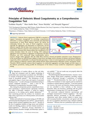
(T0). Permittivity of blood changed dynamically with blood
coagulation. The typical result is shown in the plot of
normalized permittivity as a function of time and frequency
in Figure 1a. Among that data, the permittivity at 10 MHz
passes through a shallow minimum in the early stage and then
increases until it reaches a nearly constant value, while at 1
MHz, there are two local maxima and a subsequent decrease
(Figure 1b). Microscopic observation of blood suggested that
the decrease at 10 MHz and increase at 1 MHz soon after the
start of the measurement are due to rouleau formation (Figure
S2). This was confirmed by the dielectric measurements of
blood under the anticoagulation state as shown in Figure 2,
Figure 1. Typical DBCM response of spontaneous blood coagulation.
The changes in permittivity that occur with coagulation are normalized
by data 1 min after recalcification. Panel (a) is a 3D image showing the
changes in all measured frequencies. Characteristic changes are visible
in the MHz band, corresponding to erythrocytes interfacial polar-
ization. Panel (b) is an extract from panel (a) showing the changes at 1
and 10 MHz. At 1 MHz, a two-step peak is observed, but at 10 MHz,
the value reaches a minimum at an early stage before increasing to an
almost constant value.
Analytical Chemistry Article
DOI: 10.1021/acs.analchem.5b02723
Anal. Chem. 2015, 87, 10072−10079
10074
4. where the reversibility of rouleau formation is reflected by the
permittivity changes induced by agitation.
Observations with SEM/TEM. The SEM images in Figure
3a−c show that the clot surface is covered with a dense fibrin
network. Cines et al.34
reported similar images but with less
dense networks probably because of the different experimental
conditions. These images indicate that erythrocytes clumped
together in a fibrin network change shape and become
polyhedrocytes as a result of the large compressive force
from platelet clot retraction. In our SEM images, on the other
hand, only a limited degree of shape transformation is observed,
probably because the erythrocytes located near fissures in the
clot suffer from reduced compressive force. Indeed, TEM
images (Figure 3d) show that many cell cross sections are not
consistent with normal erythrocyte shape, suggesting eryth-
rocyte aggregation and shape modification in the central
portion of clots. We note that TEM images of clots in the
presence of a platelet inhibitor show less erythrocytes
aggregation and transformation (Figure S3).
Effects of Blood Coagulation/Fibrinolysis Activators
and Inhibitors (T2 to T6). The permittivity curves at 10 and 1
MHz for extrinsic activation (T2) and spontaneous clotting
(T1) assays are compared in Figure 4a,b, respectively. At 10
MHz, the curve shifted toward shorter times upon activation,
while the curve shape changed at 1 MHz.
Figure 5a shows the extrinsic activation curves (10 MHz) at
different CyD concentrations corresponding to different
platelet function levels (T3). The amplitude from the minimum
value decreased with increasing CyD concentrations and
leveled off above 5 μM. This convergence was similarly
observed in a thromboelastography study.38
On the other hand,
no significant change in the minimum position was observed.
As discussed later, this is consistent with the fact that CyD does
not affect thrombin production and fibrin polymerization.
Corresponding data at 1 MHz are shown in Figure S4 as a
typical example of the clot strength influence at this frequency.
Different fibrin polymerization levels were reproduced in
vitro by varying H-Gly-Pro-Arg-Pro-OH concentration (T4) in
an extrinsic activation assay. Figure 5b shows that the amplitude
and rate of the increase from the minimum value decreased
with increasing concentrations of H-Gly-Pro-Arg-Pro-OH. No
significant prolongation of the time corresponding to the
minimum value was observed below 1.0 mg/mL. This agrees
Figure 2. Rouleau formation and dielectric response. Panel (a) shows
the dielectric spectrum right after pipet mixing and 5 min after mixing
stopped. Panels (b) and (c) show the dielectric changes at 10 and 1
MHz, respectively. These changes were measured as mixing was
stopped and started again, three times in a row. The arrows indicate
the point at which mixing was stopped.
Figure 3. SEM and TEM images of a clot. On the SEM image, the clot
surface covered with fibrin is observed as well as erythrocytes seen
through surface fissures (a, b, c). On the TEM image, erythrocytes that
are clumped together and have deformed shapes are visible in the clot
interior (d).
Analytical Chemistry Article
DOI: 10.1021/acs.analchem.5b02723
Anal. Chem. 2015, 87, 10072−10079
10075
5. with the argument of Chakroun et al.38
that, though H-Gly-Pro-
Arg-Pro-OH inhibits fibrin polymerization, it has almost no
effect on the upstream of blood coagulation cascade, before
thrombin production is started.
Figure 6a,b shows the extrinsic activation curves with the
addition of tissue plasminogen activator (tPA) and urokinase,
respectively (T5), as well as the results of fibrinolysis inhibition
by aprotinin (T6). In the absence of aprotinin, the permittivity
at 10 MHz first increased with clot formation, then settled at a
constant value, and finally decreased as fibrinolysis settled in.
When fibrinolysis is inhibited by aprotinin, the final decrease
disappeared. This demonstrates that with DBCM it is possible
to observe thrombolysis as a decrease in permittivity at 10 MHz
after clot formation. We note that fibrinolysis induced changes
at 1 MHz, as well (Figure S5).
The contribution of heparin is generally observed as an
extension of the coagulation onset time of the intrinsic
activation assay (T7).14
Figure 6c shows that the DBCM
response was prolonged due to the addition of heparin (T8)
and that this prolongation was lost upon heparin neutralization
with the addition of heparinase (T9). Figure S6 shows the
corresponding results at 1 MHz.
■ DISCUSSION
Analysis of Dielectric Spectra. A dielectric spectrum is
modeled by a function, appropriately chosen to represent the
physical mechanism behind the spectrum, which contains,
among others, relaxation strength Δε and frequency fc (see
Figure 7a,b for definition) as fitting parameters.27−29,31,36
Following well-established dielectric spectroscopy theory of
cell suspensions, we assumed a Cole−Cole function and a
constant phase angle impedance to model erythrocytes
interfacial polarization and electrode polarization, respectively.
The curved lines in Figure 7a,b are the best-fit results to the
actual data, while the broken lines show only the relevant
contribution from erythrocytes. Changes of Δε and fc with
Figure 4. Results of extrinsic activation. The changes at 10 MHz (a)
and 1 MHz (b) are shown using permittivity normalized by the
minimum value and compared with samples without tissue factor.
Figure 5. Changes due to platelet or fibrin polymerization inhibition.
The changes in permittivity at 10 MHz normalized by the minimum
value are shown with error bars indicating levels of uncertainty due to
the normalization by the minimum values. (a) The amplitude
diminution as cytochalasin D concentration increases is visible. In
the inset, the normalized permittivity 15 min after the start of
measurements is plotted against cytochalasin D concentration. (b)
The amplitude diminution as the fibrin polymerization inhibitor
concentration increases is visible.
Figure 6. Responses to fibrinolysis activation and residual heparin.
The changes in permittivity at 10 MHz normalized by the minimum
value are shown in panels (a) and (b) with error bars indicating levels
of uncertainty due to the normalization by the minimum value. The
sample of panels (a) and (b) are made by addition of tPA and
urokinase to the blood specimen, respectively. Responses in the
presence and absence of a fibrinolysis inhibitor (aprotinin) are shown.
The experiment using tPA (a) and urokinase (b) were carried out
using specimens from separate healthy people. In panel (c), the
changes in permittivity at 10 MHz normalized by both the minimum
value and the value 60 min after the start of measurements are shown.
Upon heparin addition to the blood specimen, a significant delay in
the response was observed in comparison with intact blood. This delay
disappeared with the neutralization of heparin by heparinase, and a
response similar to that of intact blood was observed.
Analytical Chemistry Article
DOI: 10.1021/acs.analchem.5b02723
Anal. Chem. 2015, 87, 10072−10079
10076
6. proceeding spontaneous clotting (T1) are shown in Figure
7c,d.
Here, we would like to review the relationship between data
at 1 or 10 MHz presented in Figures 2b,c, and 4−6 and the
dielectric relaxation parameters Δε and fc. In a dielectric
measurement of whole blood, permittivity at 1 MHz is sensitive
to Δε changes and less sensitive to shifts of fc. In comparison,
permittivity at 10 MHz is sensitive to both Δε changes and
small shifts of fc. For a numerical example using the Cole−Cole
function presented in Figure 7a, a 20% increase of Δε induces
19% and 15% increases of the permittivity at 1 and 10 MHz,
respectively, while a 20% increase of fc induces 10% and 14%
increases at those frequencies. Therefore, monitoring of 1 and
10 MHz allows us to access information about Δε and fc
changes with blood coagulation. To realize DBCM as a medical
device, data analysis using specific frequencies is easier to
implement than nonlinear curve fitting and suitable for an
automated system.
Phase 1: Rouleau Formation Process. The results of
rouleau formation measurements presented in Figure 2 suggest
that the initial increase in Δε and decrease in fc for several
minutes after the start of measurements in Figure 7c,d,
hereafter referred to as phase 1 (P1), are attributable to the
rouleau formation process. Referring to past reports,43,44
we
suppose that P1 covers the whole stepwise rouleau formation
process and regroups linear erythrocyte aggregation spanning
several seconds, incorporation of aggregates in a three-
dimensional network during several minutes, and final
transformation into spherical aggregates.
Phase 2: Erythrocytes Aggregation Occurring with
Coagulation. Whole blood recalcification time was manually
measured (Figure S7). The time until blood loses its fluidity,
CT, was determined visually as CT = 330 ± 30 s and was found
to agree with the start of phase 2 (P2) in Figure 7c,d. P2 is
characterized by the increase of Δε and minimal changes in fc
observed approximately 7 min from the start. The fact that the
coagulation-induced DBCM response starts from CT is
consistent with reports by Chernysh et al.45,46
that fibrin
network formation continues after CT and a report by Rand et
al.47
that no more than 2% of prothrombin had become α-
thrombin and, moreover, that only approximately 15% of it was
acting on fibrinogen at CT. During P2, therefore, it is
reasonable to assume that erythrocytes move with considerable
freedom so that they continue to form aggregates trapped in
fibrin networks.34
We think that the original rouleau structure is
conserved while further aggregation proceeds during the early
phase of coagulation, which is in line with arguments suggesting
the production of soluble fibrin polymers is the main force
promoting aggregation in this phase.48,49
There may also be an
increase in heterogeneity from the rearrangement of the
aggregate structures. From a numerical simulation based on the
dielectric response theory of heterogeneous systems, the
permittivity near 1 MHz was reported to increase with cell
aggregation.21
Even when the aggregation level is assumed
constant, the permittivity near 1 MHz would increase when
hollow (foam-structured) elements are formed from the
rearrangement of the aggregate structures. Almost no change
in fc was observed in that theoretical analysis.21
These
observations appear coherent with the behaviors of Δε and fc
observed in P2.
Phase 3: Change in Erythrocyte Shape with Blood
Coagulation. Phase 3 (P3) in Figure 7c,d is characterized by
the decrease in Δε and increase in fc approximately 15 min
from the start. Both a SEM study by Cines et al.34
and our
TEM images (Figure 3d) demonstrate the shape trans-
formation of erythrocytes from native discocytes to polyhedral
erythrocytes during clot formation. A systematic study of the
influence of erythrocytes shape on the dielectric spectra has
demonstrated that, at constant volume fraction, Δε decreases
while fc increases as the native shape is transformed into other
shapes (Figures S8 and S9).31
It is possible that, in addition to
this, the partial disappearance of the rouleau structure within
aggregates also contributes to the observed changes. A careful
observation of TEM images (Figure 3d) indeed suggests that
rouleau structures after coagulation appear disturbed compared
to noncoagulated blood observed using an optical microscope
(Figure S2).
Effects from Blood Coagulation Activators. As shown
in Figure 4, in assays accelerated by tissue factor, not only does
the response time shorten but also the amplitude and shape of
the change (especially 1 MHz, Figure 4b) differ in comparison
with spontaneous coagulation. Such behavior was repeatedly
observed for different individuals (data not shown). Therefore,
it seems to be the general response to activation of blood
coagulation. This is thought to be because the response deemed
to be the erythrocyte aggregation phase (P2) that accompanies
blood coagulation shown in Figure 7c,d shifts before it can
proceed to the next phase (the erythrocyte deformation that
occurs with blood coagulation, P3). Visually determined CT =
165 ± 15 s in the presence of tissue factor was found to agree
with the time corresponding to a permittivity minimum at 10
MHz. Ninivaggi et al.50
developed a whole blood thrombin
production assay and reported thrombin production lag times
of 4.8 and 4.2 min at tissue factor concentrations of 0.5 and 1.0
pM, respectively, and thrombin production maximum at 6.9
Figure 7. Dielectric relaxation analysis and blood coagulation phase
identification. Panel (a) shows the real part and panel (b) the
imaginary part of the complex permittivity. The broken red lines are
the result of an analysis of the interfacial polarization phenomenon
according to a Cole−Cole type dielectric relaxation function. This
function is characterized by the relaxation strength Δε corresponding
to changes in ε′ (a) and the relaxation frequency fc that corresponds to
the peak of this function observed in the imaginary part of permittivity,
ε″ (b). The curved lines in the figure are the sum of all the assumed
contributions in the analysis, and agree well with the experimental
values. Spontaneous whole blood coagulation data shown in Figure 1
were analyzed using a Cole−Cole type dielectric relaxation modeling.
The changes in relaxation strength and relaxation frequency are shown
in panel (c) and (d), respectively. In P1, immediately after the start of
measurements the response to rouleau formation is observed. It is
followed by the response to blood coagulation induced erythrocyte
aggregation in the P2 phase and the response to erythrocytes
deformation corresponding to clot retraction in the P3 phase.
Analytical Chemistry Article
DOI: 10.1021/acs.analchem.5b02723
Anal. Chem. 2015, 87, 10072−10079
10077
7. and 6.3 min, respectively. Because the final concentration of
tissue factors in our extrinsic activation assay was 0.6−0.7 pM,
within the above test concentrations, the start of P3 at 4 min
and the maximal increase rate at 6 min are seen to be coherent
with the time scale reported by Ninivaggi et al.50
Platelet inhibition adversely affects clot retraction and thus
limits erythrocyte deformation and disturbance of rouleau
structures within aggregates, so that the amplitude of P3
diminishes with increasing inhibitor concentration, as shown in
Figure 5a. Yet, the stagnation of P3 amplitude with excess
inhibitor concentrations suggests that erythrocyte deformation
and aggregates disappearance occur, though in a limited way,
even under platelet total inhibition. Conversely, when fibrin
formation is inhibited significantly, it appears that almost no
erythrocyte deformation occurs even when there is no effect on
platelet activity (Figure 5b). This agrees with the fact that clot
retraction does not occur unless fibrin networks are formed.
Fibrinolytic System Monitoring and Evaluation of
Residual Heparin. In the clot lysis process, the dielectric
response approaches the value before blood coagulation
(Figure 6a,b). This is thought to be because erythrocytes
recover their native shape as the clot compressive force is
relaxed with the loss of the fibrin network.
Heparin remaining in blood forms complexes with
antithrombin III and effectively inhibits thrombin, producing
a delay in the blood coagulation reaction (Figure 6c). When
heparinase is added, this delay disappears. Thus, it is possible to
determine whether the reason for the coagulation delay is due
to residual heparin or to other coagulation abnormalities by
comparing measurement results under the two conditions, with
and without heparinase.
■ CONCLUSIONS
For effective thrombosis and hemostasis treatment in the
perioperative period, a comprehensive test that can evaluate the
coagulation and fibrinolytic capacity of whole blood samples
simply and quantitatively is effective. In the field of internal
medicine as well, there are clear needs in the management of
new anticoagulant therapies using direct thrombin inhibitors or
factor Xa inhibitors. DBCM meets these needs by sensitively
measuring and analyzing changes in permittivity with blood
coagulation. The main dielectric response on DBCM is
produced by the aggregation and shape transformation of
erythrocytes during coagulation. This response was useful to
evaluate the activation of the extrinsic and intrinsic blood
coagulation reaction pathways. Decreases in the amount of
change were also observed to be dependent on the
concentration of platelet inhibitors and fibrin aggregation
inhibitors. Examples of measurements were also shown for
fibrinolysis activity and residual heparin evaluation, which are
important as perioperative tests. According to these results,
proofs of concept for multiple assays have been established to
realize comprehensive coagulation testing with an automated
point-of-care device. Further evaluation of DBCM is now
ongoing through clinical studies to statistically demonstrate the
reliability of the multiple parameters quantitatively extracted
from the dielectric response, such as CT, the clotting
amplitude, and clotting velocity. It is a new blood coagulation
test method that is promising for the achievement of
personalized medicine in the coming years.
■ ASSOCIATED CONTENT
*S Supporting Information
The Supporting Information is available free of charge on the
ACS Publications website at DOI: 10.1021/acs.anal-
chem.5b02723.
Diagram of the disposable cartridge, optical microscope
and TEM images, and other data (PDF)
■ AUTHOR INFORMATION
Corresponding Author
*Tel.: +81 3 3811 8970. Fax: +81 3 5803 4790. E-mail:
Yoshihito.Hayashi@jp.sony.com.
Notes
The authors declare the following competing financial
interest(s): Y.H., M.-A.B. and K.M. are employees of Sony
Corp. M.N. declares no competing financial interests.
■ ACKNOWLEDGMENTS
This work was partially supported by the Medical Research and
Development Programs Focused on Technology Transfer:
Development of Advanced Measurement and Analysis Systems
(SENTAN) from Japan Agency for Medical Research and
Development, AMED (Y.H.). A part of this study for SEM and
TEM observations was supported by the Project at Tokyo
Medical and Dental University for Creation of Research
Platforms and Sharing of Advanced Research Infrastructure of
the Ministry of Education, Culture, Sports, Science and
Technology of Japan. We would like to thank Ms. Aya Murata,
Ms. Seungmin Lee, and Ms. Kaori Kawaguchi, from Sony
Corp., for their helpful discussion.
■ REFERENCES
(1) Marder, V. J., Aird, W. C., Bennett, J. S., Schulman, S., White, G.
C., Eds. Hemostasis and Thrombosis: Basic Principles and Clinical
Practice, 6th ed.; Lippincott Williams & Wilkins: Philadelphia, 2013.
(2) Greenblatt, D. J.; von Moltke, L. L. J. Clin. Pharmacol. 2005, 45,
127−132.
(3) Connolly, S. J.; Ezekowitz, M. D.; Yusuf, S.; Eikelboom, J.;
Oldgren, J.; Parekh, A.; Pogue, J.; Reilly, P. A.; Themeles, E.; Varrone,
J.; Wang, S.; Alings, M.; Xavier, D.; Zhu, J.; Diaz, R.; Lewis, B. S.;
Darius, H.; Diener, H. C.; Joyner, C. D.; Wallentin, L. N. Engl. J. Med.
2009, 361, 1139−1151.
(4) EINSTEIN Investigators; Bauersachs, R.; Berkowitz, S. D.;
Brenner, B.; Buller, H. R.; Decousus, H.; Gallus, A. S.; Lensing, A. W.;
Misselwitz, F.; Prins, M. H.; Raskob, G. E.; Segers, A.; Verhamme, P.;
Wells, P.; Agnelli, G.; Bounameaux, H.; Cohen, A.; Davidson, B. L.;
Piovella, F.; Schellong, S. N. Engl. J. Med. 2010, 363, 2499−2510.
(5) Patel, M. R.; Mahaffey, K. W.; Garg, J.; Pan, G.; Singer, D. E.;
Hacke, W.; Breithardt, G.; Halperin, J. L.; Hankey, G. J.; Piccini, J. P.;
Becker, R. C.; Nessel, C. C.; Paolini, J. F.; Berkowitz, S. D.; Fox, K. A.;
Califf, R. M. N. Engl. J. Med. 2011, 365, 883−891.
(6) Gachet, C.; Aleil, B. Eur. Heart J. Suppl. 2008, 10, A28−A34.
(7) Lenk, E.; Spannagl, M. J. Int. Fed. Clin. Chem. 2013, 24, 1−7.
(8) Cohoon, K. P.; Heit, J. A. Clin. Chem. 2013, 59, 1299−1300.
(9) Lawson, J. H.; Murphy, M. P. Semin. Hematol. 2004, 41, 55−64.
(10) Kroll, M. H. Clin. Lab. News 2010, 36, 8−10.
(11) Bischof, D.; Dalbert, S.; Zollinger, A.; Ganter, M. T.; Hofer, C.
K. Minerva Anestesiol. 2010, 76, 131−137.
(12) CRASH-2 trial collaborators; Shakur, H.; Roberts, I.; Bautista,
R.; Caballero, J.; Coats, T.; Dewan, Y.; El-Sayed, H.; Gogichaishvili, T.;
Gupta, S.; Herrera, J.; Hunt, B.; Iribhogbe, P.; Izurieta, M.; Khamis, H.;
Komolafe, E.; Marrero, M. A.; Mejía-Mantilla, J.; Miranda, J.; Morales,
C.; Olaomi, O.; Olldashi, F.; Perel, P.; Peto, R.; Ramana, P. V.; Ravi, R.
R.; Yutthakasemsunt, S. Lancet 2010, 376, 23−32.
Analytical Chemistry Article
DOI: 10.1021/acs.analchem.5b02723
Anal. Chem. 2015, 87, 10072−10079
10078
8. (13) Johansson, P. I.; Stensballe, J.; Oliveri, R.; Wade, C. E.;
Ostrowski, S. R.; Holcomb, J. B. Blood 2014, 124, 3052−3058.
(14) Anderson, L.; Quasim, I.; Soutar, R.; Steven, M.; Macfie, A.;
Korte, W. Transfus. Med. 2006, 16, 31−39.
(15) Tanaka, K. A.; Key, N. S.; Levy, J. H. Anesth. Analg. 2009, 108,
1433−1446.
(16) Ferraris, V. A.; Ferraris, S. P.; Saha, S. P.; Hessel, E. A., II; Haan,
C. K.; Royston, B. D.; Bridges, C. R.; Higgins, R. S.; Despotis, G.;
Brown, J. R.; Spiess, B. D.; Shore-Lesserson, L.; Stafford-Smith, M.;
Mazer, C. D.; Bennett-Guerrero, E.; Hill, S. E.; Body, S. Ann. Thorac.
Surg. 2007, 83, S27−S86.
(17) McMichael, M. A.; Smith, S. A. Vet. Clin. Pathol. 2011, 40, 140−
153.
(18) Park, M. S.; Martini, W. Z.; Dubick, M. A.; Salinas, J.; Butenas,
S.; Kheirabadi, B. S.; Pusateri, A. E.; Vos, J. A.; Guymon, C. H.; Wolf,
S. E.; Mann, K. G.; Holcomb, J. B. J. Trauma 2009, 67, 266−276.
(19) Chitlur, M.; Sorensen, B.; Rivard, G. E.; Young, G.; Ingerslev, J.;
Othman, M.; Nugent, D.; Kenet, G.; Escobar, M.; Lusher, J.
Haemophilia 2011, 17, 532−537.
(20) Evans, P. A.; Hawkins, K.; Williams, P. R. Rheol. Rev. 2006,
255−291.
(21) Hayashi, Y.; Katsumoto, Y.; Omori, S.; Yasuda, A.; Asami, K.;
Kaibara, M.; Uchimura, I. Anal. Chem. 2010, 82, 9769−9774.
(22) Kaibara, M. J. Biorheol. 2009, 23, 2−10.
(23) Debye, P. Polar Molecules; Chemical Catalog Co.: New York,
1929.
(24) Grant, E. H.; Sheppard, R. J.; South, G. P. Dielectric Behaviour of
Biological Molecules in Solution; Oxford University Press: Oxford, 1978.
(25) Foster, K. R.; Schwan, H. P. Crit. Rev. Biomed. Eng. 1989, 17,
25−104.
(26) Pethig, R. Dielectric and Electronic Properties of Biological
Materials; John Wiley & Sons: New York, 1979.
(27) Takashima, S. Electrical Properties of Biopolymers and Membranes;
IOP Publishing Ltd: Philadelphia, 1989.
(28) Asami, K. Prog. Polym. Sci. 2002, 27, 1617−1659.
(29) Feldman, Y.; Ermolina, I.; Hayashi, Y. IEEE Trans. Dielectr.
Electr. Insul. 2003, 10, 728−753.
(30) Chiba, S.; Uchibori, K.; Fujiwara, T.; Ogata, T.; Yamauchi, S.;
Shirai, T.; Masuo, M.; Okamoto, T.; Tateishi, T.; Furusawa, H.; Fujie,
T.; Sakashita, H.; Tsuchiya, K.; Tamaoka, M.; Miyazaki, Y.; Inase, N.;
Sumi, Y. J. Sci. Res. Rep. 2015, 4, 180−188.
(31) Hayashi, Y.; Oshige, I.; Katsumoto, Y.; Omori, S.; Yasuda, A.;
Asami, K. Phys. Med. Biol. 2008, 53, 2553−2564.
(32) Irimajiri, A.; Ando, M.; Matsuoka, R.; Ichinowatari, T.;
Takeuchi, S. Biochim. Biophys. Acta, Gen. Subj. 1996, 1290, 207−209.
(33) Asami, K.; Sekine, K. J. Phys. D: Appl. Phys. 2007, 40, 2197−
2204.
(34) Cines, D. B.; Lebedeva, T.; Nagaswami, C.; Hayes, V.;
Massefski, W.; Litvinov, R. I.; Rauova, L.; Lowery, T. J.; Weisel, J.
W. Blood 2014, 123, 1596−1603.
(35) Kasahara, K.; Kaneda, M.; Miki, T.; Iida, K.; Sekino-Suzuki, N.;
Kawashima, I.; Suzuki, H.; Shimonaka, M.; Arai, M.; Ohno-Iwashita,
Y.; Kojima, S.; Abe, M.; Kobayashi, T.; Okazaki, T.; Souri, M.;
Ichinose, A.; Yamamoto, N. Blood 2013, 122, 3340−3348.
(36) Bordi, F.; Cametti, C.; Gili, T. Bioelectrochemistry 2001, 54, 53−
61.
(37) Kremer, F.; Schönhals, A. Broadband Dielectric Spectroscopy;
Springer: Berlin, 2003.
(38) Chakroun, T.; Gerotziafas, G. T.; Seghatchian, J.; Samama, M.
M.; Hatmi, M.; Elalamy, I. Thromb. Haemost. 2006, 95, 822−828.
(39) Cooper, J. A. J. Cell Biol. 1987, 105, 1473−1478.
(40) Cerecedo, D.; Stock, R.; González, S.; Reyes, E.; Mondragón, R.
Haematologica 2002, 87, 1165−1176.
(41) Laudano, A. P.; Doolittle, R. F. Proc. Natl. Acad. Sci. U. S. A.
1978, 75, 3085−3089.
(42) Rijkers, D. T.; Wielders, S. J.; Béguin, S.; Hemker, H. C.
Thromb. Res. 1998, 89, 161−169.
(43) Dobbe, J. G.; Streekstra, G. J.; Strackee, J.; Rutten, M. C.;
Stijnen, J. M.; Grimbergen, C. A. IEEE Trans. Biomed. Eng. 2003, 50,
97−106.
(44) Fabry, T. L. Blood 1987, 70, 1572−1576.
(45) Chernysh, I. N.; Weisel, J. W. Blood 2008, 111, 4854−4861.
(46) Chernysh, I. N.; Nagaswami, C.; Weisel, J. W. Blood 2011, 117,
4609−4614.
(47) Rand, M. D.; Lock, J. B.; van’t Veer, C.; Gaffney, D. P.; Mann,
K. G. Blood 1996, 88, 3432−3445.
(48) Hunter, R. L.; Papadea, C.; Gallagher, C. J.; Finlayson, D. C.;
Check, I. J. Thromb. Haemost. 1990, 63, 6−12.
(49) van Gelder, J. M.; Nair, C. H.; Dhall, D. P. Biorheology 1994, 31,
259−275.
(50) Ninivaggi, M.; Apitz-Castro, R.; Dargaud, Y.; de Laat, B.;
Hemker, H. C.; Lindhout, T. Clin. Chem. 2012, 58, 1252−1259.
Analytical Chemistry Article
DOI: 10.1021/acs.analchem.5b02723
Anal. Chem. 2015, 87, 10072−10079
10079