The document discusses techniques of mandibular anesthesia, primarily focusing on local anesthetics, nerve blocks, and related anatomical considerations. It details various methods such as the inferior alveolar nerve block, Gow-Gates technique, and Vazirani-Akinosi technique, including their indications, advantages, disadvantages, and potential complications. It also emphasizes the importance of accurate anatomical localization and technique to minimize risks during anesthesia procedures.
![TECHNIQUES OF MANDIBULAR
ANESTHESIA
PRATHIBA . E[ FINAL YEAR ]
DPT OF OMFS](https://image.slidesharecdn.com/techniquesofmandibularanesthesia-240605152404-edf52930/75/Techniques-of-mandibular-anesthesia-pptx-1-2048.jpg)


![COMPOSITION OF LOCAL ANESTHESIA
Local
anesthetic
agent ;
Lignocaine
HCI 2%(20
mg/ml)
Reducing
agent;
Sodium
meta-
bisulphite
[0.5mg]
Vasoconstr
ictor;
Adrenaline
[0.012mg]
Isotonic
solution ;
Ringer’s
solution[6
mg]
Sodium
hydroxide
– to adjust
pH
VASOCONSTRICTORS OF L.A ;
- Decrease blood flow to the site of
injection – better visualization
- Reduced systemic toxicity
- More LA enters into nerve-
Increase duration of action](https://image.slidesharecdn.com/techniquesofmandibularanesthesia-240605152404-edf52930/75/Techniques-of-mandibular-anesthesia-pptx-5-2048.jpg)


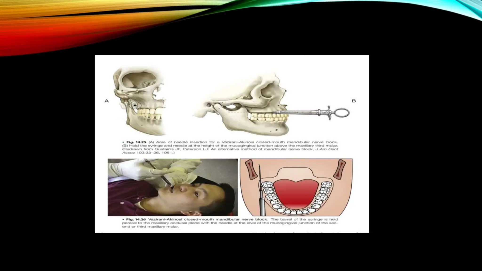

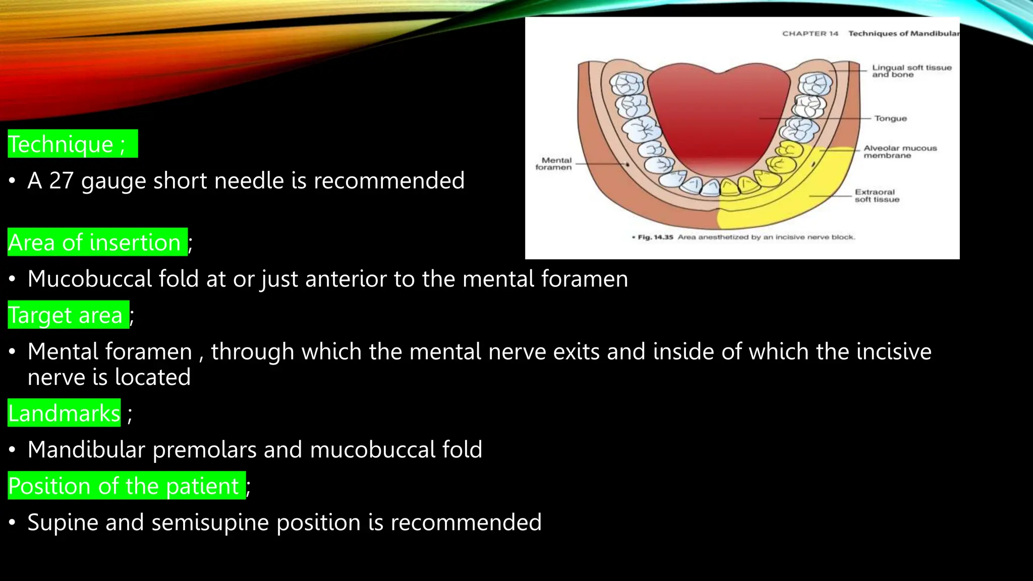
![Technique
A 25 or 27 gauge short needle recommended
Area of insertion
• mucobuccal fold at or just anterior to the mental foramen
Target area;
mental nerve as it exits the mental foramen [ usually located between the apices of the
first and second premolars]
Landmarks ;
mandibular premolars and mucobuccal fold
Position of the patient ;
supine position is recommended](https://image.slidesharecdn.com/techniquesofmandibularanesthesia-240605152404-edf52930/75/Techniques-of-mandibular-anesthesia-pptx-24-2048.jpg)