1) Solid state NMR spectroscopy can distinguish between leathers tanned with different vegetable tanning agents based on characteristic spectral fingerprints. Condensed tannins from mimosa and quebracho leave distinct fingerprints in leather spectra related to their chemical structures.
2) Hydrolyzable tannins from chestnut and tara also leave identifiable fingerprints in leather spectra. NMR can differentiate between leathers tanned with condensed versus hydrolyzable tannins.
3) Chromium tanned leather shows selective disappearance of collagen signals, including from acidic aspartyl and glutamyl residues likely bound to chromium. This indicates chromium binding to acidic residues in the collagen matrix.
![Molecules 2011, 16 1241
Keywords: vegetable tannins; polyphenols; chromium; aluminium; glutaraldehyde
1. Introduction
Tanning is an essential phase in one of civilization’s oldest processes, the transformation of hide
and skins into leather [1], and vegetable tannins were probably the earliest used reagents [2]. In spite
of the development of numerous synthetic fabrics, leather remains indispensible in many applications
because of its distinctive properties: toughness, non-flammability, resistance to heat, impermeability to
water, and permeability to air and water vapour. However, partly perhaps because the know-how built
up over time is still able to produce satisfactory finished material, the fundamental molecular events
underlying leather production are still not completely understood. Apart from the vegetable tannins,
other reagents used in tanning include salts of chromium (III) and aluminium (III), and the bifunctional
organic reagent glutaraldehyde [2]. Characterization of the nature of intermediates and the final
product in any tanning process would clearly be of importance in mechanism-based attempts to
enhance tanning efficiency or finished leather quality. Currently the industry uses certain standards
based on the physical characteristics of intermediate and final product such as shrinkage and shrinkage
temperature, and thermal properties, which give no direct clue as to any underlying chemical and
physicochemical transformations. Detailed understanding of these could improve tanning methodology,
finished product quality, and process control. It is also conceivable that chemical “signatures” could
prove valuable in assessing and demonstrating the authenticity of high value
leather products.
Nuclear magnetic resonance spectroscopy (NMR) is increasingly used in analysis and quality
assurance of industrial samples of biological origin, such as fats, oils, and juices [3], material of wood
origin [4], and biopharmaceuticals [5]. A major factor underlying this is that NMR seldom requires
significant sample manipulation. In industry this is attractive because information-rich analyses can be
carried out on raw materials, process intermediates or final products without possible confounding
effects arising from sample manipulation, extraction, derivatization, or contamination. This can be a
considerable advantage in internal process and quality control, and in presentation of data to external
regulators. NMR has another significant strength: data is easily interpreted in chemical terms because
each atom in a distinct environment gives rise to only a single signal, the frequency and characteristics
of which are predictable consequences of molecular structure and environment. Thus many pure
industrial substances, mixtures and composites produce unique and readily recognised spectroscopic
“signatures”. Moreover with certain simple precautions signal intensity can be related to molecular
abundance in a way not possible with more sensitive vibrational spectroscopy, mass spectrometry, and
diffraction techniques. Finally, limited access to expensive NMR equipment is no longer the constraint
to industrial applications that it once was, because high performance spectrometers are proliferating
through academia and within certain industries themselves.
NMR is useful in in situ characterization of tannic substances in wood derived materials [6], and
changes in tannin content of woody materials under various transformation processes [7-9]. NMR can
distinguish the solid tannins extracted from Acacia mangium [10] and maritime pine [11], and has been
proposed as a general method of quantifying polyphenols, including tannins, in plant material [12,13].](https://image.slidesharecdn.com/ad59e445-bbd5-4161-88bd-8a82e018293a-160921085144/85/Tannin-Article-molecules-16-01240-2-320.jpg)
![Molecules 2011, 16 1242
Tannins leave a clear spectroscopic fingerprint in the 13
C-NMR spectra of industrial materials in which
they are used in adhesives [14,15], which moreover is sensitive to chemical changes brought about by
processing [16]. NMR can also quantify the degree of extraction of tannins from bark [17], with the
NMR results validating against conventional extractive gravimetric techniques.
Solid state NMR has also been used to study leather [18,19], and in a single case to infer tannin
content [20]. The latter paper anticipates the general usefulness of NMR in characterization of leather
tanned with vegetable tannins by emphasizing the convenient 13
C-NMR spectral “window” between
resonance frequencies of ca. 71 ppm and ca. 165 ppm which contains few signals from leather collagen
protein but numerous signals from tannins. Tanning with other reagents has also been studied by NMR.
Spectroscopic changes induced in model collagen peptides by Cr (III) [21] and Al (III) [22] are
interpreted in terms of metal ion recognition by acidic protein groups.
Here we extend this work to a systematic study of the effects exerted by vegetable tanning and other
common tanning procedures on the NMR characteristics of whole leather, leading to the identification
of spectroscopic changes characteristic of some of these. The study also identifies effects that certain
tanning methods exert on the protein structures in leather, which may hold clues to the molecular
events underlying different processes.
2. Results and Discussion
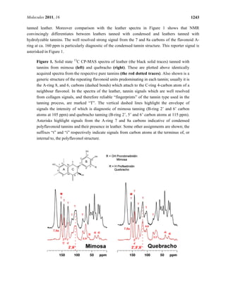
2.1. Tanning with condensed tannins
Spectra of leathers tanned with two types of condensed tannin, from mimosa and quebracho species,
are shown in Figure 1. Each is accompanied by a spectrum of the respective pure tannin. Condensed
tannins are complex oligomers of flavan-3-ol monomers of general structures as shown in the figure.
The spectra of both pure condensed tannins are consistent with structure and with literature
assignments of whole tannins and constituent monomers [23-26]. Moreover the condensed tannins
from the two species are clearly distinguishable by their 13
C-NMR spectral fingerprints. Most
importantly each tannin spectrum is faithfully recapitulated in the spectra of the leathers tanned by
them. Mimosa tannin is predominantly a prorobinetinidin polymer (resorcinol type A-ring and
pyrogallol type B-ring) [27,28] while quebracho tannin is predominantly a profisetinidin (resorcinol
type A- and B-ring). The B-ring C-H carbons (2', 5', 6') of quebracho tannin resonate conspicuously at
ca. 115 and those of mimosa (2', 6' carbons) at 105 ppm, and are effective diagnostics of condensed
tannin type in whole leather. The envelope of signals in which these markers are dominant are
highlighted by vertical dashed lines in Figure 1.
2.2. Tanning with hydrolyzable tannins
Similarly, spectra of leathers tanned with two hydrolyzable tannins, from chestnut and tara, are
shown in Figure 2. Hydrolyzable tannins are complex mixtures of sugar monomers or polymers
esterified with polyphenols such as gallic acid and its derivatives and closely related compounds
[29-31]; typical constituent structures are exemplified in the figure.
As in the condensed tannin cases, pure hydrolyzable tannin spectra reflect the chemistry of each
[32-35]. Again the fingerprint of each tannin manifests itself clearly in the spectrum of each respective](https://image.slidesharecdn.com/ad59e445-bbd5-4161-88bd-8a82e018293a-160921085144/85/Tannin-Article-molecules-16-01240-3-320.jpg)
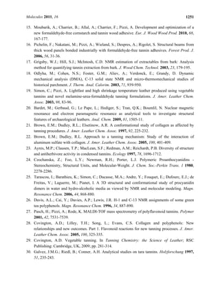
![Molecules 2011, 16 1244
Figure 2. Solid state 13
C CP-MAS spectra of leather tanned with tannins from chestnut
(left) and tara (right) (black traces) plotted above spectra of the pure tannin corresponding
to the respective leather sample. Also shown are some chemical structural formulae of
typical constituents of hydrolysable tannin, and spectral assignments. The asterisk (*)
indicates a signal due to non-sugar sp3
carbons in compounds such as the chebulic acid
depicted here.
In principle it is possible to measure leather:tannin ratios from solid state NMR spectra and thereby
infer the tannin load in the product. When cross polarization [36] from 1
H to 13
C (the standard 13
C
solid state observation technique) is used to enhance the inherently weak 13
C signal, relating signal
intensity to absolute molecular abundance is complicated by the fact that cross polarization efficiency
is a function of molecular structure and must be established for different signals by examining their
intensity dependence on cross polarization times. Although we have not carried out the necessary
detailed studies it is nevertheless possible on the basis of a few simple assumptions to infer
approximate leather collagen:tannin ratios from the data as presented here. If one assumes that cross
polarization dynamics of non-protonated carbons are likely to be approximately similar to eachother,
the well resolved signal from collagen amide carbons at ca. 175 p.p.m. is a measure of protein content,
and the signal at ca. 160 p.p.m. (quaternary 7 and 8a carbons) is a measure of condensed tannin content.
The ratio of the integrals of these two signals for both the mimosa and quebracho tanned materials is](https://image.slidesharecdn.com/ad59e445-bbd5-4161-88bd-8a82e018293a-160921085144/85/Tannin-Article-molecules-16-01240-5-320.jpg)
![Molecules 2011, 16 1245
ca. 0.4 implying a molar ratio of collagen amino acid residues:tannin repeat units of ca. 0.2. Assuming
an average molecular mass of 100 and 200 for collagen amino acid residues and tannin monomers,
respectively, this molar ratio corresponds to a mass ratio of collagen:tannin of ca. 0.4. Quantification
of hydrolysable tannins in leather is complicated by overlap between tannin carboxylate and ester
carbons and collagen amide carbons, but tannin loadings seem to be quite similar to those resulting
from the condensed tannin processes.
Once the details of cross polarization dynamics have been established for a given material, however,
more accurate quantification would be straightforward. Quantification by NMR will probably prove a
more robust measure of tannin loading than other spectroscopic, or extractive, approaches, and likely
informative in studies correlating leather properties with tannin loading and type. NMR should also
yield useful information about the molecular events underlying processes using multiple reagents such
as chrome, tannins, and other metal salts and organic tanning agents.
2.3. Chromium (III) tanning
Having shown that NMR was able to clearly recognize different vegetable tanning agents in final
product materials, it was of interest to compare these leathers with leathers produced by processes
which use other reagents. In Figure 3, a 13
C-NMR spectrum of chromium (III) tanned leather is
superimposed on that of untanned hide. The latter is very similar to that of pure collagen, and
assignments [37] of some signals to specific amino acid residues or functional groups are shown. The
13
C spectrum of the Cr (III) tanned leather is broadly similar, but it is notable that a number of signals
in this spectrum are reduced in intensity relative to their counterparts in the native material. Cr (III),
with three unpaired 3d electrons in its outer valence shell, is strongly paramagnetic. The large electron
magnetic moment associated with the paramagnetic state can increase NMR relaxation rates, and/or
shift the NMR frequencies, of nuclei which are close in space to the paramagnetic centre [38]. In the
former case (relaxation enhancement) signals from atoms close to the paramagnetic centre broaden,
and can become so broad that they actually become unobservable. It is very likely that the loss of
signal intensity in the Cr (III) tanned leather is due to signals from atoms close to hydrated chromium
ions bound into the collagen matrix thus becoming unobservably broad. Each signal in the collagen
spectrum is due to overlap of a few, or numerous, signals from chemically equivalent, or similar,
atoms in the large collagen molecule of ca. 1,000 amino acid residues, so it is impossible to ascribe
chromium induced signal broadening to binding to any particular portion of the collagen structure.
Moreover the precise magnitude of chromium induced signal broadening (or shifting) is a complex
function of the geometric relationship of chromium ions and affected atoms, and of their sharing of
unpaired paramagnetic electron density through direct bonding. For this reason it is impossible to
pinpoint interactions at specific amino acid residues or locations on the collagen triple helix.
Nevertheless it is significant that there is marked broadening of the envelope of signals centred at ca.
53 p.p.m.; this region contains signals from the α-carbons of acidic aspartate and glutamate residues
(among others). Also strongly broadened is the shoulder centred at ca. 180 ppm to high frequency of
the amide carbon signal, comprising signals from the carboxylate carbons of the same acidic residues
[21]. It is these acidic residues which are likely to play a significant role in binding Cr (III) into the
collagen network.](https://image.slidesharecdn.com/ad59e445-bbd5-4161-88bd-8a82e018293a-160921085144/85/Tannin-Article-molecules-16-01240-6-320.jpg)
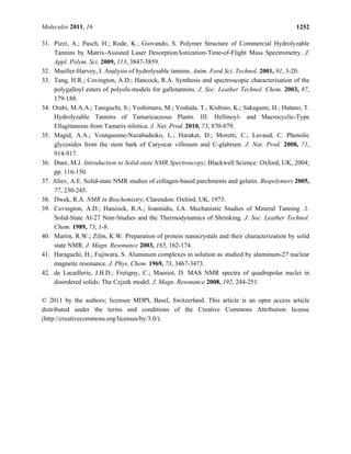
![Molecules 2011, 16 1246
Figure 3. Solid state 13
C CP-MAS spectrum of leather tanned with chromium (III) (the
black solid trace) superimposed on a spectrum of untanned hide (red dotted trace). Some
assignments to carbon atoms in specific collagen amino acid residues or residue types are
also indicated. The asterisk marks the envelope of overlapped signals from inter alia the α-
carbons of several types of residues, including aspartate and glutamate. Ala – alanine, Gly
– glycine, Hyp – hydroxyproline, Pro – proline.
2.4. Tanning with aluminium (III) and glutaraldehyde
Spectra of leathers tanned with aluminium sulphate, and with glutaraldehyde, are compared with a
spectrum of an equivalent sample of untanned material in Figure 4. Also shown (inset) is an 27
Al-NMR
spectrum acquired from the Al (III) tanned material [39]. The chemical shift of the 27
Al signal is close
to that of aqueous hydrated Al3+
at 0 ppm, and the linewidth at half peak height is ca. 1.8 kHz. Neither
the diamagnetic Al (III) ion nor the organic glutaraldehyde reagent produce the large changes in peak
intensity caused by the paramagnetic Cr (III) ion. This is entirely to be expected because the ability of
these diamagnetic species to exert long range effects on the NMR properties of the leather is much
more limited than paramagnetic Cr (III). Nevertheless the response of the spectral linewidths of leather
to tanning with either material is notable. Both tanned leathers exhibit signals which are generally
much sharper than those of the parent untanned leather. In solid state NMR a significant cause of
signal broadening is environmental heterogeneity resulting from partial or complete molecular disorder.
Thus the signal sharpening seen with both processes is likely attributable to an increase in molecular
order [40] brought about by whatever interactions, or reactions, occur between the tanning agents and
the collagen matrix. These two chemically dissimilar reagents produce very similar changes in the
collagen spectrum arguing for similar molecular ordering, although almost certainly via different
underlying chemical processes.](https://image.slidesharecdn.com/ad59e445-bbd5-4161-88bd-8a82e018293a-160921085144/85/Tannin-Article-molecules-16-01240-7-320.jpg)
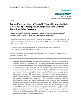
![Molecules 2011, 16 1247
Figure 4. Solid state 13
C CP-MAS spectra of untanned hide (a, red dotted trace), and
identically acquired spectra of aluminium sulphate tanned leather (b, black trace), and
glutaraldehyde tanned leather in which the resolved backbone amide glycyl and
prolyl/hydroxyprolyl signals are marked (c, blue trace). The inset is a 27
Al Hahn spin echo
spectrum of the Al (III) tanned material.
In the case of the Al (III) tanned leather the NMR properties of the 27
Al isotope (100% natural
abundance, gyromagnetic ratio, γ, about 1.04 times that of 13
C) provide an extra NMR probe of the
chemistry of the tanning process. The chemical shift of the 27
Al signal argues for octahedral
coordination by six oxygen ligands (as opposed to the other common tetrahedral geometry resulting
from Al (III) coordination by four ligands) [41]. The quadrupolar nature of the 27
Al nucleus with a
nuclear spin I = 5/2 means that the 27
Al resonance lineshape is responsive to electric field gradients at
the nucleus, which are a function of its bonded and non-bonded environment, particularly the nature](https://image.slidesharecdn.com/ad59e445-bbd5-4161-88bd-8a82e018293a-160921085144/85/Tannin-Article-molecules-16-01240-8-320.jpg)
![Molecules 2011, 16 1248
and symmetry of the inner and outer coordination spheres. The lineshape is potentially amenable to
modelling in terms of Al (III) coordination environment and the distribution of these environments [42]
but we have not attempted this.
In the case of the glutaraldehyde tanned leather there are no clear signals attributable to the carbon
atoms of the tanning reagent. There are several possible explanations; the incorporated glutaraldehyde
molecules may be too mobile to cross polarize, their signals may be unobservably broad due to factors
such as environmental heterogeneity, or the glutaraldehyde content required to produce a functional
leather may simply be below the detection threshold of NMR. Much lower levels of reagent are used in
the glutaraldehyde tanning process than in the vegetable tanning processes (see Section 3.1), probably
reflecting the different molecular mechanisms of the covalently interacting aldehyde and the weaker
non-covalently binding tannins. Additionally the signals of the covalently bound glutaraldehyde
reaction products in the glutaraldehyde tanned leather would overlap with collagen signals further
compromising the ability of NMR to detect and resolve them.
The effects of these two reagents, one a metal ion and the other a bifunctional reactive organic
dialdehyde, on the spectrum of leather collagen are rather different from those of the vegetable tannins.
While the sharpening of signals induced by Al (III) and glutaraldehyde is quite general throughout the
leather collagen spectrum, it is best appreciated by considering the envelope of signals centred at ca.
175 p.p.m. due to the collagen amide carbons. In the Al (III) and glutaraldehyde tanned materials it is
possible to resolve three distinct signals from amide carbons of (in order of increasing resonance
frequency) glycyl, prolyl/hydroxyprolyl, and other, residues. This resolution is not observed in spectra
of the untanned or vegetable tannin tanned materials, in which these three signals are broadened into a
single envelope.
3. Experimental Section
3.1. The tanning processes
UK bovine hides were prepared for tanning using standard industrial processes, as follows: hides
were soaked, washed, limed to remove hair, fleshed to remove unwanted flesh, connective tissue and
fat, split to a substance of 3.0 mm, and weighed. All further processing percentages were based on the
limed weight. The hides were then washed, delimed with ammonium sulphate and formic acid to a pH
of 8.3, bated to remove unwanted inter-fibrillary proteins, washed again and pickled using salt, and
formic and sulphuric acids to a pH of ca. 2.8 (chrome), 3.0–3.3 (alum, glutaraldehyde) or ca. 4.5
(vegetable tannin extracts), and the penetration of pickle ensured to be complete.
Hides were tanned with vegetable tannin extracts by adding 2% tannin and drumming for
30 minutes, followed by 10% tannin for 60 minutes, another 10% tannin (60 minutes) and a further
10% which was then drummed until penetrated. After tannage, a small percentage of EDTA
(ethylenediamine tetraacetic acid) was added to sequester any iron present, followed by acidification
with formic acid to a pH of 3.5. The leather was then given a light wash and dried (henceforth the
leather is termed veg tanned).
Chrome tanning was effected by adding 6–8% basic chromium sulfate (25% Cr2O3, 33% basicity),
running for 2–4 hours until penetrated and then basifying with magnesium oxide or soda ash or sodium](https://image.slidesharecdn.com/ad59e445-bbd5-4161-88bd-8a82e018293a-160921085144/85/Tannin-Article-molecules-16-01240-9-320.jpg)
![Molecules 2011, 16 1249
bicarbonate to a pH of 3.8–4.2, followed by draining and washing (the resulting leather is now referred
to as wet blue).
Hides were alum tanned by adding 4% salt and then 15% aluminium sulphate and running for ca.
5 hours, then adding 4% soda ash and running until penetrated. Glutaraldehyde tanning was carried out
by adding 1.5–1.8% of masked glutaraldehyde and running for 5–8 hours until penetrated at pH
3.9–4.1. Hides were removed from the tannage bath without washing, piled and dried (the leather is
now referred to as wet white).
3.2. Solid state NMR
Dry leather samples were prepared for NMR by first cutting them into small pieces about 1 to 2 mm
in size with a scalpel, and ball milling these for ca. 1 minute to a powder after freezing in liquid
nitrogen, using a Sartorius Mikrodismembrator at 3,000 r.p.m.. Tannin powders were analyzed as
received. All samples were packed into 4 mm outer diameter zirconia rotors (Bruker, Karlsruhe,
Germany) and 13
C-NMR spectra obtained using a Bruker AVANCE-400 9.4 Tesla wide bore
spectrometer equipped with a standard dual channel broad band probe, at a magic angle spinning rate
of 14 kHz and radio frequencies of 400.1 MHz (1
H), 100.5 MHz (13
C) and 104.2 MHz (27
Al)
respectively. 13
C signal intensity was enhanced using standard cross polarization [36] (CP) MAS
techniques (1
H π/2 pulse length 2.5 μs, 1
H cross polarization field 70 kHz, 1
H-13
C cross-polarization
contact time 2.5 ms, broadband TPPM15 decoupling during signal acquisition at a 1
H field strength of
100 kHz, recycle time 2 s, typical number of scans accumulated per spectrum ca. 3,000). Chemical
shifts were referenced to the methylene signal from solid glycine at 43.1 p.p.m. relative to
tetramethylsilane at 0 p.p.m.. 27
Al spectra were acquired using a rotor synchronized Hahn spin echo
(τ = 1 rotor period, 71 μs, π/2 pulse 3 μs, π pulse 6 μs, number of scans accumulated ca. 50,000) and
0.2 s recycle delay, and no broadband decoupling, which had been established to have no effect on the
27
Al-NMR signal characteristics. Chemical shifts were referenced to the resonance of aqueous ca. 1 M
AlCl3 in ca. 1 M HNO3 at 0 p. p. m..
4. Conclusions
Vegetable tanning agents all leave a distinctive spectroscopic signature in the tanned leather product
by which the origin and type of the tannin used may be inferred. Several other commonly used tanning
reagents also leave readily distinguishable spectroscopic fingerprints in leather products. Paramagnetic
Cr (III) causes the loss of considerable signal intensity from the spectrum of the collagen proteins
constituting leather without significantly changing the linewidths of remaining signals. The
diamagnetic reagents Al (III) and glutaraldehyde cause considerable collagen signal sharpening
indicative of an increase in collagen molecular order. It is to be expected that processes using mixtures
of tanning reagents will leave equally distinctive fingerprints in the leather product. The fingerprint
reflects not only the process chemistry but also underlying molecular mechanisms whereby tanning
converts unprocessed leather into a commercial product. As such NMR potentially represents a
powerful tool in understanding and improving tanning processes.](https://image.slidesharecdn.com/ad59e445-bbd5-4161-88bd-8a82e018293a-160921085144/85/Tannin-Article-molecules-16-01240-10-320.jpg)