Moyamoya disease (MMD) is a rare and unique cerebrovascular disease. The term “moyamoya” is Japanese and refers to a hazy puff of smoke or cloud. In people with moyamoya disease, this is how the blood vessels appear in the angiogram. MMD is characterized by the progressive stenosis of the distal internal carotid artery (ICA) resulting in a hazy network of basal collaterals called moyamoya vessels. This may be a consequence of Mutations in a few genes. In addition, MMD is also associated with many genetically transmitted disorders, including neurofibromatosis, Down syndrome, Sickle cell anemia, and Collagen vascular disease. It follows bimodal age distribution. Younger populations present with ischaemic symptoms, whereas adults show hemorrhagic symptoms The exact cause remains unknown. Immune, genetic and other factors contribute to this disease. It follows complex pathophysiology resulting in neovascularization as a compensatory mechanism. Diagnosis is based on cerebral angiography using the DSA scale. Treatment involves managing symptoms with medicine or surgery, improving blood flow to the brain, and controlling seizures. Revascularization helps to rebuild the blood supply to the underside of the brain.
Moyamoya disease (MMD) is a rare and unique cerebrovascular disease. The term “moyamoya” is Japanese and refers to a hazy puff of smoke or cloud. In people with moyamoya disease, this is how the blood vessels appear in the angiogram. MMD is characterized by the progressive stenosis of the distal internal carotid artery (ICA) resulting in a hazy network of basal collaterals called moyamoya vessels. This may be a consequence of Mutations in a few genes. In addition, MMD is also associated with many genetically transmitted disorders, including neurofibromatosis, Down syndrome, Sickle cell anemia, and Collagen vascular disease. It follows bimodal age distribution. Younger populations present with ischaemic symptoms, whereas adults show hemorrhagic symptoms The exact cause remains unknown. Immune, genetic and other factors contribute to this disease. It follows complex pathophysiology resulting in neovascularization as a compensatory mechanism. Diagnosis is based on cerebral angiography using the DSA scale. Treatment involves managing symptoms with medicine or surgery, improving blood flow to the brain, and controlling seizures. Revascularization helps to rebuild the blood supply to the underside of the brain.
Title: Sense of Smell
Presenter: Dr. Faiza, Assistant Professor of Physiology
Qualifications:
MBBS (Best Graduate, AIMC Lahore)
FCPS Physiology
ICMT, CHPE, DHPE (STMU)
MPH (GC University, Faisalabad)
MBA (Virtual University of Pakistan)
Learning Objectives:
Describe the primary categories of smells and the concept of odor blindness.
Explain the structure and location of the olfactory membrane and mucosa, including the types and roles of cells involved in olfaction.
Describe the pathway and mechanisms of olfactory signal transmission from the olfactory receptors to the brain.
Illustrate the biochemical cascade triggered by odorant binding to olfactory receptors, including the role of G-proteins and second messengers in generating an action potential.
Identify different types of olfactory disorders such as anosmia, hyposmia, hyperosmia, and dysosmia, including their potential causes.
Key Topics:
Olfactory Genes:
3% of the human genome accounts for olfactory genes.
400 genes for odorant receptors.
Olfactory Membrane:
Located in the superior part of the nasal cavity.
Medially: Folds downward along the superior septum.
Laterally: Folds over the superior turbinate and upper surface of the middle turbinate.
Total surface area: 5-10 square centimeters.
Olfactory Mucosa:
Olfactory Cells: Bipolar nerve cells derived from the CNS (100 million), with 4-25 olfactory cilia per cell.
Sustentacular Cells: Produce mucus and maintain ionic and molecular environment.
Basal Cells: Replace worn-out olfactory cells with an average lifespan of 1-2 months.
Bowman’s Gland: Secretes mucus.
Stimulation of Olfactory Cells:
Odorant dissolves in mucus and attaches to receptors on olfactory cilia.
Involves a cascade effect through G-proteins and second messengers, leading to depolarization and action potential generation in the olfactory nerve.
Quality of a Good Odorant:
Small (3-20 Carbon atoms), volatile, water-soluble, and lipid-soluble.
Facilitated by odorant-binding proteins in mucus.
Membrane Potential and Action Potential:
Resting membrane potential: -55mV.
Action potential frequency in the olfactory nerve increases with odorant strength.
Adaptation Towards the Sense of Smell:
Rapid adaptation within the first second, with further slow adaptation.
Psychological adaptation greater than receptor adaptation, involving feedback inhibition from the central nervous system.
Primary Sensations of Smell:
Camphoraceous, Musky, Floral, Pepperminty, Ethereal, Pungent, Putrid.
Odor Detection Threshold:
Examples: Hydrogen sulfide (0.0005 ppm), Methyl-mercaptan (0.002 ppm).
Some toxic substances are odorless at lethal concentrations.
Characteristics of Smell:
Odor blindness for single substances due to lack of appropriate receptor protein.
Behavioral and emotional influences of smell.
Transmission of Olfactory Signals:
From olfactory cells to glomeruli in the olfactory bulb, involving lateral inhibition.
Primitive, less old, and new olfactory systems with different path
Title: Sense of Taste
Presenter: Dr. Faiza, Assistant Professor of Physiology
Qualifications:
MBBS (Best Graduate, AIMC Lahore)
FCPS Physiology
ICMT, CHPE, DHPE (STMU)
MPH (GC University, Faisalabad)
MBA (Virtual University of Pakistan)
Learning Objectives:
Describe the structure and function of taste buds.
Describe the relationship between the taste threshold and taste index of common substances.
Explain the chemical basis and signal transduction of taste perception for each type of primary taste sensation.
Recognize different abnormalities of taste perception and their causes.
Key Topics:
Significance of Taste Sensation:
Differentiation between pleasant and harmful food
Influence on behavior
Selection of food based on metabolic needs
Receptors of Taste:
Taste buds on the tongue
Influence of sense of smell, texture of food, and pain stimulation (e.g., by pepper)
Primary and Secondary Taste Sensations:
Primary taste sensations: Sweet, Sour, Salty, Bitter, Umami
Chemical basis and signal transduction mechanisms for each taste
Taste Threshold and Index:
Taste threshold values for Sweet (sucrose), Salty (NaCl), Sour (HCl), and Bitter (Quinine)
Taste index relationship: Inversely proportional to taste threshold
Taste Blindness:
Inability to taste certain substances, particularly thiourea compounds
Example: Phenylthiocarbamide
Structure and Function of Taste Buds:
Composition: Epithelial cells, Sustentacular/Supporting cells, Taste cells, Basal cells
Features: Taste pores, Taste hairs/microvilli, and Taste nerve fibers
Location of Taste Buds:
Found in papillae of the tongue (Fungiform, Circumvallate, Foliate)
Also present on the palate, tonsillar pillars, epiglottis, and proximal esophagus
Mechanism of Taste Stimulation:
Interaction of taste substances with receptors on microvilli
Signal transduction pathways for Umami, Sweet, Bitter, Sour, and Salty tastes
Taste Sensitivity and Adaptation:
Decrease in sensitivity with age
Rapid adaptation of taste sensation
Role of Saliva in Taste:
Dissolution of tastants to reach receptors
Washing away the stimulus
Taste Preferences and Aversions:
Mechanisms behind taste preference and aversion
Influence of receptors and neural pathways
Impact of Sensory Nerve Damage:
Degeneration of taste buds if the sensory nerve fiber is cut
Abnormalities of Taste Detection:
Conditions: Ageusia, Hypogeusia, Dysgeusia (parageusia)
Causes: Nerve damage, neurological disorders, infections, poor oral hygiene, adverse drug effects, deficiencies, aging, tobacco use, altered neurotransmitter levels
Neurotransmitters and Taste Threshold:
Effects of serotonin (5-HT) and norepinephrine (NE) on taste sensitivity
Supertasters:
25% of the population with heightened sensitivity to taste, especially bitterness
Increased number of fungiform papillae
Ethanol (CH3CH2OH), or beverage alcohol, is a two-carbon alcohol
that is rapidly distributed in the body and brain. Ethanol alters many
neurochemical systems and has rewarding and addictive properties. It
is the oldest recreational drug and likely contributes to more morbidity,
mortality, and public health costs than all illicit drugs combined. The
5th edition of the Diagnostic and Statistical Manual of Mental Disorders
(DSM-5) integrates alcohol abuse and alcohol dependence into a single
disorder called alcohol use disorder (AUD), with mild, moderate,
and severe subclassifications (American Psychiatric Association, 2013).
In the DSM-5, all types of substance abuse and dependence have been
combined into a single substance use disorder (SUD) on a continuum
from mild to severe. A diagnosis of AUD requires that at least two of
the 11 DSM-5 behaviors be present within a 12-month period (mild
AUD: 2–3 criteria; moderate AUD: 4–5 criteria; severe AUD: 6–11 criteria).
The four main behavioral effects of AUD are impaired control over
drinking, negative social consequences, risky use, and altered physiological
effects (tolerance, withdrawal). This chapter presents an overview
of the prevalence and harmful consequences of AUD in the U.S.,
the systemic nature of the disease, neurocircuitry and stages of AUD,
comorbidities, fetal alcohol spectrum disorders, genetic risk factors, and
pharmacotherapies for AUD.
NVBDCP.pptx Nation vector borne disease control programSapna Thakur
NVBDCP was launched in 2003-2004 . Vector-Borne Disease: Disease that results from an infection transmitted to humans and other animals by blood-feeding arthropods, such as mosquitoes, ticks, and fleas. Examples of vector-borne diseases include Dengue fever, West Nile Virus, Lyme disease, and malaria.
Recomendações da OMS sobre cuidados maternos e neonatais para uma experiência pós-natal positiva.
Em consonância com os ODS – Objetivos do Desenvolvimento Sustentável e a Estratégia Global para a Saúde das Mulheres, Crianças e Adolescentes, e aplicando uma abordagem baseada nos direitos humanos, os esforços de cuidados pós-natais devem expandir-se para além da cobertura e da simples sobrevivência, de modo a incluir cuidados de qualidade.
Estas diretrizes visam melhorar a qualidade dos cuidados pós-natais essenciais e de rotina prestados às mulheres e aos recém-nascidos, com o objetivo final de melhorar a saúde e o bem-estar materno e neonatal.
Uma “experiência pós-natal positiva” é um resultado importante para todas as mulheres que dão à luz e para os seus recém-nascidos, estabelecendo as bases para a melhoria da saúde e do bem-estar a curto e longo prazo. Uma experiência pós-natal positiva é definida como aquela em que as mulheres, pessoas que gestam, os recém-nascidos, os casais, os pais, os cuidadores e as famílias recebem informação consistente, garantia e apoio de profissionais de saúde motivados; e onde um sistema de saúde flexível e com recursos reconheça as necessidades das mulheres e dos bebês e respeite o seu contexto cultural.
Estas diretrizes consolidadas apresentam algumas recomendações novas e já bem fundamentadas sobre cuidados pós-natais de rotina para mulheres e neonatos que recebem cuidados no pós-parto em unidades de saúde ou na comunidade, independentemente dos recursos disponíveis.
É fornecido um conjunto abrangente de recomendações para cuidados durante o período puerperal, com ênfase nos cuidados essenciais que todas as mulheres e recém-nascidos devem receber, e com a devida atenção à qualidade dos cuidados; isto é, a entrega e a experiência do cuidado recebido. Estas diretrizes atualizam e ampliam as recomendações da OMS de 2014 sobre cuidados pós-natais da mãe e do recém-nascido e complementam as atuais diretrizes da OMS sobre a gestão de complicações pós-natais.
O estabelecimento da amamentação e o manejo das principais intercorrências é contemplada.
Recomendamos muito.
Vamos discutir essas recomendações no nosso curso de pós-graduação em Aleitamento no Instituto Ciclos.
Esta publicação só está disponível em inglês até o momento.
Prof. Marcus Renato de Carvalho
www.agostodourado.com
Flu Vaccine Alert in Bangalore Karnatakaaddon Scans
As flu season approaches, health officials in Bangalore, Karnataka, are urging residents to get their flu vaccinations. The seasonal flu, while common, can lead to severe health complications, particularly for vulnerable populations such as young children, the elderly, and those with underlying health conditions.
Dr. Vidisha Kumari, a leading epidemiologist in Bangalore, emphasizes the importance of getting vaccinated. "The flu vaccine is our best defense against the influenza virus. It not only protects individuals but also helps prevent the spread of the virus in our communities," he says.
This year, the flu season is expected to coincide with a potential increase in other respiratory illnesses. The Karnataka Health Department has launched an awareness campaign highlighting the significance of flu vaccinations. They have set up multiple vaccination centers across Bangalore, making it convenient for residents to receive their shots.
To encourage widespread vaccination, the government is also collaborating with local schools, workplaces, and community centers to facilitate vaccination drives. Special attention is being given to ensuring that the vaccine is accessible to all, including marginalized communities who may have limited access to healthcare.
Residents are reminded that the flu vaccine is safe and effective. Common side effects are mild and may include soreness at the injection site, mild fever, or muscle aches. These side effects are generally short-lived and far less severe than the flu itself.
Healthcare providers are also stressing the importance of continuing COVID-19 precautions. Wearing masks, practicing good hand hygiene, and maintaining social distancing are still crucial, especially in crowded places.
Protect yourself and your loved ones by getting vaccinated. Together, we can help keep Bangalore healthy and safe this flu season. For more information on vaccination centers and schedules, residents can visit the Karnataka Health Department’s official website or follow their social media pages.
Stay informed, stay safe, and get your flu shot today!
ARTIFICIAL INTELLIGENCE IN HEALTHCARE.pdfAnujkumaranit
Artificial intelligence (AI) refers to the simulation of human intelligence processes by machines, especially computer systems. It encompasses tasks such as learning, reasoning, problem-solving, perception, and language understanding. AI technologies are revolutionizing various fields, from healthcare to finance, by enabling machines to perform tasks that typically require human intelligence.
- Video recording of this lecture in English language: https://youtu.be/lK81BzxMqdo
- Video recording of this lecture in Arabic language: https://youtu.be/Ve4P0COk9OI
- Link to download the book free: https://nephrotube.blogspot.com/p/nephrotube-nephrology-books.html
- Link to NephroTube website: www.NephroTube.com
- Link to NephroTube social media accounts: https://nephrotube.blogspot.com/p/join-nephrotube-on-social-media.html
Tom Selleck Health: A Comprehensive Look at the Iconic Actor’s Wellness Journeygreendigital
Tom Selleck, an enduring figure in Hollywood. has captivated audiences for decades with his rugged charm, iconic moustache. and memorable roles in television and film. From his breakout role as Thomas Magnum in Magnum P.I. to his current portrayal of Frank Reagan in Blue Bloods. Selleck's career has spanned over 50 years. But beyond his professional achievements. fans have often been curious about Tom Selleck Health. especially as he has aged in the public eye.
Follow us on: Pinterest
Introduction
Many have been interested in Tom Selleck health. not only because of his enduring presence on screen but also because of the challenges. and lifestyle choices he has faced and made over the years. This article delves into the various aspects of Tom Selleck health. exploring his fitness regimen, diet, mental health. and the challenges he has encountered as he ages. We'll look at how he maintains his well-being. the health issues he has faced, and his approach to ageing .
Early Life and Career
Childhood and Athletic Beginnings
Tom Selleck was born on January 29, 1945, in Detroit, Michigan, and grew up in Sherman Oaks, California. From an early age, he was involved in sports, particularly basketball. which played a significant role in his physical development. His athletic pursuits continued into college. where he attended the University of Southern California (USC) on a basketball scholarship. This early involvement in sports laid a strong foundation for his physical health and disciplined lifestyle.
Transition to Acting
Selleck's transition from an athlete to an actor came with its physical demands. His first significant role in "Magnum P.I." required him to perform various stunts and maintain a fit appearance. This role, which he played from 1980 to 1988. necessitated a rigorous fitness routine to meet the show's demands. setting the stage for his long-term commitment to health and wellness.
Fitness Regimen
Workout Routine
Tom Selleck health and fitness regimen has evolved. adapting to his changing roles and age. During his "Magnum, P.I." days. Selleck's workouts were intense and focused on building and maintaining muscle mass. His routine included weightlifting, cardiovascular exercises. and specific training for the stunts he performed on the show.
Selleck adjusted his fitness routine as he aged to suit his body's needs. Today, his workouts focus on maintaining flexibility, strength, and cardiovascular health. He incorporates low-impact exercises such as swimming, walking, and light weightlifting. This balanced approach helps him stay fit without putting undue strain on his joints and muscles.
Importance of Flexibility and Mobility
In recent years, Selleck has emphasized the importance of flexibility and mobility in his fitness regimen. Understanding the natural decline in muscle mass and joint flexibility with age. he includes stretching and yoga in his routine. These practices help prevent injuries, improve posture, and maintain mobilit
Ozempic: Preoperative Management of Patients on GLP-1 Receptor Agonists Saeid Safari
Preoperative Management of Patients on GLP-1 Receptor Agonists like Ozempic and Semiglutide
ASA GUIDELINE
NYSORA Guideline
2 Case Reports of Gastric Ultrasound
Ozempic: Preoperative Management of Patients on GLP-1 Receptor Agonists
Stroke lancet 2020
1. Seminar
www.thelancet.com Vol 396 July 11, 2020 129
Stroke
Bruce CV Campbell, Pooja Khatri
Stroke is a major cause of death and disability globally. Diagnosis depends on clinical features and brain imaging to
differentiate between ischaemic stroke and intracerebral haemorrhage. Non-contrast CT can exclude haemorrhage,
but the addition of CT perfusion imaging and angiography allows a positive diagnosis of ischaemic stroke versus
mimics and can identify a large vessel occlusion target for endovascular thrombectomy. Management of ischaemic
stroke has greatly advanced, with rapid reperfusion by use of intravenous thrombolysis and endovascular
thrombectomy shown to reduce disability. These therapies can now be applied in selected patients who present late to
medical care if there is imaging evidence of salvageable brain tissue. Both haemostatic agents and surgical
interventions are investigational for intracerebral haemorrhage. Prevention of recurrent stroke requires an
understanding of the mechanism of stroke to target interventions, such as carotid endarterectomy, anticoagulation
for atrial fibrillation, and patent foramen ovale closure. However, interventions such as lowering blood pressure,
smoking cessation, and lifestyle optimisation are common to all stroke subtypes.
Introduction
Stroke is a common disease, with one in four people
affected over their lifetime, and is the second leading
cause of death and third leading cause of disability in
adults worldwide.1
Substantial advances in therapy have
occurred in the past 5 years, particularly for the acute
treatment of ischaemic stroke. New strategies for
preventing recurrence have also been identified. This
Seminar outlines the diagnosis and management of
ischaemic stroke and intracerebral haemorrhage in
contemporary stroke units.
Definition of stroke
Stroke is defined as a neurological deficit attributed to an
acute focal injury of the CNS (ie, brain, retina, or spinal
cord) by a vascular cause.2
Most strokes are ischaemic due
to reduced blood flow, generally resulting from arterial
occlusion. A rarer type of ischaemic stroke is venous
infarction due to occlusion of cerebral veins or venous
sinuses. The remaining 10–40% of stroke presentations,
depending on regional epidemiology, are haemorrhagic
and result from the rupture of cerebral arteries.3,4
These
haemorrhages can be intracerebral or subarachnoid;
subarachnoid haemorrhages typically result from a
ruptured aneurysm and are out of the scope of this
Seminar. Ischaemic stroke is differentiated from transient
ischaemic attack by the presence of an infarct on brain
imaging. Patients diagnosed with transient ischaemic
attack, by use of former clinical definitions that were
based on symptom resolution within 24 h, have evidence
of infarction on diffusion-weighted MRI in approximately
40% of cases and represent a group who are at high risk
for recurrent stroke.5
Diagnosis of stroke and mimics
The key clinical feature of stroke is the sudden onset of a
focal neurological deficit. The timing of this sudden
onset can be masked if the patient awakens with stroke
symptoms or if the onset is unwitnessed and the patient
is unable to communicate or does not have the insight to
recognise the timing of deficit. The time of stroke onset
is therefore defined as the time that the patient was last
known to be well.
Knowledge of neuroanatomical structures and vascular
territories allows localisation and estimation of the size
of the affected territory; patterns, such as right hemi
paresis with aphasia due to occlusion of the left middle
cerebral artery, are common and well recognised. Stroke
symptoms that are under-recognised, such as nausea,
vomiting, vertigo, and decreased level of consciousness,
are more common in the setting of occlusions in the
posterior circulation.6
Sudden onset of neurological deficits generally
indicates a vascular cause, although seizures, specifically
focal impaired awareness seizures or a postictal state,
can also produce sudden onset of symptoms. Add
itionally, migraine with aura or hemiplegic migraine
can also lead to sudden onset of focal neurological
symptoms, but this should be a diagnosis of exclusion.
Functional (psychogenic) deficits, such as conversion
disorder, can mimic stroke. Occasionally, space-
occupying lesions can present suddenly if they cause a
seizure or bleeding. Other mimics, which would
typically not show abrupt onset with an adequate
patient history, include toxic metabolic derangements
Lancet 2020; 396: 129–42
Department of Medicine and
Neurology, Melbourne Brain
Centre, Royal Melbourne
Hospital andThe Florey
Institute of Neuroscience and
Mental Health, University of
Melbourne, Parkville,VIC,
Australia
(Prof B C V Campbell PhD); and
Department of Neurology,
University of Cincinnati,
Cincinnati, OH, USA
(Prof P Kahtri MD)
Correspondence to:
Prof Bruce Campbell,
Department of Medicine and
Neurology, Melbourne Brain
Centre, Royal Melbourne
Hospital, Parkville,VIC 3050,
Australia
bruce.campbell@mh.org.au
Search strategy and selection criteria
We searched the Cochrane Library, MEDLINE, and Embase for
articles published in English between Jan 1, 2015, and
Dec 31, 2019. We used the search terms “ischaemic/ischemic
stroke” or “intracerebral haemorrhage/hemorrhage”,
and “clinical trial” or “meta-analysis”. We also searched the
reference lists of articles identified by this search strategy
and selected those that we judged to be relevant. We largely
selected publications in the past 5 years but did not exclude
commonly referenced and highly regarded publications that
were older. Review articles are cited to provide readers with
more details and references than this Seminar is able to.
Our reference list was modified on the basis of comments
from peer reviewers.
2. Seminar
130 www.thelancet.com Vol 396 July 11, 2020
(eg, hypoglycaemia). Particularly in patients with a
previous history of stroke, the previous neurological
deficit can return with intercurrent illness.7
Brain imaging (CT scan or MRI) crucially comple
ments the clinical examination to differentiate the
stroke subtype and mechanism. Clinical symptoms and
signs alone cannot reliably differentiate ischaemic
stroke from intracerebral haemorrhage, and manage
ment sharply diverges between these two conditions. In
ischaemic stroke, the presence of a large vessel
occlusion, defined as occlusion in the internal carotid
artery, proximal middle cerebral artery, or basilar artery,
determines the most appropriate reperfusion therapy.
The secondary prevention of large artery atherosclerotic
disease versus cardioembolic stroke also differs
substantially. Imaging of the brain and its vascular tree
is therefore one of the most urgent priorities when
patients present to hospital with suspected stroke
(figure 1).
Epidemiology and risk factors
The 2016 Global Burden of Disease data that were
published in 2019 indicate that one in four people will
have a stroke in their lifetime.3
There are estimated
to be 9·6 million ischaemic strokes and 4·1 million
haemorrhagic strokes (including intracerebral and
subarachnoid haemorrhage) globally each year, with a
relatively stable incidence adjusted for age in high-income
countries but an increasing incidence in low-income and
middle-income countries.3
The absolute incidence is
expected to increase with an ageing population.
Estimations indicate that approximately 90% of
strokes are attributable to modifiable risk factors.8
Stroke shares many risk factors with other cardio
vascular diseases although their relative importance
varies. The most potent risk factor for stroke is high
blood pressure, which applies to both ischaemic stroke
and intracerebral haemorrhage. Smoking, diabetes,
hyperlipidaemia, and physical inactivity are also
significant risks and require interventions that are
regulatory and are based in the community to alter
lifestyle and the environment, as well as individual
treatment.9
Atrial fibrillation, a specific risk factor for
ischaemic stroke, is increasing in detection and preva
lence.10
Strokes related to atrial fibrillation tend to be
larger and more disabling than are strokes due to other
mechanisms.11
Pathophysiology
Ischaemic stroke
Most ischaemic stroke is due to embolism, either from
atherosclerotic plaque in the aortic arch or in the
cervical arteries or from the heart (panel, figure 2).
Intracranial atherosclerosis with in-situ thrombosis is
also an important mechanism of stroke, particularly in
Asian and Black ethnic groups.12
Small vessel disease
causes small subcortical infarcts (ie, lacunar stroke)
and deep intracerebral haemorrhage. Cervical artery
dissection is one of the common causes of stroke
in younger patients (eg, <60 years), and arterial
Figure 1: Stroke diagnosis using neuroimaging
(A) Ischaemic stroke established in the territory of the right middle cerebral artery 12 h after onset (arrowhead).
A patient with left hemiparesis onset 2 h before scan (B–G) showing subtle loss of differentiation between grey and
white matter (arrowhead) in the basal ganglia (B) and hyperdense thrombus (arrowhead) in the right middle
cerebral artery (C). CT perfusion showing reduced CBV (arrowhead) in the right insular region (D) corresponding to
region of diffusion restriction (arrowhead; most likely irreversibly injured) on MRI (E). CT perfusion showing delayed
Tmax (arrowhead; substantially delayedTmax [ie, >6 s] indicates brain tissue that is critically hypoperfused,
functionally impaired, and potentially at risk of infarction in the absence of reperfusion) in the right middle cerebral
artery territory (F) corresponding to intracranial occlusion of the right middle cerebral artery (arrowhead) on CT
angiogram (G). (H) Diffusion MRI lesions (arrowhead) in a patient with two 5 min episodes of aphasia that fully
resolved—now defined as ischaemic stroke rather than transient ischaemic attack. (I) Focal subarachnoid
haemorrhage (arrowhead) related to amyloid angiopathy presenting as transient parasthesias on the left side
(differential diagnosis of transient ischaemic attack). Lobar intracerebral haemorrhage (arrowhead) in a patient with
amyloid angiopathy (J) and intracerebral haemorrhage (arrowhead) in the right basal ganglia most likely resulting
from deep perforating vasculopathy (K). (L)Thrombosis in the cerebral venous sinus with hyperdense sagittal sinus
(arrow) and haemorrhagic venous infarction (arrowhead). CBV=cerebral blood volume.Tmax=time to maximum.
A C
E
B
D F
HG I
KJ L
CBV (mL/100 g) Tmax (s)
3. Seminar
www.thelancet.com Vol 396 July 11, 2020 131
inflammation can also cause stroke (eg, inflammatory
arteriopathy after infection is a major cause of
paediatric stroke and can also occur after herpes zoster
in adults).
When a cerebral artery is occluded and blood flow
decreases below a critical level, neuronal electrical func
tion ceases and a clinical deficit develops.13
If cerebral
blood flow is severely reduced, then irreversible tissue
injury will ensue rapidly. However, in many patients,
collateral blood supply via leptomeningeal anastomoses
or the circle of Willis can be sufficient to maintain
cellular viability for a period of time. These hibernating,
but potentially salvageable, brain regions are termed the
ischaemic penumbra. Reperfusion therapies restore
blood flow to the ischaemic penumbra and substantially
reduce disability after ischaemic stroke. The salvageable
ischaemic penumbra can be identified non-invasively
by use of the mismatch between the ischaemic core,
which is irreversibly injured (estimated using diffusion
MRI or severely reduced cerebral blood flow on CT
perfusion), and the critically hypoperfused region
(estimated as the region of substantially delayed blood
flow arrival).14,15
This estimation of salvageable tissue by
use of perfusion imaging has been successfully used to
identify patients who would benefit from reperfusion
therapies beyond the standard time windows of 6·0 h
for endovascular thrombectomy and 4·5 h for intra
venous thrombolysis.16,17
Ischaemia and reperfusion can cause secondary injury.
Although reperfusion injury is well described in animal
models, it has been less easily recognised in humans
since the benefits of reperfusion usually far outweigh
the detrimental effects.18
However, symptomatic haem
orrhagic transformation and malignant oedema are
two clinical manifestations of reperfusion injury.
Glutamate excitotoxicity, free radical injury, and matrix
metalloprotease degradation of the blood–brain barrier
are just some of the described mechanisms of secondary
injury after reperfusion.19
Intracerebral haemorrhage
The most common cause of intracerebral haemorrhage
is deep perforating vasculopathy related to high blood
pressure,20
with cerebral microbleeds (seen on MRI) and
clinical haemorrhages most often affecting the basal
ganglia, cerebellum, pons, or thalamus. Another major
cause is amyloid angiopathy; these haemorrhages are
typically lobar and occur in older patients (ie, aged
>55 years but most frequently in patients aged
70–80 years) with MRI evidence of cortical microbleeds
and superficial haemosiderosis. Vascular malformations
(eg, arteriovenous malformations, cavernous malfor
mations, and dural arteriovenous fistulae) and mass
lesions (eg, metastatic tumours) should be ruled out
with neuroimaging, especially in younger patients
(eg, <60 years) or without evidence of vasculopathy
on MRI.
The detrimental effects of intracerebral haemorrhage
due to mass effect from the haematoma itself are readily
recognised. The oedema that subsequently develops
(and can increase for up to 2 weeks) also contributes
to injury from mass effect, and toxicity from thrombin
and iron are thought to be key contributors to the
development of oedema.20
Acute management
Acute management of patients with stroke should occur
in a stroke unit that is organised and geographically
defined. Care in a stroke unit has been clearly shown to
increase survival without disability for patients of all
ages, severities, and stroke subtypes,21
and comprises an
expert integrated medical, nursing, and allied health
team applying evidence-based clinical protocols (table).
Care in a stroke unit is the foundation on which acute
stroke interventions can be delivered. The aims are to
reduce complications, such as aspiration pneumonia,
venous thromboembolism, and pressure sores; com
mence early rehabilitation; and institute targeted
secondary prevention. Protocolised nursing management
of fever, glucose, and swallowing reduced mortality in
one cluster-randomised trial.32
However, a 2019 trial did
not show any benefit of more intensive glucose control
versus standard management after stroke.33
Panel: Major causes of stroke
Atherosclerosis:
• Aortic arch or cervical arteries
• Intracranial arteries
Cardioembolism:
• Atrial fibrillation
• Akinetic myocardial segment
• Patent foramen ovale
• Endocarditis
Small vessel disease
Other causes:
• Other arterial diseases (eg, dissection, vasculitis)
• Haematological diseases (eg, antiphospholipid syndrome,
polycythaemia rubra vera, essential thrombocytosis)
Figure 2: Determining stroke mechanism
(A) CT angiography showing atherosclerosis of the internal carotid artery. (B) Intracranial atherosclerotic disease.
(C) Fat saturatedT1 MRI showing intramural hyperintensity diagnostic of carotid artery dissection.
A CB
4. Seminar
132 www.thelancet.com Vol 396 July 11, 2020
Acute treatments for ischaemic stroke
Intravenous thrombolysis with recombinant human
tissue plasminogen activator (alteplase) aims to reperfuse
the ischaemic brain by converting plasminogen (PLG) to
plasmin, which can dissolve the thrombus that is causing
the stroke. Alteplase was first shown to reduce disability
in the NINDS part A and B trials34
when administered
within 3·0 h of stroke onset. The treatment window was
subsequently extended to 4·5 h, although the benefit
reduces rapidly with increasing time after stroke onset
(table).22
When alteplase is delivered within 3·0 h of
onset, approximately one in four patients have reduced
disability, which decreases to one in seven patients
between 3·0 h and 4·5 h.35
This benefit includes the
effect of the approximately 2% absolute risk of fatal
intracerebral haemorrhage. A large meta-analysis of
individual patient data established that, although age and
clinical severity measured by use of the National
Institutes of Health Stroke Scale are strongly prognostic,
the treatment benefit of alteplase is preserved across
the spectrum of these variables.22
Patients with mild
but disabling symptoms benefit from thrombolysis.
However, the prematurely terminated 2018 PRISMS
trial36
showed no evidence of benefit in patients with
symptoms that were judged to be non-disabling at
presentation, and who were selected on the basis of non-
contrast CT brain imaging and clinical characteristics.
Trials selecting patients with non-disabling symptoms
but with arterial occlusion or perfusion abnormality are
ongoing (eg, TEMPO-2, NCT02398656).
In 2019, the use of CT or perfusion MRI was
established to select patients between 4·5 h and 9·0 h
from the time that they were last seen to be well (or
within 9·0 h of the midpoint of sleep if they awoke with
stroke) if they had imaging evidence of salvageable
brain tissue.17,37
This subset of patients derives at least as
much benefit with similar risk of fatal intracerebral
haemorrhage to those treated 0·0–3·0 h after stroke
onset, and the ability to select patients using CT-based
imaging puts this treatment approach within reach of
most hospitals that are capable of thrombolysis inter
vention. Thrombolysis also improved outcomes in
patients with unknown time of stroke onset (including
those waking up with stroke) in whom MRI showed
diffusion lesions that were not yet hyperintense on
fluid-attenuated inversion recovery (FLAIR).38
This
diffusion-FLAIR mismatch indicates that the patient is
likely to be within 4·5 h of stroke onset. Compared
with CT perfusion, MRI diffusion-FLAIR mismatch is
more likely to detect patients with lacunar stroke, and
who could benefit from thrombolysis.39
However, the
requirement for urgent MRI reduces the applicability of
the MRI diffusion-FLAIR technique in many regions.
The 0·9 mg/kg licensed dose of alteplase in most
regions was based on data from small studies, and the
licensed dose in Japan is 0·6 mg/kg.40
A randomised trial
comparing these two doses did not show non-inferiority
Treatment
patients with
outcome, %
Control
patients with
outcome, %
Odds ratio
(95% CI)
Absolute
difference, %
Care in a stroke unit
Death or dependency
(mRS 3–6)21
52·4% 60·9% 0·75 (0·66–0·85) 8·5%
Ischaemic stroke
Alteplase thrombolysis, non-contrast CT brain selection22,23
mRS 0–1 in patients treated
0·0–3·0 h after stroke onset
32·9% 23·1% 1·75 (1·35–2·27) 9·8%
mRS 0–1 in patients treated
3·0–4·5 h after stroke onset
35·3% 30·1% 1·26 (1·05–1·51) 5·2%
SICH 3·7% 0·6% 6·67 (4·11–10·84) 3·1%
Fatal SICH 2·7% 0·4% 7·14 (3·98–12·79) 2·3%
Mortality in patients treated
0·0–3·0 h after stroke onset
22·2% 21·8% 1·00 (0·81−1·24) 0·4% (p=0·70)
Mortality in patients treated
3·0–4·5 h after stroke onset
16·9% 15·9% 1·14 (0·95−1·36) 1·0% (p=0.96)
Alteplase thrombolysis >4·5 h after stroke onset in patients selected by use of perfusion imaging17
mRS 0–1 36·2% 25·8% 2·06 (1·17–3·62) 10·4%
mRS 0–2 50·7% 39·7% 2·22 (1·25–3·94) 11·0%
SICH 4·6% 0·7% 7·29 (0·88–60·18) 3·9% (p=0·067)
Mortality 13·2% 10·5% 1·28 (0·60–2·73) 2·7% (p=0·52)
Endovascular thrombectomy initiated 0·0-6·0 h after stroke onset24
mRS 0–1 26·9% 12·9% 2·72 (1·99–3·71) 14·0%
mRS 0–2 46·0% 26·5% 2·71 (2·07–3·55) 19·5%
SICH 4·4% 4·3% 1·07 (0·62–1·84) 0·1% (p=0·81)
Mortality 15·3% 18·9% 0·73 (0·47–1·13) 3·6% (p=0·16)
Endovascular thrombectomy initiated 6·0–24·0 h after stroke onset in patients selected by the use of
perfusion imaging25
mRS 0–2 46·7% 14·8% 5·01 (3·07–8·17) 31·9%
SICH 6·0% 3·7% 1·67 (0·64–4·35) 2·3% (p=0·29)
Mortality 16·6% 21·7% 0·71 (0·34–1·51) 5·1% (p=0·38)
Hemicraniectomy26
mRS 4–6 56·9% 78·6% 0·33 (0·13–0·86) 21·7%
Mortality 21·6% 71·4% 0·10 (0·04–0·27) 49·8%
Aspirin administered <48·0 h after stroke onset27
mRS 0–2 54·4% 53·1% 1·05 (1·01–1·10) 1·3%
Intracerebral haemorrhage
Intensive blood pressure lowering28
mRS 3–6 52·0% 55·6% 0·87 (0·75–1·01) 3·6% (p=0·059)
Surgical evacuation overall29
Death or disability 59·4% 67·4% 0·72 (0·61–0·84) 8·0%
Mortality 27·3% 31·8% 0·82 (0·69–0·97) 4·5%
Surgery commenced within 0·0–8·0 h of stroke onset29
Death or disability* 70·3% 79·2% 0·59 (0·42–0·84) 8·9%
Minimally invasive surgery30
Death or disability† 47·4% 65·4% 0·59 (0·42–0·84) 18·0%
SICH is defined as parenchymal haematoma occupying >30% of the infarcted territory with substantial mass effect
combined with an increase of ≥4 points in National Institutes of Health Stroke Scale score, as used in the Safe
Implementation ofThrombolysis in Stroke-Monitoring Study.31
mRS=modified Rankin scale. SICH=symptomatic
intracerebral haemorrhage. *Component studies used different outcomes: composite of death, vegetative or severe
disability outcome on Glasgow Outcomes Score; mRS ≥3; or Barthel Index ≤90 in the 0·0–8·0 h analysis. †Component
studies used different outcomes: composite of mRS ≥3; or Barthel Index ≤60.
Table: Patient outcomes following acute interventions for stroke
5. Seminar
www.thelancet.com Vol 396 July 11, 2020 133
of 0·6 mg/kg, although symptomatic intracerebral
haemorrhage was reduced from 2% to 1%.41
There are
plans to explore the lower dose of 0·6 mg/kg further in
patients who are felt to be at high risk of bleeding (eg,
concurrent antiplatelet use). However, as of June, 2020,
all guidelines outside of those in Japan recommend
0·9 mg/kg alteplase (maximum 90·0 mg) delivered as a
10% bolus followed by 90% infused over 1 h.
Tenecteplase is a genetically modified form of alteplase
that has a longer half-life, permitting a single bolus
administration (rather than bolus and 1 h infusion of
alteplase) and greater fibrin specificity and resistance to
plasminogen activator inhibitors than does alteplase.42
Randomised trial data suggest that tenecteplase is at least
as safe and as effective in patients with stroke43
and that
patients with large vessel occlusion had higher rates of
reperfusion with tenecteplase versus with alteplase.44,45
Although 0·25 mg/kg and 0·40 mg/kg have been used
in trials, the EXTEND-IA TNK part 2 trial46
indicated
that there was no advantage in increasing the dose from
0·25 mg/kg to 0·40 mg/kg. Because many patients
now require transfer between hospitals, single bolus
administration simplifies the transport process and
ensures that the full dose of thrombolytic agent is given,
since alteplase infusions could be interrupted in transit.
Tenecteplase use also avoids the common gap between
administration of the bolus and infusion of alteplase,
which, given the short half-life of alteplase, can mean that
the desired plasma concentration is not sustained.
Although tenecteplase has entered guidelines in Europe,47
the USA,48
and Australia49
as a possible alternative to
alteplase, it is not currently licensed for use as a stroke
treatment outside of India, where a generic form of
tenecteplase was licensed on the basis of non-ran
domised data,50
and its biosimilarity is debated.51
Ongoing
phase 3 trials of tenecteplase aim to definitively establish
the role of tenecteplase for patients with stroke
(TASTE,ACTRN12613000243718;ATTEST2,NCT02814409;
ACT, NCT03889249), including potentially combining
tenecteplase with endovascular thrombectomy in patients
presenting later than 4·5 h after stroke onset (TIMELESS,
NCT03785678; ETERNAL, NCT04454788).
Intravenous thrombolysis is the most accessible
reperfusion therapy for stroke because endovascular
thrombectomy is restricted to major stroke centres and
completely unavailable in many parts of the world. Several
trials are testing whether thrombolysis can be safely
omitted for patients with large vessel occlusion who present
directly to a centre that is capable of thrombectomy.
The first trial to report results showed similar outcomes
between groups that narrowly met a generous 20% non-
inferiority margin (DIRECT-MT)52
and further studies are
ongoing (SWIFT DIRECT, NCT03192332; DIRECT SAFE,
NCT03494920; MR CLEAN-NO IV, ISRCTN80619088).
The current standard of care is to give thrombolysis and
proceed to thrombectomy as quickly as possible. Therefore,
approaches to improve the effectiveness of intravenous
thrombolysis have great potential value. Current trials are
exploring the addition of adjuvant agents (eg, eptifibatide
or argatroban in the MOST trial, NCT03735979). Novel
drugs that dissolve clots and target other structural
components of thrombi (eg, von Willebrand factor and
neutrophil extracellular traps), inhibitors of fibrinolysis
(eg, CPB2 and α2-AP), and mechanical strategies, such as
sonothrombolysis, are also under investigation.53
Endovascular thrombectomy is another type of reper
fusion therapy. After several trials did not show any
benefit with endovascular thrombectomy in 2013,54–56
five trials published in 2015 established endovascular
thrombectomy as one of the most powerful treatments to
reduce disability in any specialty of medicine (table).57–61
The benefits shown by these five trials were driven by a
combination of improved devices (which allowed faster,
more effective reperfusion), improved patient selection
(requiring at least a documented large vessel occlusion
on non-invasive angiography), and faster treatment
workflow. A meta-analysis of individual patient data
emphasised the importance of time, with one in
100 patients worse off for every 4 min delay in reperfusion
after arriving in the emergency department.62
In what might superficially appear to contradict this
crucial relationship between time to treatment and func
tional outcome, trials in 2018 established the benefits of
endovascular thrombectomy up to 24 h after the time
that the patient was last known to be well, if perfusion
imaging was favourable.16,63
The key point is that the
proportion of patients who have favourable perfusion
imaging decreases over time, and so the urgency to
evaluate and treat rapidly still exists; fast treatment will
maximise the proportion of patients who have salvageable
brain tissue. However, if a patient is unavoidably delayed
in presenting to hospital and they still have favourable
imaging, they will derive at least as much treatment
benefit as patients who receive treatment within 0–6 h of
stroke onset (table).
Endovascular thrombectomy, analogous to thrombo
lysis, is of generalised benefit across the spectrum of age
and clinical severity.24
The benefit of thrombectomy is
uncertain in patients who are mildly affected (only
14 patients with National Institutes of Health Stroke
Scale score <6 were enrolled in completed trials), and
ongoing trials are addressing the use of endovascular
thrombectomy in this population (eg, ENDO-LOW,
NCT04167527; MOSTE, NCT03796468). Patients with
occlusions in the internal carotid artery and proximal
middle cerebral artery (M1 segment; figure 3) benefit
from endovascular thrombectomy, including those with
tandem cervical internal carotid artery and intracranial
occlusion. Once the middle cerebral artery has bifurcated
(M2 segments), the benefit is potentially reduced because
a smaller territory is at risk and there is an increased
effect of thrombolysis. Additionally, the risk of arterial
injury might be increased because of increased technical
difficulty of thrombectomy in smaller, more tortuous
6. Seminar
134 www.thelancet.com Vol 396 July 11, 2020
vessels. Data indicate the benefit of thrombectomy in
patients with occlusions in the large, more proximal M2
branches, who have clinically significant neurological
deficits, but treatment decisions need to be individualised
for these patients.26
Technology continues to evolve and
thrombectomy in more distal vessels will consequently
require further evaluation.
Thrombectomy in the basilar artery is recommended by
guidelines47–49
but convincing randomised data are scarce.
The Chinese BEST trial64
reported a benefit of approxi
mately 20% in an as-treated analysis but this result was
confounded by a high crossover rate from control to
intervention. The BASICS trial65
has been reported in an
abstract and overall results were neutral. The more
severely affected subgroup of patients (National Institutes
of Health Stroke Scale score ≥10) did appear to benefit
from thrombectomy. A second Chinese trial (BAOCHE,
NCT02737189) is due to be completed soon.
Whether endovascular thrombectomy benefits patients
with large areas of irreversibly injured brain (ischaemic
core) is uncertain. The ischaemic core can be estimated
(in order of increasing precision) using hypodensity
on non-contrast CT (loss of the normal differentiation
between grey and white matter), severely reduced
blood flow on CT perfusion, or diffusion restriction on
MRI. Increasing ischaemic core volume is undoubtedly
associated with a worse prognosis. However, a crucial
question is whether a meaningful treatment benefit from
thrombectomy exists in patients with a large ischaemic
core volume (eg, >70 mL or >100 mL). Data indicate that
functional improvement is noted for at least a proportion
of patients with a large ischaemic core,66,67
although few
of these patients were included in the pivotal trials.
Several ongoing trials are attempting to address this issue
(eg, TENSION, NCT03094715; SELECT-2, NCT03876457;
TESLA, NCT03805308; LASTE, NCT03811769). One
challenge is that futile treatment has sometimes been
defined as not enabling a return to independence. This
definition ignores clinically and economically meaningful
shifts from death or disability that requires residence in a
nursing home to requiring a moderate level of assistance
that is compatible with living at home.
Notably, even after successful endovascular thromb
ectomy, approximately half of patients with large vessel
occlusion do not regain independent function.24
This issue
has spurred a new generation of trials in neuroprotection
and recovery enhancement. Before trials showing the
benefit of thrombectomy, many neuroprotection trials did
not translate the seemingly potent effects in animal
models to humans, which created intense scepticism
about neuroprotection in humans. The Stroke Therapy
Academic Industry Roundtable (STAIR) criteria were
created in 1999 in response to preclinical studies that did
not show translation to humans and were updated in 2009
with the aim of introducing greater rigour into preclinical
research.68
One of the first completed phase 3 trials that
followed the STAIR pathway, and also capitalised on the
new era of endovascular thrombectomy, was the ESCAPE
NA-1 trial69
of nerinetide, a PSD95 (DLG4) inhibitor that
aims to reduce glutamate excitotoxicity. Nerinetide was
tested in multiple preclinical models, including primates,
using randomisation and blinding.70
A subsequent phase 2
clinical trial71
showed that nerinetide significantly reduced
incidental diffusion lesions in patients undergoing endo
vascular aneurysm repair. The phase 3 trial69
enrolled
1105 patients and, although not statistically significant
overall, the results suggested reduced disability in
patients who had not also received alteplase. Alteplase
treatment was associated with substantially lower serum
concentrations of nerinetide than in patients who did not
receive alteplase, due to protease activation.
Another example of adjuvant therapy is the treatment
of malignant oedema in patients with large hemispheric
infarction. Hemicraniectomy reduces death and
disability in these patients, mostly younger than 60 years,
who are at risk of transtentorial herniation leading to
brainstem compression due to large infarcts in the
Figure 3: Intracranial vasculature
The evidence supports endovascular thrombectomy in the internal carotid artery,
M1 segment of the middle cerebral artery, and selected patients with proximal
M2 segment occlusion (approximated by the dotted red line). Distal vessels could
become more accessible with technological developments. ICA=internal carotid
artery.
M3
M4
M2
M1
ICA
7. Seminar
www.thelancet.com Vol 396 July 11, 2020 135
middle cerebral artery territory.72
Intravenous gliben
clamide inhibits SUR-1 (ABCC8) and a phase 3 trial
(CHARM, NCT02864953) is underway to test this
pharmacological approach to oedema. In patients with
large cerebellar infarcts, posterior fossa craniectomy is a
life-saving procedure to decompress the brainstem and
the fourth ventricle.48
Acute treatments for intracerebral haemorrhage
Other than care in a stroke unit, intensive lowering
of blood pressure at an early stage to approximately
140 mm Hg systolic is the only evidence-based treatment
for intracerebral haemorrhage.28
Even then, the extent of
absolute reduction in disability was 3·6% and the primary
outcome of the trial was not significant (table). Lowering
the blood pressure more intensively to 120 mm Hg was
not beneficial and led to increased renal adverse events.73
Although pooled as-treated analysis showed improved
outcomes with a reduction to 120 mm Hg, this result
could have been confounded by incomplete adjustment
of prognostic variables.74
Reversal of antithrombotic medications is another acute
treatment for intracerebral haemorrhage. The effects of
warfarin can be reversed with prothrombin factor concen
trate and vitamin K. Unfractionated heparin can be
reversed with protamine. Dabigatran can be reversed
almost instantaneously with idarucizumab, and low
molecular weight heparin and the anti-Xa direct oral
anticoagulants apixaban and rivaroxaban can be reversed
using andexanet alfa. Some data indicate that faster
normalisation of coagulation status by use of prothombin
factor concentrate, rather than fresh frozen plasma,
in patients treated with warfarin is associated with
less haematoma expansion and improved outcomes.75
However, platelet transfusion for patients taking anti
platelet agents and not undergoing surgery worsened
outcomes, which is thought to be related to immune
activation.76
Haemostatic therapies have also been trialled for
acute treatment of intracerebral haemorrhage. Trials of
tranexamic acid77
and recombinant activated factor VII78,79
in intracerebral haemorrhage patients with normal
coagulation have not shown a significant effect. Further
trials of earlier treatment with these agents are ongoing
(eg,STOP-MSU,NCT03385928;FASTEST,NCT03496883).
Tranexamic acid, an inexpensive drug, has shown
encouraging results in traumatic intracerebral haemor
rhage, reducing mortality in patients treated within 3 h
of injury.80
Surgical interventions are another option for acute
treatment. Surgical evacuation of the haematoma has
been assessed in multiple trials, which were often
confounded by high crossover rates from control to
intervention. Heterogeneous results have prevented
mainstream adoption of surgical treatment, although a
meta-analysis29
suggests an overall benefit (table). In
some countries, such as Japan, minimally invasive
surgery is routine and a meta-analysis30
has suggested
promising results. The MISTIE III trial81
involved
inserting a catheter into the haematoma after showing
stable volume on serial CT scans (median 46 h after
onset) and instilling alteplase. This treatment reduced the
volume of the haematoma over approximately 4 days.
Overall, the trial did not show a significant effect, but the
subgroup with successful haematoma removal to less
than 15 mL residual volume did have better functional
outcomes than those of standard care. MISTIE III offers
hope that surgical evacuation techniques that are
consistently effective might translate to improved patient
outcomes, and several trials are underway (ENRICH,
NCT02880878; MIND, NCT03342664; EVACUATE,
NCT04434807). Hemicraniectomy is also being explored
for intracerebral haemorrhage (SWITCH, NCT02258919).
Acute systems of care
Faster treatment would deliver the greatest benefit from
reperfusion therapies.22,62
Implementing fast treatment
requires system engineering across the prehospital and
emergency department continuum of care, with the
prehospital setting comprising the largest component of
time delay between stroke onset and reperfusion.62
Increasing community recognition of stroke reduces the
time taken for a patient to present to medical care. The
Face, Arm, Speech, Time to act (known as the FAST
mnemonic) message is used internationally to teach the
general public about the signs of stroke and emphasise the
need to call an ambulance immediately. Approximately
89% of patients with stroke will have face, arm, or speech
affected.82
The aim of prehospital care by paramedics is to
recognise stroke with high sensitivity and rapidly transport
the patient to an appropriate hospital that is equipped to
deal with stroke. Paramedics should give prenotification
to the receiving emergency department to allow the stroke
team to meet the patient at the door and proceed directly
to CT scan.83
Ideally, paramedics would also use severity-
based triage tools84–86
to identify suspected large vessel
occlusion and transport those patients directly to a centre
capable of endovascular thrombectomy, provided that the
travel time is not excessive (eg, is less than 30 min) and
the additional time taken will not disqualify the patient
from thrombolysis.87
These triage tools combine various
elements of face, arm, speech, and hemispatial inattention
examination findings and detect most patients with
large vessel occlusion. However, a proportion of patients
with intracerebral haemorrhage and some patients with
ischaemic stroke who require only thrombolysis would
also be taken to a centre capable of endovascular
thrombectomy, rather than their nearest stroke centre.
Modelling has indicated that in most metropolitan
geographies, bypass to a hospital capable of endovascular
thrombectomy should deliver net benefit.88
Randomised
trials are ongoing to test this concept (RACECAT,
NCT02795962; TRIAGE, NCT03542188).
8. Seminar
136 www.thelancet.com Vol 396 July 11, 2020
The mobile stroke unit, an ambulance equipped with a
CT scanner and personnel with stroke expertise, is
another prehospital innovation that aims to reduce delays
in treatment.89
Approximately 30 units are currently
operating worldwide, predominantly in metropolitan
environments with high availability of resources. The
ability to exclude intracerebral haemorrhage, com
mence thrombolysis in the field, and accurately triage
patients with large vessel occlusion to hospitals capable
of endovascular thrombectomy saves considerable
time compared with assessment in the emergency
department.90
The B_PROUD part 1 study based in Berlin
(NCT02869386) has been reported in an abstract and
showed improved functional outcomes in patients
treated on the mobile stoke unit.91
B_PROUD part 2
(NCT03931616) and BEST-MSU (NCT02190500) are
underway, aiming to show definitive improvement in
clinical outcomes and cost savings.
Prenotification from paramedics needs to be passed
on to the emergency department and the stroke team.
Transporting the patient directly to a CT scanner on the
ambulance stretcher prioritises the rate-limiting step in
decision making and saves approximately 30 min in
most studies compared with offloading the patient in
an emergency department cubicle and then organising
the CT scan.92
However, an ongoing controversy in
stroke care is the optimal imaging approach for fast but
accurate treatment. A non-contrast CT brain image is
all that is required for thrombolysis within 4·5 h and
occlusion on CT angiogram is all that is required for
thrombectomy within 6·0 h.48
Reperfusion therapies
should not be delayed for the sake of additional
imaging. However, when treatment decisions are
complicated by diagnostic uncertainty, mild deficits, or
patient comorbidities, there can be diagnostic and
prognostic advantages to gaining additional information
from CT perfusion imaging, even within 6·0 h
(figure 1).66,93
Beyond 4·5 h, selection of patients who
might benefit from thrombolysis requires data from CT
perfusion, MR perfusion-diffusion, or MR diffusion-
FLAIR imaging. Beyond 6·0 h, all trials establishing
the benefit of thrombectomy have required perfusion
data to identify patients who would benefit from
delayed reperfusion.
Transfers between hospitals also need to be opti
mised. Globally, most patients who receive endovascular
thrombectomy have been transferred after initially
presenting to a hospital that does not offer endovascular
thrombectomy. These patients have worse outcomes
than those who present directly to a hospital capable of
endovascular thrombectomy, largely because of delays in
reperfusion.94
Holding the original paramedic crew until
after the CT scan has been done, to establish whether a
secondary transfer is required, and streamlining referral
pathways to a stroke centre capable of endovascular
thrombectomy can reduce the time spent at the initial
hospital (door-in door-out time).95
Secondary prevention
Ischaemic stroke and transient ischaemic attack
The general principles of secondary stroke prevention
involve an approach to absolute cardiovascular risk with
treatment of all risk factors in a patient who is, as a
result of having had a stroke, at high risk of recurrent
stroke and cardiovascular disease. However, secondary
prevention also needs to be tailored to the specific
mechanism of the incident stroke, and this requires
thorough investigation for causative factors.
CT angiography from aortic arch to cerebral vertex is
the favoured modality to assess atherosclerotic burden,
cervical arterial dissection, and other arteriopathies. CT
venography is required if there is suspicion of venous
sinus thrombosis. Electrocardiogram (ECG) monitoring
is required to detect atrial fibrillation, which is often
paroxysmal and therefore difficult to capture.
The traditional approach of Holter monitoring for 24 h
is inadequate and monitoring for a longer term signifi
cantly increases the diagnostic yield.96
Loop recorders can
be implanted to continuously monitor heart rhythm for
3 years, and simulation studies suggest that most atrial
fibrillation that is detected occurs beyond the first month
in which monitoring with non-invasive ECG might be
applied.97
An ECG can provide clues to atrial fibrillation (eg, left
atrial enlargement) and abnormalities might suggest
akinetic left ventricular segments that pose a risk for
mural thrombus. In patients younger than 60 years
with no other identified cause of stroke, patent foramen
ovale is now an accepted and treatable cause of stroke.
Percutaneous closure of the patent foramen ovale has
been shown to reduce recurrent stroke risk by appro
ximately 1% per annum.98
This risk appears to be
cumulative year after year, leading to a significant
reduction in absolute risk for young patients. A
transthoracic echocardiogram or transcranial Doppler
ultrasound with intravenous injection of agitated saline
and Valsalva manoeuvre has high sensitivity to detect
intracardiac (or intrapulmonary) shunting.99
Trans
oesophageal echocardiography is then indicated to
confirm the anatomical abnormality and plan closure.
Lowering blood pressure is crucial and epidemiological
studies suggest that there is no lower limit to the
benefit.100
A reduction of approximately 9 mm Hg in
systolic blood pressure was associated with a 23%
(95% CI 10–35) relative reduction in ischaemic stroke
risk.101
Although targeting systolic blood pressure of less
than 120 mm Hg versus less than 140 mm Hg reduced
the risk of stroke in one trial, patients with a history of
stroke were excluded.102
More intensive lowering of blood
pressure to less than 130 mm Hg systolic in patients with
small subcortical strokes showed that recurrent stroke
might be reduced103
but further trial data are awaited that
are specific to stroke. The optimal timing to lower blood
pressure is undefined. Starting medication within 30 h of
stroke did not improve outcomes104
but commencing
9. Seminar
www.thelancet.com Vol 396 July 11, 2020 137
medication before discharge is advisable to improve
adherence and outcomes.105
The amount that the blood
pressure is lowered by appears to be more important
than the class of medication used, although β blockers
are not recommended as first-line medication106
and can
increase blood pressure variability, which is associated
with increased risk of stroke.107
Weight loss, physical
activity, decreased dietary sodium intake, a diet rich in
fruits, vegetables, and low-fat dairy, and low alcohol
consumption are also recommended.108
High dose, high potency statins are indicated for most
patients with ischaemic stroke.109
This recommendation
particularly relates to atherosclerotic mechanisms,
although patients with atrial fibrillation might also
benefit from statins.110
A target of less than 1·8 mmol/L
versus 2·3–2·8 mmol/L reduced the number of
subsequent cardiovascular events.111
In patients who are
intolerant of statins, ezetimibe can be used, although
data for cardiovascular outcome are weaker. PCSK9
inhibitors have strong evidence from trial data and are
starting to be used in clinical practice but are expensive
and require subcutaneous injection.112
Antiplatelet agents are indicated after ischaemic stroke
unless there is atrial fibrillation, in which case anti
coagulation is required. Trials of anticoagulation in
patients with an embolic stroke caused by an uncertain
source did not show a significant effect.113,114
However,
there is ongoing interest in whether atrial cardiopathy
might be a risk factor for stroke in the absence of atrial
fibrillation, and whether the atrial fibrillation could be an
epiphenomenon (ARCADIA, NCT03192215).115
A combination of aspirin and clopidogrel in the short
term, commenced with loading doses within 24 h and
continued for 3 weeks, has been shown to reduce recurrent
stroke after minor stroke and high risk transient ischaemic
attack.116,117
Dual antiplatelet therapy over a longer term
increased the risk of bleeding without a significant benefit
in stroke prevention. Aspirin is still an acceptable first-
line agent, with clopidogrel118
or aspirin–dipyridamole119
being slightly more effective. There is ongoing interest
in ticagrelor, particularly given pharmacogenomic
variation among patients in the activation of clopidogrel
to its active form.
For patients with non-valvular atrial fibrillation (ie, no
mechanical prosthetic valve or moderate to severe mitral
stenosis) and adequate renal function, the direct oral
anticoagulants are generally preferred over warfarin
because of convenience and the reduced risk of
intracerebral haemorrhage.120
Anticoagulation remains
underused, leading to many preventable strokes. Perceived
risk of bleeding might be overestimated, for example, in
patients who have experienced falls. Many risk factors for
bleeding are also risk factors for ischaemic stroke and so
the risks tend to run in parallel with preserved net
treatment benefit.121
Perioperative management is also
often suboptimal with excessive periods of withholding
anticoagulation because of application of warfarin
protocols to direct oral anticoagulants (which generally
only need 24–48 h cessation preoperatively120
) or post
ponement of surgery time.
Carotid endarterectomy is the preferred procedure for
symptomatic carotid stenosis of 70–99% with smaller
but still significant benefit in patients with 50–69%
symptomatic stenosis.122
Surgery should typically be done
within 2 weeks of the index stroke or transient ischaemic
attack and the benefits rapidly decrease with elapsed
time. The benefits reported in endarterectomy trials
might be reduced in clinical practice because of improved
medical management; ongoing trials are seeking to
refine risk stratification in the context of intensive
medical therapy. Although an early trial suggested a
benefit of endarterectomy in selected patients with
asymptomatic carotid stenosis,123
this trial did not reflect
contemporary intensive medical management, which
should be the cornerstone of management. An ongoing
trial is assessing intervention in the setting of maximal
medical management (CREST2, NCT03385928).124
Carotid stenting has a role in patients with unfavourable
anatomy, restenosis of endarterectomy, high perioperative
risk, previous radiotherapy, or other factors that would
increase the risk of endarterectomy. To date, stenting in
patients who are also eligible for endarterectomy has
shown consistently higher risk of periprocedural stroke
than endarterectomy,125,126
with possible exception of
patients aged younger than 70 years.127
Stenting is mainly
used in the context of emergency endovascular throm
bectomy. However, transcarotid stenting, which involves
direct percutaneous access to the common carotid
(avoiding traversing the aortic arch), and flow reversal
before crossing the stenosis, appeared to have lower
perioperative stroke risk in initial observational data than
did transfemoral carotid stenting128
and could become a
preferred approach.
Percutaneous closure of a patent foramen ovale for
patients younger than 60 years with no other identified
cause of stroke is now supported by the results of multiple
randomised trials.98
The coexistence of an atrial septal
aneurysm (hypermobile interatrial septum) portends a
higher risk of recurrent stroke than for a patent foramen
ovale without this aneurysm.129
The main risk of closure is
atrial fibrillation, which occurs in approximately 2·4% of
patients but is usually transient.98
Patients are prescribed
aspirin and clopidogrel for 3–6 months to reduce the risk
of device thrombus pending endothelialisation.
Mechanical occlusion of the left atrial appendage might
be beneficial in some patients with atrial fibrillation
and a genuine contraindication to anticoagulation.
Approximately 90% of thromboemboli in atrial fibrillation
originate from the left atrial appendage. Randomised
trials have suggested similar stroke prevention to
anticoagulation.130
In these trials, patients still required
anticoagulation in the periprocedural period, although
dual antiplatelet therapy with aspirin and clopidogrel has
been used in practice.131
Left atrial appendage occlusion
10. Seminar
138 www.thelancet.com Vol 396 July 11, 2020
does, however, provide an alternative to long-term
anticoagulation.
Intracerebral haemorrhage
Distinguishing the specific mechanism of intracerebral
haemorrhage is increasingly recognised as clinically
relevant, rather than accepting a classification as primary
intracerebral haemorrhage.20
A CT angiogram can rapidly
exclude most aneurysms and arteriovenous malforma
tions. MRI with contrast done approximately 3 months
after intracerebral haemorrhage is helpful to ensure the
expected evolution of haematoma and exclude an
underlying mass lesion or a vascular malformation that
was initially compressed by the haematoma. MRI can
also show an underlying deep perforating vasculopathy or
amyloid angiopathy. In the absence of evidence of
angiopathy, further investigation with catheter angiog
raphy might be warranted to exclude small vascular
malformations.
Lowering blood pressure is the mainstay of secondary
prevention after intracerebral haemorrhage. A reduction
of approximately 9 mm Hg in systolic blood pressure was
associated with a 50% (95% CI 26–67) relative reduction
in risk of intracerebral haemorrhage,101
with no lower
threshold for benefit identified.132
Although it might seem logical to avoid antithrombotics
after intracerebral haemorrhage, atherosclerosis often
coexists and there is a competing risk of ischaemic events.
The RESTART trial133
randomly assigned patients who
had previous ischaemic heart disease or cerebrovascular
disease to cease versus restart antiplatelet medications
after they had an intracerebral haemorrhage. Importantly,
restarting antiplatelets was associated with a non-
significant reduction in recurrent intracerebral haemor
rhage events (adjusted hazard ratio 0·51 [95% CI
0·25–1·03], p=0·060). Thus, a substantial increase in
bleeding related to antiplatelet use appears unlikely.
There are ongoing randomised trials of restarting
anticoagulation in patients with atrial fibrillation who
have intracerebral haemorrhage, and also substantial
risk of ischaemic stroke (eg, ASPIRE, NCT03907046). A
meta-analysis of observational studies suggested that the
balance of risk might favour anticoagulation overall.134
Clearly these data could be confounded by factors that
influenced physicians’ decisions whether to restart
anticoagulation. Importantly, there was no benefit of
using an antiplatelet agent rather than anticoagulation.
The risk equation can most likely be refined by
separating patients with deep perforating vasculopathy
from those with amyloid angiopathy, which has a higher
risk of recurrent intracerebral haemorrhage.135
Within
patients with amyloid angiopathy, a large number of
microbleeds and particularly cortical haemosiderosis
on MRI most likely indicates a group at particularly
high risk for restarting anticoagulation.136
Importantly,
percutaneous occlusion of the left atrial appendage
offers an alternative to long-term anticoagulation in
these patients130
and has not been included as a
comparator in studies to date. If anticoagulation is to
be restarted in these patients, use of a direct oral
anticoagulant might be preferable on the basis of the
lower risk of cerebral bleeding versus warfarin in other
contexts. Timing for starting anticoagulation after an
intracerebral haemorrhage is also based on scarce data
but 4–8 weeks might be reasonable.137
In patients with mechanical heart valves, recommence
ment of anticoagulation is especially crucial as the risk of
ischaemic stroke is higher, but availability of data to
guide the timing of restarting anticoagulation is scarce.
The largest observational study suggested that the
optimal balance of risks occurred with recommencement
1–2 weeks following intracerebral haemorrhage.138
Rehabilitation and recovery
For people who have had stroke, the ability to return to
work and social functions is the key priority. Structured
rehabilitation is the accepted practice in most high-
income countries but is non-existent in many low-
income or middle-income regions where the family are
responsible for postacute care. Developing evidence for
rehabilitation interventions has been challenging. Most
randomised trials have not shown a benefit of the
intervention of interest. For example, the largest trial for
stroke rehabilitation showed a harmful effect of early
intensive mobilisation within 24 h of stroke onset.139
Constraint-induced movement therapy is one of the
few rehabilitation interventions that is supported by
evidence from randomised trials, improving limb
function but not significantly reducing disability.140–142
There is increasing recognition of the heterogeneity
among patients with stroke and the influence of
spontaneous recovery trajectories. Researchers in the
field are actively investigating biomarkers to better
select and stratify patients for future physical and
pharmacological strategies to enhance recovery after
stroke. Robotics and other approaches to increase task
repetition and the daily dose of physical therapies are
investigational, as are the use of stem cells and other
pharmacological approaches to induce a microenvir
onment that promotes recovery.
Conclusions
Care for patients with stroke has transformed over the
past 5 years, particularly with reperfusion therapies for
ischaemic stroke and improved secondary prevention,
although large gaps between evidence and practice still
exist. Interventions for intracerebral haemorrhage might
similarly revolutionise our approach to that condition in
the future. There is reinvigorated interest in the fields of
cytoprotection and recovery enhancement. Improved
implementation of our existing knowledge about
prevention and rapid treatment of patients with stroke
could substantially reduce the major global burden of
disability related to stroke.
11. Seminar
www.thelancet.com Vol 396 July 11, 2020 139
Contributors
BCVC drafted the Seminar. PK edited the Seminar.
Declaration of interests
BCVC declares grants from the National Health and Medical Research
Council of Australia (1043242, 1035688, 1113352, 1111972) and National
Heart Foundation of Australia (100782). PK declares grants from the
National Institutes of Health, Nervive, and Cerenovus to her department,
and payments to her department from Bayer and Genentech for her role
as principal investigator for clinical trials.
References
1 Feigin VL, Nguyen G, Cercy K, et al. Global, regional, and country-
specific lifetime risks of stroke, 1990 and 2016. N Engl J Med 2018;
379: 2429–37.
2 Sacco RL, Kasner SE, Broderick JP, et al. An updated definition of
stroke for the 21st century: a statement for healthcare professionals
from the American Heart Association/American Stroke
Association. Stroke 2013; 44: 2064–89.
3 GBD 2016 Stroke Collaborators. Global, regional, and national
burden of stroke, 1990–2016: a systematic analysis for the Global
Burden of Disease Study 2016. Lancet Neurol 2019; 18: 439–58.
4 Zhang LF, Yang J, Hong Z, et al. Proportion of different subtypes
of stroke in China. Stroke 2003; 34: 2091–96.
5 Calvet D, Touze E, Oppenheim C, Turc G, Meder JF, Mas JL.
DWI lesions and TIA etiology improve the prediction of stroke after
TIA. Stroke 2009; 40: 187–92.
6 Arch AE, Weisman DC, Coca S, Nystrom KV, Wira CR 3rd,
Schindler JL. Missed ischemic stroke diagnosis in the emergency
department by emergency medicine and neurology services. Stroke
2016; 47: 668–73.
7 Hand PJ, Kwan J, Lindley RI, Dennis MS, Wardlaw JM.
Distinguishing between stroke and mimic at the bedside: the Brain
Attack Study. Stroke 2006; 37: 769–75.
8 O’Donnell MJ, Xavier D, Liu L, et al. Risk factors for ischaemic
and intracerebral haemorrhagic stroke in 22 countries
(the INTERSTROKE study): a case-control study. Lancet 2010;
376: 112–23.
9 Pandian JD, Gall SL, Kate MP, et al. Prevention of stroke: a global
perspective. Lancet 2018; 392: 1269–78.
10 Chugh SS, Havmoeller R, Narayanan K, et al. Worldwide
epidemiology of atrial fibrillation: a Global Burden of Disease 2010
Study. Circulation 2014; 129: 837–47.
11 Tu HT, Campbell BC, Christensen S, et al. Pathophysiological
determinants of worse stroke outcome in atrial fibrillation.
Cerebrovasc Dis 2010; 30: 389–95.
12 Kim JS, Kim YJ, Ahn SH, Kim BJ. Location of cerebral
atherosclerosis: why is there a difference between East and West?
Int J Stroke 2018; 13: 35–46.
13 Astrup J, Siesjo BK, Symon L. Thresholds in cerebral ischemia—
the ischemic penumbra. Stroke 1981; 12: 723–25.
14 Warach S, Dashe JF, Edelman RR. Clinical outcome in ischemic
stroke predicted by early diffusion-weighted and perfusion
magnetic resonance imaging: a preliminary analysis.
J Cereb Blood Flow Metab 1996; 16: 53–59.
15 Campbell BCV, Christensen S, Levi CR, et al. Comparison of
computed tomography perfusion and magnetic resonance imaging
perfusion-diffusion mismatch in ischemic stroke. Stroke 2012;
43: 2648–53.
16 Albers GW, Marks MP, Kemp S, et al. Thrombectomy for stroke at
6 to 16 hours with selection by perfusion imaging. N Engl J Med
2018; 378: 708–18.
17 Campbell BCV, Ma H, Ringleb P, et al. Extending thrombolysis
to 4·5–9 hours and wake-up stroke using perfusion imaging:
a systematic review and meta-analysis of individual patientdata.
Lancet 2019; 394: 139–47.
18 Bai J, Lyden PD. Revisiting cerebral postischemic reperfusion
injury: new insights in understanding reperfusion failure,
hemorrhage, and edema. Int J Stroke 2015; 10: 143–52.
19 George PM, Steinberg GK. Novel stroke therapeutics: unraveling
stroke pathophysiology and its impact on clinical treatments.
Neuron 2015; 87: 297–309.
20 Cordonnier C, Demchuk A, Ziai W, Anderson CS. Intracerebral
haemorrhage: current approaches to acute management. Lancet
2018; 392: 1257–68.
21 Langhorne P, Kamachandra S. Organised inpatient (stroke unit)
care for stroke: network meta-analysis. Cochrane Database Syst Rev
2020; 4: CD000197.
22 Emberson J, Lees KR, Lyden P, et al. Effect of treatment delay, age,
and stroke severity on the effects of intravenous thrombolysis with
alteplase for acute ischaemic stroke: a meta-analysis of individual
patient data from randomised trials. Lancet 2014; 384: 1929–35.
23 Whiteley WN, Emberson J, Lees KR, et al. Risk of intracerebral
haemorrhage with alteplase after acute ischaemic stroke:
a secondary analysis of an individual patient data meta-analysis.
Lancet Neurol 2016; 15: 925–33.
24 Goyal M, Menon BK, van Zwam WH, et al. Endovascular
thrombectomy after large-vessel ischaemic stroke: a meta-analysis
of individual patient data from five randomised trials. Lancet 2016;
387: 1723–31.
25 Snelling B, Mccarthy DJ, Chen S, et al. Extended window for stroke
thrombectomy. J Neurosci Rural Pract 2019; 10: 294–300.
26 Menon BK, Hill MD, Davalos A, et al. Efficacy of endovascular
thrombectomy in patients with M2 segment middle cerebral artery
occlusions: meta-analysis of data from the HERMES collaboration.
J Neurointerv Surg 2019; 11: 1065–69.
27 Chen Z, Sandercock P, Pan H, et al. Indications for early aspirin
use in ischemic stroke—a combined analysis of 40 000 randomised
patients from the Chinese Acute Stroke Trial and the International
Stroke Trial. Stroke 2000; 31: 1240–49.
28 Anderson CS, Heeley E, Huang Y, et al. Rapid blood-pressure
lowering in patients with acute intracerebral hemorrhage.
N Engl J Med 2013; 368: 2355–65.
29 Gregson BA, Broderick JP, Auer LM, et al. Individual patient data
subgroup meta-analysis of surgery for spontaneous supratentorial
intracerebral hemorrhage. Stroke 2012; 43: 1496–504.
30 Scaggiante J, Zhang X, Mocco J, Kellner CP. Minimally invasive
surgery for intracerebral hemorrhage. Stroke 2018; 49: 2612–20.
31 Wahlgren N, Ahmed N, Davalos A, et al. Thrombolysis with
alteplase for acute ischaemic stroke in the Safe Implementation of
Thrombolysis in Stroke-Monitoring Study (SITS-MOST):
an observational study. Lancet 2007; 369: 275–82.
32 Middleton S, McElduff P, Ward J, et al. Implementation of evidence-
based treatment protocols to manage fever, hyperglycaemia,
and swallowing dysfunction in acute stroke (QASC): a cluster
randomised controlled trial. Lancet 2011; 378: 1699–706.
33 Johnston KC, Bruno A, Pauls Q, et al. Intensive vs standard
treatment of hyperglycemia and functional outcome in patients
with acute ischemic stroke: the SHINE randomized clinical trial.
JAMA 2019; 322: 326–35.
34 The National Institute of Neurological Disorders and Stroke rt-PA
Stroke Study Group. Tissue plasminogen activator for acute
ischemic stroke. N Engl J Med 1995; 333: 1581–87.
35 Lansberg MG, Schrooten M, Bluhmki E, Thijs VN, Saver JL.
Treatment time-specific number needed to treat estimates for tissue
plasminogen activator therapy in acute stroke based on shifts over the
entire range of the modified Rankin Scale. Stroke 2009; 40: 2079–84.
36 Khatri P, Kleindorfer DO, Devlin T, et al. Effect of alteplase vs
aspirin on functional outcome for patients with acute ischemic
stroke and minor nondisabling neurologic deficits: the PRISMS
randomized clinical trial. JAMA 2018; 320: 156–66.
37 Ma H, Campbell BCV, Parsons MW, et al. Thrombolysis guided by
perfusion imaging up to 9 hours after onset of stroke. N Engl J Med
2019; 380: 1795–803.
38 Thomalla G, Simonsen CZ, Boutitie F, et al. MRI-guided
thrombolysis for stroke with unknown time of onset. N Engl J Med
2018; 379: 611–22.
39 Barow E, Boutitie F, Cheng B, et al. Functional outcome of
intravenous thrombolysis in patients with lacunar infarcts in
the WAKE-UP trial. JAMA Neurol 2019; 76: 641–49.
40 Yamaguchi T, Mori E, Minematsu K, et al. Alteplase at 0·6 mg/kg
for acute ischemic stroke within 3 hours of onset: Japan alteplase
clinical trial (J-ACT). Stroke 2006; 37: 1810–15.
41 Anderson CS, Robinson T, Lindley RI, et al. Low-dose versus
standard-dose intravenous alteplase in acute ischemic stroke.
N Engl J Med 2016; 374: 2313–23.
42 Tanswell P, Modi N, Combs D, Danays T. Pharmacokinetics and
pharmacodynamics of tenecteplase in fibrinolytic therapy of acute
myocardial infarction. Clin Pharmacokinet 2002; 41: 1229–45.
12. Seminar
140 www.thelancet.com Vol 396 July 11, 2020
43 Burgos AM, Saver JL. Evidence that tenecteplase is noninferior
to alteplase for acute ischemic stroke. Stroke 2019; 50: 2156–62.
44 Bivard A, Huang X, Levi CR, et al. Tenecteplase in ischemic stroke
offers improved recanalization: analysis of 2 trials. Neurology 2017;
89: 62–67.
45 Campbell BCV, Mitchell PJ, Churilov L, et al. Tenecteplase versus
alteplase before thrombectomy for ischemic stroke. N Engl J Med
2018; 378: 1573–82.
46 Campbell BCV, Mitchell PJ, Churilov L, et al. Effect of intravenous
tenecteplase dose on cerebral reperfusion before thrombectomy in
patients with large vessel occlusion ischemic stroke:
the EXTEND-IA TNK part 2 randomized clinical trial. JAMA 2020;
323: 1257–65.
47 Turc G, Bhogal P, Fischer U, et al. European Stroke Organisation
(ESO)—European Society for Minimally Invasive Neurological
Therapy (ESMINT) guidelines on mechanical thrombectomy in
acute ischaemic stroke endorsed by Stroke Alliance for Europe
(SAFE). Eur Stroke J 2019; 4: 6–12.
48 Powers WJ, Rabinstein AA, Ackerson T, et al. Guidelines for
the early management of patients with acute ischemic stroke:
2019 update to the 2018 guidelines for the early management of
acute ischemic stroke: a guideline for healthcare professionals
from the American Heart Association/American Stroke Association.
Stroke 2019; 50: e344–418.
49 Stroke Foundation (Australia). Clinical guidelines for stroke
management. 2019. https://informme.org.au/Guidelines (accessed
Dec 20, 2019).
50 Ramakrishnan TCR, Kumaravelu S, Narayan SK, et al. Efficacy and
safety of intravenous tenecteplase bolus in acute ischemic stroke:
results of two open-label, multicenter trials. Am J Cardiovasc Drugs
2018; 18: 387–95.
51 Kliche W, Krech I, Michel MC, Sangole NV, Sathaye S. Comparison
of clot lysis activity and biochemical properties of originator
tenecteplase (Metalyse) with those of an alleged biosimilar.
Front Pharmacol 2014; 5: 7.
52 Yang P, Zhang Y, Zhang L, et al. Endovascular thrombectomy with
or without intravenous alteplase in acute stroke. N Engl J Med 2020;
382: 1981–93.
53 Martinod K, Wagner DD. Thrombosis: tangled up in NETs. Blood
2014; 123: 2768–76.
54 Broderick JP, Palesch YY, Demchuk AM, et al. Endovascular therapy
after intravenous t-PA versus t-PA alone for stroke. N Engl J Med
2013; 368: 893–903.
55 Ciccone A, Valvassori L, Nichelatti M, et al. Endovascular
treatment for acute ischemic stroke. N Engl J Med 2013;
368: 904–13.
56 Kidwell CS, Jahan R, Gornbein J, et al. A trial of imaging selection
and endovascular treatment for ischemic stroke. N Engl J Med 2013;
368: 914–23.
57 Berkhemer OA, Fransen PS, Beumer D, et al. A randomized trial of
intraarterial treatment for acute ischemic stroke. N Engl J Med 2015;
372: 11–20.
58 Campbell BC, Mitchell PJ, Kleinig TJ, et al. Endovascular therapy
for ischemic stroke with perfusion-imaging selection. N Engl J Med
2015; 372: 1009–18.
59 Goyal M, Demchuk AM, Menon BK, et al. Randomized assessment
of rapid endovascular treatment of ischemic stroke. N Engl J Med
2015; 372: 1019–30.
60 Saver JL, Goyal M, Bonafe A, et al. Stent-retriever thrombectomy
after intravenous t-PA vs t-PA alone in stroke. N Engl J Med 2015;
372: 2285–95.
61 Jovin TG, Chamorro A, Cobo E, et al. Thrombectomy within 8 hours
after symptom onset in ischemic stroke. N Engl J Med 2015;
372: 2296–306.
62 Saver JL, Goyal M, van der Lugt A, et al. Time to treatment with
endovascular thrombectomy and outcomes from ischemic stroke:
a meta-analysis. JAMA 2016; 316: 1279–88.
63 Nogueira RG, Jadhav AP, Haussen DC, et al. Thrombectomy
6 to 24 hours after stroke with a mismatch between deficit and
infarct. N Engl J Med 2018; 378: 11–21.
64 Liu X, Xu G, Liu Y, et al. Acute basilar artery occlusion:
endovascular interventions versus standard medical treatment
(BEST) trial-design and protocol for a randomized, controlled,
multicenter study. Int J Stroke 2017; 12: 779–85.
65 BASICS study group. A randomised acute stroke trial of
endovascular therapy in acute basilar artery occlusion. May, 2020.
https://eso-wso-conference.org/eso-wso-may-webinar (accessed
June 26, 2020).
66 Campbell BCV, Majoie C, Albers GW, et al. Penumbral imaging and
functional outcome in patients with anterior circulation ischaemic
stroke treated with endovascular thrombectomy versus medical
therapy: a meta-analysis of individual patient-level data.
Lancet Neurol 2019; 18: 46–55.
67 Roman LS, Menon BK, Blasco J, et al. Imaging features and safety
and efficacy of endovascular stroke treatment: a meta-analysis of
individual patient-level data. Lancet Neurol 2018; 17: 895–904.
68 Fisher M, Feuerstein G, Howells DW, et al. Update of the stroke
therapy academic industry roundtable preclinical
recommendations. Stroke 2009; 40: 2244–50.
69 Hill MD, Goyal M, Menon BK, et al. Efficacy and safety of
nerinetide for the treatment of acute ischaemic stroke
(ESCAPE-NA1): a multicentre, double-blind, randomised controlled
trial. Lancet 2020; 395: 878–87.
70 Cook DJ, Teves L, Tymianski M. Treatment of stroke with a PSD-95
inhibitor in the gyrencephalic primate brain. Nature 2012; 483: 213–17.
71 Hill MD, Martin RH, Mikulis D, et al. Safety and efficacy of NA-1 in
patients with iatrogenic stroke after endovascular aneurysm repair
(ENACT): a phase 2, randomised, double-blind, placebo-controlled
trial. Lancet Neurol 2012; 11: 942–50.
72 Vahedi K, Hofmeijer J, Juettler E, et al. Early decompressive surgery
in malignant infarction of the middle cerebral artery: a pooled
analysis of three randomised controlled trials. Lancet Neurol 2007;
6: 215–22.
73 Qureshi AI, Palesch YY, Barsan WG, et al. Intensive blood-pressure
lowering in patients with acute cerebral hemorrhage. N Engl J Med
2016; 375: 1033–43.
74 Moullaali TJ, Wang X, Martin RH, et al. Blood pressure control and
clinical outcomes in acute intracerebral haemorrhage: a preplanned
pooled analysis of individual participant data. Lancet Neurol 2019;
18: 857–64.
75 Steiner T, Poli S, Griebe M, et al. Fresh frozen plasma versus
prothrombin complex concentrate in patients with intracranial
haemorrhage related to vitamin K antagonists (INCH):
a randomised trial. Lancet Neurol 2016; 15: 566–73.
76 Baharoglu MI, Cordonnier C, Salman RA, et al. Platelet transfusion
versus standard care after acute stroke due to spontaneous cerebral
haemorrhage associated with antiplatelet therapy (PATCH):
a randomised, open-label, phase 3 trial. Lancet 2016; 387: 2605–13.
77 Sprigg N, Flaherty K, Appleton JP, et al. Tranexamic acid for
hyperacute primary intracerebral haemorrhage (TICH-2):
an international randomised, placebo-controlled, phase 3
superiority trial. Lancet 2018; 391: 2107–15.
78 Mayer SA, Brun NC, Begtrup K, et al. Efficacy and safety of
recombinant activated factor VII for acute intracerebral
hemorrhage. N Engl J Med 2008; 358: 2127–37.
79 Mayer SA, Brun NC, Begtrup K, et al. Recombinant activated factor
VII for acute intracerebral hemorrhage. N Engl J Med 2005;
352: 777–85.
80 The CRASH-3 Trial Collaborators. Effects of tranexamic acid on
death, disability, vascular occlusive events and other morbidities in
patients with acute traumatic brain injury (CRASH-3):
a randomised, placebo-controlled trial. Lancet 2019; 394: 1713–23.
81 Hanley DF, Thompson RE, Rosenblum M, et al. Efficacy and safety
of minimally invasive surgery with thrombolysis in intracerebral
haemorrhage evacuation (MISTIE III): a randomised, controlled,
open-label, blinded endpoint phase 3 trial. Lancet 2019; 393: 1021–32.
82 Kleindorfer DO, Miller R, Moomaw CJ, et al. Designing a message
for public education regarding stroke: does FAST capture enough
stroke? Stroke 2007; 38: 2864–68.
83 Meretoja A, Weir L, Ugalde M, et al. Helsinki model cut stroke
thrombolysis delays to 25 minutes in Melbourne in only 4 months.
Neurology 2013; 81: 1071–76.
84 Perez de la Ossa N, Carrera D, Gorchs M, et al. Design and validation
of a prehospital stroke scale to predict large arterial occlusion:
the rapid arterial occlusion evaluation scale. Stroke 2014; 45: 87–91.
85 Noorian AR, Sanossian N, Shkirkova K, et al. Los Angeles motor
scale to identify large vessel occlusion: prehospital validation and
comparison with other screens. Stroke 2018; 49: 565–72.
13. Seminar
www.thelancet.com Vol 396 July 11, 2020 141
86 Zhao H, Pesavento L, Coote S, et al. Ambulance clinical triage for
acute stroke treatment: paramedic triage algorithm for large vessel
occlusion. Stroke 2018; 49: 945–51.
87 Panagos P, Schwamm L. Mission lifeline: severity-based stroke
triage algorithm for EMS. 2020. https://www.heart.org/en/
professional/quality-improvement/mission-lifeline/mission-
lifeline-stroke (accessed April 7, 2020).
88 Holodinsky JK, Williamson TS, Demchuk AM, et al. Modeling
stroke patient transport for all patients with suspected large-vessel
occlusion. JAMA Neurol 2018; 75: 1477–86.
89 Walter S, Kostopoulos P, Haass A, et al. Diagnosis and treatment of
patients with stroke in a mobile stroke unit versus in hospital:
a randomised controlled trial. Lancet Neurol 2012; 11: 397–404.
90 Ebinger M, Winter B, Wendt M, et al. Effect of the use of
ambulance-based thrombolysis on time to thrombolysis in acute
ischemic stroke: a randomized clinical trial. JAMA 2014;
311: 1622–31.
91 Audebert H, Ebinger M, Siegerink B, et al. The effects of mobile
stroke units on functional outcome after acute cerebral ischemia.
2020. https://professional.heart.org/idc/groups/ahamah-public/@
wcm/@sop/@scon/documents/downloadable/ucm_505623.pdf
(accessed June 30, 2020).
92 Fonarow GC, Zhao X, Smith EE, et al. Door-to-needle times for
tissue plasminogen activator administration and clinical outcomes
in acute ischemic stroke before and after a quality improvement
initiative. JAMA 2014; 311: 1632–40.
93 Campbell BC, Weir L, Desmond PM, et al. CT perfusion improves
diagnostic accuracy and confidence in acute ischaemic stroke.
J Neurol Neurosurg Psychiatry 2013; 84: 613–18.
94 Froehler MT, Saver JL, Zaidat OO, et al. Interhospital transfer
before thrombectomy is associated with delayed treatment and
worse outcome in the STRATIS registry (systematic evaluation
of patients treated with neurothrombectomy devices for acute
ischemic stroke). Circulation 2017; 136: 2311–21.
95 Choi PMC, Tsoi AH, Pope AL, et al. Door-in-door-out time of
60 minutes for stroke with emergent large vessel occlusion at a
primary stroke center. Stroke 2019; 50: 2829–34.
96 Sanna T, Diener HC, Passman RS, et al. Cryptogenic stroke and
underlying atrial fibrillation. N Engl J Med 2014; 370: 2478–86.
97 Ziegler PD, Rogers JD, Ferreira SW, et al. Long-term detection of
atrial fibrillation with insertable cardiac monitors in a real-world
cryptogenic stroke population. Int J Cardiol 2017; 244: 175–79.
98 Turc G, Calvet D, Guerin P, et al. Closure, anticoagulation,
or antiplatelet therapy for cryptogenic stroke with patent foramen
ovale: systematic review of randomized trials, sequential meta-
analysis, and new insights from the CLOSE study. J Am Heart Assoc
2018; 7: e008356.
99 Palazzo P, Ingrand P, Agius P, Belhadj Chaidi R, Neau JP.
Transcranial doppler to detect right-to-left shunt in cryptogenic
acute ischemic stroke. Brain Behav 2019; 9: e01091.
100 Prospective Studies Collaboration. Age-specific relevance of usual
blood pressure to vascular mortality: a meta-analysis of individual
data for one million adults in 61 prospective studies. Lancet 2002;
360: 1903–13.
101 PROGRESS Collaborative Group. Randomised trial of a perindopril-
based blood-pressure-lowering regimen among 6105 individuals
with previous stroke or transient ischaemic attack. Lancet 2001;
358: 1033–41.
102 Wright JT Jr, Williamson JD, Whelton PK, et al. A randomized trial
of intensive versus standard blood-pressure control. N Engl J Med
2015; 373: 2103–16.
103 The SPS3 Study Group. Blood-pressure targets in patients with recent
lacunar stroke: the SPS3 randomised trial. Lancet 2013; 382: 507–15.
104 Sandset EC, Bath PM, Boysen G, et al. The angiotensin-receptor
blocker candesartan for treatment of acute stroke (SCAST):
a randomised, placebo-controlled, double-blind trial. Lancet 2011;
377: 741–50.
105 Andrew NE, Kim J, Thrift AG, et al. Prescription of antihypertensive
medication at discharge influences survival following stroke.
Neurology 2018; 90: e745–53.
106 National Institute for Health and Care Excellence. Hypertension in
adults: diagnosis and management. 2019. https://www.nice.org.uk/
guidance/ng136/chapter/Recommendations (accessed
March 30, 2020).
107 Webb AJ, Fischer U, Mehta Z, Rothwell PM. Effects of
antihypertensive-drug class on interindividual variation in blood
pressure and risk of stroke: a systematic review and meta-analysis.
Lancet 2010; 375: 906–15.
108 Kernan WN, Ovbiagele B, Black HR, et al. Guidelines for the
prevention of stroke in patients with stroke and transient ischemic
attack: a guideline for healthcare professionals from the American
Heart Association/American Stroke Association. Stroke 2014;
45: 2160–236.
109 Amarenco P, Bogousslavsky J, Callahan A 3rd, et al. High-dose
atorvastatin after stroke or transient ischemic attack. N Engl J Med
2006; 355: 549–59.
110 Choi KH, Seo WK, Park MS, et al. Effect of statin therapy on
outcomes of patients with acute ischemic stroke and atrial
fibrillation. J Am Heart Assoc 2019; 8: e013941.
111 Amarenco P, Kim JS, Labreuche J, et al. A comparison of two LDL
cholesterol targets after ischemic stroke. N Engl J Med 2020;
382: 9.
112 Sabatine MS, Giugliano RP, Pedersen TR. Evolocumab in patients
with cardiovascular disease. N Engl J Med 2017; 377: 787–88.
113 Hart RG, Sharma M, Mundl H, et al. Rivaroxaban for stroke
prevention after embolic stroke of undetermined source.
N Engl J Med 2018; 378: 2191–201.
114 Diener HC, Sacco RL, Easton JD, et al. Dabigatran for prevention of
stroke after embolic stroke of undetermined source. N Engl J Med
2019; 380: 1906–17.
115 Jalini S, Rajalingam R, Nisenbaum R, Javier AD, Woo A, Pikula A.
Atrial cardiopathy in patients with embolic strokes of unknown
source and other stroke etiologies. Neurology 2019; 92: e288–94.
116 Wang Y, Zhao X, Liu L, et al. Clopidogrel with aspirin in acute
minor stroke or transient ischemic attack. N Engl J Med 2013;
369: 11–19.
117 Johnston SC, Elm JJ, Easton JD, et al. Time course for benefit and
risk of clopidogrel and aspirin after acute transient ischemic attack
and minor ischemic stroke: a secondary analysis from the POINT
randomized trial. Circulation 2019; 140: 658–64.
118 CAPRIE Steering Committee. A randomised, blinded, trial of
clopidogrel versus aspirin in patients at risk of ischaemic events
(CAPRIE). Lancet 1996; 348: 1329–39.
119 ESPRIT Steering Group, Halkes PH, van Gijn J, Kappelle LJ,
Koudstaal PJ, Algra A. Aspirin plus dipyridamole versus aspirin
alone after cerebral ischaemia of arterial origin (ESPRIT):
randomised controlled trial. Lancet 2006; 367: 1665–73.
120 Kirchhof P, Benussi S, Kotecha D, et al. 2016 ESC guidelines for
the management of atrial fibrillation developed in collaboration
with EACTS. Eur Heart J 2016; 37: 2893–962.
121 Friberg L, Rosenqvist M, Lip GY. Net clinical benefit of warfarin in
patients with atrial fibrillation: a report from the Swedish atrial
fibrillation cohort study. Circulation 2012; 125: 2298–307.
122 Orrapin S, Rerkasem K. Carotid endarterectomy for symptomatic
carotid stenosis. Cochrane Database Syst Rev 2017; 6: CD001081.
123 Halliday A, Harrison M, Hayter E, et al. 10-year stroke prevention
after successful carotid endarterectomy for asymptomatic stenosis
(ACST-1): a multicentre randomised trial. Lancet 2010; 376: 1074–84.
124 Chambers BR, Donnan GA. Carotid endarterectomy for
asymptomatic carotid stenosis. Cochrane Database Syst Rev 2005;
4: CD001923.
125 International Carotid Stenting Study Investigators. Carotid artery
stenting compared with endarterectomy in patients with
symptomatic carotid stenosis (International Carotid Stenting
Study): an interim analysis of a randomised controlled trial. Lancet
2010; 375: 985–97.
126 Brott TG, Hobson RW 2nd, Howard G, et al. Stenting versus
endarterectomy for treatment of carotid-artery stenosis.
N Engl J Med 2010; 363: 11–23.
127 Carotid Stenting Trialists’ Collaboration. Short-term outcome after
stenting versus endarterectomy for symptomatic carotid stenosis:
a preplanned meta-analysis of individual patient data. Lancet 2010;
376: 1062–73.
128 Schermerhorn ML, Liang P, Eldrup-Jorgensen J, et al. Association
of transcarotid artery revascularization vs transfemoral carotid
artery stenting with stroke or death among patients with carotid
artery stenosis. JAMA 2019; 322: 2313–22.
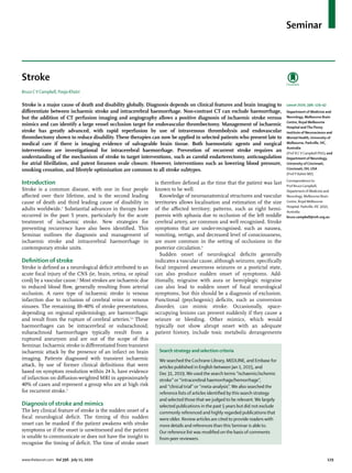
![Seminar
130 www.thelancet.com Vol 396 July 11, 2020
(eg, hypoglycaemia). Particularly in patients with a
previous history of stroke, the previous neurological
deficit can return with intercurrent illness.7
Brain imaging (CT scan or MRI) crucially comple
ments the clinical examination to differentiate the
stroke subtype and mechanism. Clinical symptoms and
signs alone cannot reliably differentiate ischaemic
stroke from intracerebral haemorrhage, and manage
ment sharply diverges between these two conditions. In
ischaemic stroke, the presence of a large vessel
occlusion, defined as occlusion in the internal carotid
artery, proximal middle cerebral artery, or basilar artery,
determines the most appropriate reperfusion therapy.
The secondary prevention of large artery atherosclerotic
disease versus cardioembolic stroke also differs
substantially. Imaging of the brain and its vascular tree
is therefore one of the most urgent priorities when
patients present to hospital with suspected stroke
(figure 1).
Epidemiology and risk factors
The 2016 Global Burden of Disease data that were
published in 2019 indicate that one in four people will
have a stroke in their lifetime.3
There are estimated
to be 9·6 million ischaemic strokes and 4·1 million
haemorrhagic strokes (including intracerebral and
subarachnoid haemorrhage) globally each year, with a
relatively stable incidence adjusted for age in high-income
countries but an increasing incidence in low-income and
middle-income countries.3
The absolute incidence is
expected to increase with an ageing population.
Estimations indicate that approximately 90% of
strokes are attributable to modifiable risk factors.8
Stroke shares many risk factors with other cardio
vascular diseases although their relative importance
varies. The most potent risk factor for stroke is high
blood pressure, which applies to both ischaemic stroke
and intracerebral haemorrhage. Smoking, diabetes,
hyperlipidaemia, and physical inactivity are also
significant risks and require interventions that are
regulatory and are based in the community to alter
lifestyle and the environment, as well as individual
treatment.9
Atrial fibrillation, a specific risk factor for
ischaemic stroke, is increasing in detection and preva
lence.10
Strokes related to atrial fibrillation tend to be
larger and more disabling than are strokes due to other
mechanisms.11
Pathophysiology
Ischaemic stroke
Most ischaemic stroke is due to embolism, either from
atherosclerotic plaque in the aortic arch or in the
cervical arteries or from the heart (panel, figure 2).
Intracranial atherosclerosis with in-situ thrombosis is
also an important mechanism of stroke, particularly in
Asian and Black ethnic groups.12
Small vessel disease
causes small subcortical infarcts (ie, lacunar stroke)
and deep intracerebral haemorrhage. Cervical artery
dissection is one of the common causes of stroke
in younger patients (eg, <60 years), and arterial
Figure 1: Stroke diagnosis using neuroimaging
(A) Ischaemic stroke established in the territory of the right middle cerebral artery 12 h after onset (arrowhead).
A patient with left hemiparesis onset 2 h before scan (B–G) showing subtle loss of differentiation between grey and
white matter (arrowhead) in the basal ganglia (B) and hyperdense thrombus (arrowhead) in the right middle
cerebral artery (C). CT perfusion showing reduced CBV (arrowhead) in the right insular region (D) corresponding to
region of diffusion restriction (arrowhead; most likely irreversibly injured) on MRI (E). CT perfusion showing delayed
Tmax (arrowhead; substantially delayedTmax [ie, >6 s] indicates brain tissue that is critically hypoperfused,
functionally impaired, and potentially at risk of infarction in the absence of reperfusion) in the right middle cerebral
artery territory (F) corresponding to intracranial occlusion of the right middle cerebral artery (arrowhead) on CT
angiogram (G). (H) Diffusion MRI lesions (arrowhead) in a patient with two 5 min episodes of aphasia that fully
resolved—now defined as ischaemic stroke rather than transient ischaemic attack. (I) Focal subarachnoid
haemorrhage (arrowhead) related to amyloid angiopathy presenting as transient parasthesias on the left side
(differential diagnosis of transient ischaemic attack). Lobar intracerebral haemorrhage (arrowhead) in a patient with
amyloid angiopathy (J) and intracerebral haemorrhage (arrowhead) in the right basal ganglia most likely resulting
from deep perforating vasculopathy (K). (L)Thrombosis in the cerebral venous sinus with hyperdense sagittal sinus
(arrow) and haemorrhagic venous infarction (arrowhead). CBV=cerebral blood volume.Tmax=time to maximum.
A C
E
B
D F
HG I
KJ L
CBV (mL/100 g) Tmax (s)](data:image/gif;base64,R0lGODlhAQABAIAAAAAAAP///yH5BAEAAAAALAAAAAABAAEAAAIBRAA7)