CODAS syndrome is a rare multi-system developmental disorder characterized by cerebral, ocular, dental, auricular, and skeletal anomalies. The researchers identified four mutations in the LONP1 gene in ten individuals with CODAS syndrome from three ancestral backgrounds. LONP1 encodes the mitochondrial Lon protease, which is involved in protein quality control and respiratory complex assembly in mitochondria. The mutations cluster in the AAA+ domain near the ATP-binding pocket and result in defects in ATP-dependent proteolysis. Lymphoblastoid cell lines from affected individuals show swollen mitochondria, aggregated cytochrome c oxidase subunit II, and reduced mitochondrial function, linking LONP1 mutations to CODAS syndrome.
![ARTICLE
CODAS Syndrome Is Associated with Mutations
of LONP1, Encoding Mitochondrial AAAþ
Lon Protease
Kevin A. Strauss,1,2,3,14,* Robert N. Jinks,3,14 Erik G. Puffenberger,1,3,14 Sundararajan Venkatesh,4
Kamalendra Singh,4,6 Iteen Cheng,5 Natalie Mikita,5 Jayapalraja Thilagavathi,4 Jae Lee,4
Stefan Sarafianos,6 Abigail Benkert,1,3 Alanna Koehler,3 Anni Zhu,3 Victoria Trovillion,3
Madeleine McGlincy,3 Thierry Morlet,7 Matthew Deardorff,8,9 A. Micheil Innes,10 Chitra Prasad,11
Albert E. Chudley,12,13 Irene Nga Wing Lee,5 and Carolyn K. Suzuki4,14
CODAS syndrome is a multi-system developmental disorder characterized by cerebral, ocular, dental, auricular, and skeletal anomalies.
Using whole-exome and Sanger sequencing, we identified four LONP1 mutations inherited as homozygous or compound-heterozygous
combinations among ten individuals with CODAS syndrome. The individuals come from three different ancestral backgrounds (Amish-
Swiss from United States, n ¼ 8; Mennonite-German from Canada, n ¼ 1; mixed European from Canada, n ¼ 1). LONP1 encodes Lon
protease, a homohexameric enzyme that mediates protein quality control, respiratory-complex assembly, gene expression, and stress
responses in mitochondria. All four pathogenic amino acid substitutions cluster within the AAAþ
domain at residues near the ATP-bind-
ing pocket. In biochemical assays, pathogenic Lon proteins show substrate-specific defects in ATP-dependent proteolysis. When ex-
pressed recombinantly in cells, all altered Lon proteins localize to mitochondria. The Old Order Amish Lon variant (LONP1
c.2161C>G[p.Arg721Gly]) homo-oligomerizes poorly in vitro. Lymphoblastoid cell lines generated from affected children have (1)
swollen mitochondria with electron-dense inclusions and abnormal inner-membrane morphology; (2) aggregated MT-CO2, the
mtDNA-encoded subunit II of cytochrome c oxidase; and (3) reduced spare respiratory capacity, leading to impaired mitochondrial pro-
teostasis and function. CODAS syndrome is a distinct, autosomal-recessive, developmental disorder associated with dysfunction of the
mitochondrial Lon protease.
Introduction
Cerebral, ocular, dental, auricular, skeletal (CODAS) syn-
drome (MIM 600373) was first described in 1991 as a
distinctive constellation of developmental delay, craniofa-
cial anomalies, cataracts, ptosis, median nasal groove,
delayed tooth eruption, anomalous cusp morphology,
malformed helices, hearing loss, short stature, delayed
epiphyseal ossification, metaphyseal hip dyplasia, and
vertebral coronal clefts.1
CODAS is a rare disease; only
three additional cases (from Brazil, Canada, and France)
were reported between 1995 and 2010.2–4
Autosomal-
recessive inheritance of CODAS syndrome was first sug-
gested by Shebib et al.,1
who identified the phenotype
within an endogamous Mennonite community, and it
was corroborated by the observations of Innes et al.3
We used whole-exome and Sanger sequencing to study
ten individuals with CODAS syndrome from three distinct
ancestral backgrounds: Amish-Swiss from the United States
(n ¼ 8), Mennonite-German from Canada (n ¼ 1), and
mixed European (German, Scottish, Irish, English) from
Canada (n ¼ 1). Our analysis revealed four different
mutations within LONP1 (RefSeq accession number
NM_004793.3 [isoform 1]; [MIM 605490]). These muta-
tions were either homozygous (LONP1 c.2161C>G
[p.Arg721Gly] and LONP1 c.2026C>T [p.Pro676Ser]) or
compound heterozygous (LONP1 c.1892C>A/c.2171C>T
[p.Ser631Tyr/p.Ala724Val]) (Table 1). In humans, LONP1
encodes the ATP-dependent mitochondrial Lon protease,
which belongs to the AAAþ
superfamily of ATPases associ-
ated with various cellular activities. AAAþ proteins share a
conserved ATPase module and mediate diverse cellular pro-
cesses such as DNA replication, membrane fusion, signal
transduction, and transcriptional regulation.5–8
Lon is an ATP-driven proteolytic machine and is highly
conserved from archaea to mammals. In human mito-
chondria, Lon is the simplest of four energy-dependent
proteolytic systems that include ClpXP, m-AAA, and
i-AAA.9
Mutations of CLPP (MIM 601119), which encodes
the proteolytic component of ClpXP, are associated with
1
Clinic for Special Children, Strasburg, PA 17579, USA; 2
Lancaster General Hospital, Lancaster, PA 17602, USA; 3
Department of Biology and Biological Foun-
dations of Behavior Program, Franklin and Marshall College, Lancaster, PA 17603, USA; 4
Department of Microbiology, Biochemistry, and Molecular
Genetics, New Jersey Medical School, Rutgers, The State University of New Jersey, Newark, NJ 07103, USA; 5
Department of Chemistry, Case Western Reserve
University, Cleveland, OH 44106, USA; 6
Department of Molecular Microbiology and Immunology, Christopher Bond Life Sciences Center, University of
Missouri, Columbia, Columbia, MO 65201, USA; 7
Auditory Physiology and Psychoacoustics Research Laboratory, duPont Hospital for Children, Wilming-
ton, DE 19803, USA; 8
Division of Human Genetics, The Children’s Hospital of Philadelphia, Philadelphia, PA 19104, USA; 9
Department of Pediatrics, Perel-
man School of Medicine, University of Pennsylvania, Philadelphia, PA 19104, USA; 10
Department of Medical Genetics and Alberta Children’s Hospital
Research Institute, Cumming School of Medicine, University of Calgary, Calgary, AB T2N 1N4, Canada; 11
Medical Genetics Program, Department of
Pediatrics, Children’s Health Research Institute and Western University, London, ON N6C 2V5, Canada; 12
Department of Pediatrics and Child Health,
University of Manitoba, Winnipeg, MB R3A 1S1, Canada; 13
Department of Biochemistry and Medical Genetics, University of Manitoba, Winnipeg,
MB R3A 1S1, Canada
14
These authors contributed equally to this work
*Correspondence: kstrauss@clinicforspecialchildren.org
http://dx.doi.org/10.1016/j.ajhg.2014.12.003. Ó2015 by The American Society of Human Genetics. All rights reserved.
The American Journal of Human Genetics 96, 121–135, January 8, 2015 121](https://image.slidesharecdn.com/d669bdfd-5c5e-47e5-b319-74b2a706a911-150320063234-conversion-gate01/85/Strauss_et_al_2015_with_supp-1-1-320.jpg)
![250 nM wild-type and mutant Lon proteins, and 0.5 mM S3. To
avoid problems from the inner filter effect, we used S3, a mixed
substrate consisting of 10% fluorescently labeled peptide S2
[Y(NO2)RGITCSGRQK(Abz)] and 90% non-fluorescent analog of
the peptide S1 (YRGITCSGRQK(Ac)). After the mixture equili-
brated at 37
C for 1 min, the reaction was initiated with 1 mM
ATP. We measured the amount of hydrolyzed peptide by deter-
mining the maximum fluorescence generated per micromole of
peptide after complete digestion by trypsin. The steady-state rate
of the reaction was determined from the tangent of the linear
portion of the time course. This rate was converted to an observed
rate constant (kobs) by division of the rate by the enzyme concen-
tration. At least three identical experiments were performed.
Peptidase assays using the fluorescent dipeptide substrate
rhodamine 110, bis-(CBZ-L-alanyl-L-alanine amide) (AA2-Rh110,
Anaspec), were performed in quadruplicate (20 ml) in 384-well
plates as previously described.39
Lon (800 nM monomer) or no-
enzyme controls were incubated in a reaction buffer (150 mM
NaCl, 50 mM HEPES [pH 8.0], and 10 mM MgCl2) containing
AA2-Rh110 (6 mM) and ATP (2 mM) for 3 hr at 37
C, after which
fluorescence was measured at an excitation and emission of
485 and 535 nm, respectively, with a Perkin Elmer Victor3
V.
The relative fluorescence units (RFU) of the background (no-
enzyme control) measurements were subtracted, and the resultant
values were normalized to percent activity of the no-drug reac-
tions. Data were fit to 4-parameter dose-response curves using
GraphPad Prism 6, and the error bars represent the SD of four repli-
cate reactions. At least three identical experiments were per-
formed. StAR and TFAM purification and degradation assays
were performed as previously described.19,41,44,45
ATPase Assay
The ATPase activity of wild-type and mutant Lon variants was
measured in quadruplicate reactions (5 ml) in a 384-well plate
via the ADP-Glo Assay (Promega) according to the manufacturer’s
recommendations. All steps were performed at room temperature.
Purified human Lon (400 nM monomer) was incubated in a
reaction buffer (40 mM Tris-HCl [pH 7.5], 20 mM MgCl2, and
0.1 mg/ml BSA) with Ultra Pure ATP (1 mM; Promega) for
60 min. ADP-GloÔ Reagent (5 ml) was added for 40 min followed
by Kinase Detection Substrate (10 ml) for 60 min, which was linear
with [ADP]. Relative luminescence units (RLU) were measured on
a Perkin Elmer Victor3
V, and background (no-enzyme control)
RLU were subtracted. At least three identical experiments were
performed.
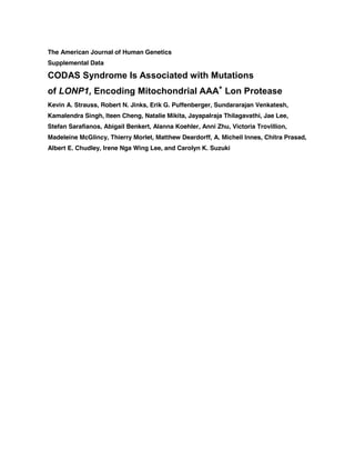
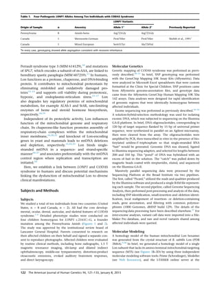
Figure 2. Radiographic Features of CODAS Syndrome
(A) Sagittal T1 (upper) and axial T2 (lower) images of an affected 4-year-old Amish child show mild diffuse cortical atrophy, an immature
pattern of myelination, and hypoplasia of the corpus callosum.
(B) Skeletal radiographs show metaphyseal dysplasia most evident at hip joints (upper) and valgus knees with both hypoplasia and
delayed ossification of epiphyses (lower).
(C) Severe scoliosis is evident early in childhood, and coronal clefts are observed at various levels of the vertebral column.
(D–G) Prenatal imaging of a 32-week-old fetus with CODAS syndrome revealed polyhydramnios, a two-vessel umbilical cord, midfacial
hypoplasia, (D) crumpled helices, (E) a balanced atrioventricular canal, (F) common atrium, omphalocele, (G, arrow) and absence of left
lower long bones, which were replaced by a relatively well-formed foot attached directly to the hip. (G, arrowhead) The asterisk in (G)
marks the left palpebral fissure for orientation.
124 The American Journal of Human Genetics 96, 121–135, January 8, 2015](https://image.slidesharecdn.com/d669bdfd-5c5e-47e5-b319-74b2a706a911-150320063234-conversion-gate01/85/Strauss_et_al_2015_with_supp-1-4-320.jpg)
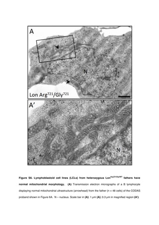
![Cell Culture
ARPE-19 cells, human embryonic kidney (HEK293T) cells, HeLa
cells (ATCC, Manassas, VA), and mouse auditory sensory epithe-
lium (UB/OC-2) cells (gift from Matthew Holley, University of
Sheffield, UK) were cultured in DMEM:F12, DMEM, Eagle’s
MEM, or MEM with GlutaMax and 50U/ml interferon-gamma
(Life Technologies), respectively, each with 10% fetal bovine
serum (FBS; Sigma or Life Technologies) at 37
C/5% CO2. Ep-
stein-Barr virus (EBV)-transformed B-lymphoblastoid cell lines
(LCLs) were generated from two Amish CODAS-syndrome-
affected probands homozygous for LONP1 c.2161CG (p.Arg721-
Gly) as well as from their respective heterozygous parents
(Lineberger Comprehensive Cancer Center, University of North
Carolina). LCLs were cultured in RPMI (ATCC formulation) with
15% FBS at 37
C and 5% CO2. Genotyping of LCLs was performed
by Sanger sequencing (The Pennsylvania State University Nucleic
Acid Facility). In brief, SuperScript III reverse transcriptase (Life
Technologies) and oligo dT primers were used for reverse transcrip-
tion of cDNA from LCL total RNA (Trizol, Life Technologies).
cDNA was then amplified by the polymerase chain reaction
(PCR) with LONP1-specific primers (Table S1) and purified for
DNA sequencing with QIAGEN PCR purification spin columns.
Lon knockdown in HeLa, PC3, and HEK293T cells was performed
as previously described46
(and as also described in Supplemental
Data).
Mitochondrial Localization
Wild-type and mutant Lon variants were overexpressed by tran-
sient transfection (FuGENE 6, Promega) in ARPE-19, HeLa, and
UB/OC-2 cells cultured on glass coverslips. After 40–48 hr, cells
were incubated with MitoTracker Red CMXRos (300 nM) at
37
C/5% CO2 for 30 min, washed with PBS (pH 7.2), and fixed
in 4% paraformaldehyde in PBS for 15 min at 37
C. Cells were per-
meabilized in acetone at À20
C for 5 min, washed with PBS, and
blocked in 1% bovine serum albumin (BSA) with 0.2% Triton X-
100 in PBS for 10 min. Immunofluorescence detection of Lon-
V5 fusion proteins was conducted as described elsewhere.35
Immunoblotting and Immunoprecipitation
Cells were lysed in the following buffers as indicated: in RIPA
buffer (50 mM Tris-HCl [pH 8.0], 150 mM NaCl, 1.0% octylphe-
noxypolyethoxyethanol CA-630, 0.5% sodium deoxycholate,
and 0.1% sodium dodecyl sulfate) supplemented with 1 mM
EDTA, 2 mM NaF, 1 mM Na3VO4, and protease-inhibitor cocktail
(Roche); Triton X-100 buffer (50 mM Tris, [pH 7.5], 300 mM
NaCl, and 0.5% Triton X-100) supplemented with Halt protease-
and phosphatase-inhibitor cocktail (ThermoScientific); or urea
buffer (50 mM triethylammonium bicarbonate buffer [TEAB,
Sigma] [pH 8.5], 8 M urea) supplemented with protease-inhibitor
cocktail and phosphatase-inhibitor cocktail (Sigma). When the
RIPA or Triton X-100 buffer was used, cells were incubated for
15 min on ice, then centrifuged at 14,000 rpm for 15 min at 4
C
so that lysates would be cleared. For the urea buffer, cells were son-
icated with microprobe for 15 s followed by a 30 s pulse, and this
was repeated 3–5 times before centrifugation at 4
C cleared
lysates.
The protein concentration of cell extracts was measured with
the Bradford assay and then normalized.42
Immunoblotting and
co-immunoprecipitation were performed as described else-
where;35
washes following immunoprecipitation contained 5%
Tween-20. The following antibodies were employed: rabbit anti-
Lon (1:400, custom made and affinity purified as previously
described)24
or rabbit anti-Lon (1:100, Novus, H00009361-
D01P); rabbit anti-ClpX (1:3,000, custom made by Dr. Irene Lee);
rabbit anti-aconitase (1:200, provided by Dr. Luke Szweda, Okla-
homa Medical Research Foundation); mouse anti-ClpP (1:4,000,
Abcam 56455); rabbit anti-Pink1 (1:500, Abcam 23707); rabbit
anti-mt-cytochrome oxidase subunit II (1:5,000, Abcam 91317);
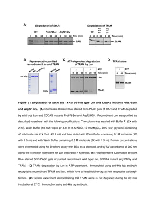
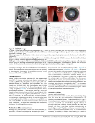
Figure 3. Location of Amino Acids That
Are Mutated within Mitochondrial Lon in
CODAS Syndrome
(A) Domain structure of the human Lon
subunit. Mitochondrial targeting sequence
(MTS), substrate recognition/binding (N)
domain, ATPase domain (AAAþ
), and a pro-
tease domain (P). Red arrows indicate the
position of pathogenic CODAS mutations.
(B) Homology model of human mitochon-
drial Lon. The model shows the position,
within a single Lon subunit (shown in
amber), of amino acids that are altered in
CODAS syndrome.
(C and D) The positions of amino acids
Pro676 and Arg721 (yellow), which are
altered in proteins encoded by CODAS
homozygous alleles. ADP is shown occu-
pying the ATP- and ADP-binding
pocket (a green dashed line represents a
proline-induced kink in the helix; a
yellow dotted line represents a salt bridge;
starbursts represent hydrophobic interac-
tions). (C) The Pro676-induced kink pro-
motes hydrophobic interactions between
Ala670, Leu667, Leu696, and Val716.
(D) The positions of Arg721 as well as
Pro676, Glu717, and the ATP- and ADP-binding site are located on the same Lon subunit (amber). Arg721 and Glu717 form salt bridges
with Glu654 and Lys517, respectively, which are located on the adjacent Lon subunit (green).
The American Journal of Human Genetics 96, 121–135, January 8, 2015 125](https://image.slidesharecdn.com/d669bdfd-5c5e-47e5-b319-74b2a706a911-150320063234-conversion-gate01/85/Strauss_et_al_2015_with_supp-1-5-320.jpg)
![OXPHOS Antibody Cocktail (1:500, Mitoscience 604); goat anti-
actin (1:2,000, 1615; Santa Cruz); mouse anti-b-actin (Sigma,
1:1,000,000); rabbit anti-mt-cytochrome B (1:1,000, Aviva
Systems Biology ARP50256_P050); mouse monoclonal anti-V5
(1:5,000, Life Technologies) or mouse anti-FLAG M2 (1:1,000
Sigma); and HRP-conjugated secondary antibodies (Cell Signaling
Technologies or Santa Cruz Biotechnology). Immunoblots were
developed by chemiluminescence.
Transmission Electron Microscopy
LCLs were prepared for transmission electron microscopy (TEM) as
described previously.39,47
In brief, cells were pelleted by centrifuga-
tion at 1,000 rpm for 5 min, washed with PBS, re-pelleted, and re-
suspended in 2.5% glutaraldehyde and 4% paraformaldehyde in
0.1 M PBS overnight at 4
C. LCLs were then post-fixed in 1%
osmium tetroxide for 1 hr at 20
C and dehydrated with ethanol;
infiltration with Spurr’s resin (Electron Microscopy Sciences) fol-
lowed. Infiltrated LCLs were collected by centrifugation and
embedded directly into BEEM capsules, which were gently centri-
fuged so that the cells would be driven to the bottom of the
capsule. Ultrathin sections (70-90 nm) were cut with glass knives,
mounted on naked copper grids (200 mesh), and stained with a
saturated solution of uranyl acetate in 50% ethanol for 20 min, fol-
lowed by Reynold’s lead citrate for 15 min. Differences in the fre-
quency of abnormal mitochondrial morphology between proband
and parent cells were analyzed by two-way ANOVA with Bonfer-
roni post-hoc correction (SPSS, IBM).
Quantitative PCR (qPCR) Determination of mtDNA
Copy Number
Genomic DNA (100 ng) from peripheral blood lymphocytes
and LCL cells was used for amplification of both the mtDNA-
encoded MT-CYB gene and the nuclear-DNA-encoded APP gene
as a reference control. Genomic DNA was amplified in reactions
(20 ml; Applied Biosystems; universal PCR master mix) with the
following Taqman primers. Forward: APP, 50
-TTTTTGTGTGCT
CTCCCAGGTCT-30
; and MT-CYB, 50
-GCCTGCCTGATCCTCCAA
AT-30
). Reverse: APP 50
-TGGTCACTGGTTGGTTGGC-30
; and MT
CYB, 50
-AAGGTAGCGGATGATTCAGCC-30
. TaqMan probes: APP,
50
-[6FAM]CCCTGAACTGCAGATCACCAATGTGGTAG[TAM]-30
;
and MT-CYB, 50
-[6FAM]CACCAGACGCCTCAACCGCCTT
[TAM]-30
An AB 7500 RT-PCR system (Applied Biosystems) and
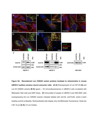
Figure 4. Enzymology, Cellular Expression, and Homo-oligomerization of Pathogenic Lon Proteins
(A) ATP-dependent degradation of the fluorogenic peptide substrate S3. The rates of S3 cleavage were determined with recombinant
Lon proteins (250 nM monomer) and 0.5 mM of S3 (0.5-fold Km) in buffer. The degradation reactions were initiated by the addition
of Mg-ATP at 37
C for 600–900 s. S3 cleavage was monitored as an increase in fluorescence at an excitation and emission of 320 nm
and 420 nm, respectively (three or more replicates). Error bars represent SEM.
(B) StAR (5.6 mM) or TFAM (5 mM) were combined in buffer with Mg-ATP, and reactions were initiated by the addition of Lon (500 nM) at
37
C. Aliquots were removed at time points, reactions were terminated with 53 reducing sample buffer, and aliquots were analyzed by
SDS-PAGE and Coomassie Brilliant Blue staining. ImageJ was used for determining the percent StAR and TFAM degraded after a 60 min
incubation period with the same Lon variant (no replicates).
(C and D) Overexpression of (C) wild-type Lon-V5 and (D) p.Arg721Gly-V5 (green ¼ V5 immunofluorescence) in ARPE-19 cells counter-
stained with Mitotracker Red (red) and DAPI (blue). Nontransfected cells display only red Mitotracker fluorescence.
(E) Coimmunoprecipitation (IP) of Lon-V5 by Lon-FLAG (co-overexpressed in HEK293T cell lysates) with anti-FLAG M2 antibody.
Immunoblotting (IB) with anti-FLAG and V5 antibodies followed.
126 The American Journal of Human Genetics 96, 121–135, January 8, 2015](https://image.slidesharecdn.com/d669bdfd-5c5e-47e5-b319-74b2a706a911-150320063234-conversion-gate01/85/Strauss_et_al_2015_with_supp-1-6-320.jpg)