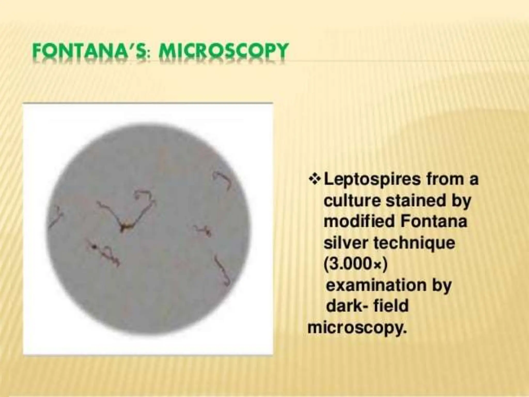
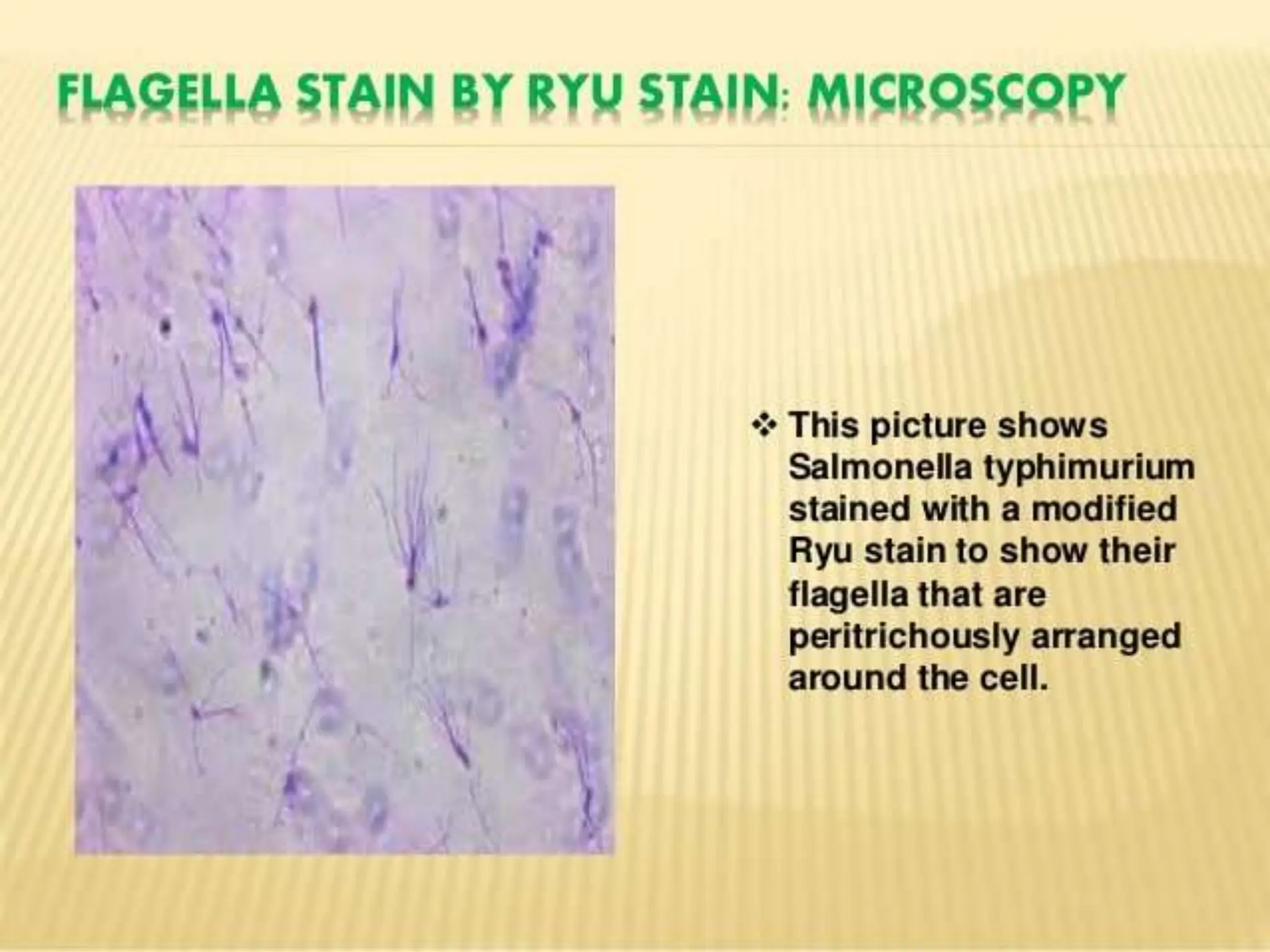
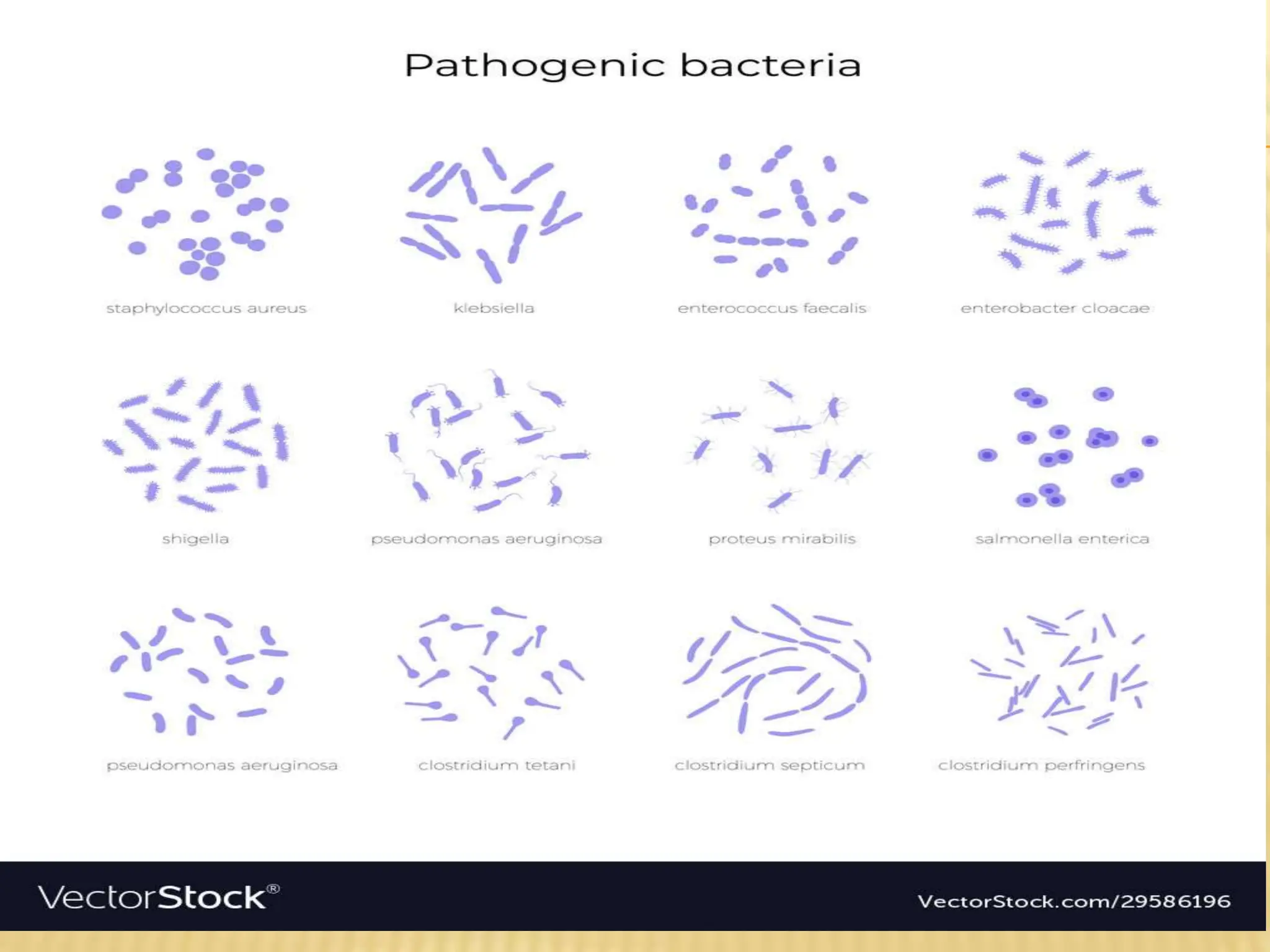
Staining is a technique used to enhance contrast in samples, generally at the microscopic level. Stains and dyes are frequently used in histology, in cytology, and in the medical fields of histopathology, hematology, and cytopathology that focus on the study and diagnoses of diseases at the microscopic level
41. VIRULENCE
Virulence is related to pathogenicity in the sense that its meaning is correlated to
the manifestation of a disease.
However, pathogenicity, in particular, is defined as the ability of a pathogen to
cause disease. An organism that harms its host and causes the disease is
referred to as a pathogen.
The capability to produce disease is associated with the
inherent characteristics of the organism in an effort to survive inside the host.
Conversely, virulence refers to the degree of pathogenicity of a particular
organism. A virulent pathogen is one that causes damage to its host to an extent
that is significantly greater than those caused by a non-pathogenic organism.
Pathogenic organisms have a different breadth of virulence. For example,
a strain of bacteria may be more virulent than the other strains of the
same species. The virulence of a pathogen is often correlated with the so-called
virulence factors. A virulence factor is defined as the factor that enables an
organism to invade a host and cause disease. It also determines the extent of
damage to the host. These factors may be secretory, membrane-associated,
or cytosolic in nature.
42. An example of a virulence factor is the ability of microbes to multiply
within their host cells. In microbiology, these factors are considered vital
to epidemiology particularly, when tracking a novel pathogenic strain.
That is because the strain is often highly virulent, and therefore more
detrimental, even fatal, to its host.
Some of the virulence factors that researchers look into are the route of
entry into its host, the pathobiological machinery employed, and its
effects on the host’s immune response. Viral virulence factors, for
instance, are chiefly proteins that are incited by the infective virus to be
produced by the host’s own protein machinery.
Bacterial virulence factors are likewise proteins that are coded for by
their own genes or by plasmids that they acquired via horizontal gene
transfer.
The damage may be compounded by the host’s overly reactive immune
response when the immune cells are so triggered by the presence of
these virulence factors that they tend to damage the host cells in an
effort to counter the infection.
43.
44. These virulence factors are, therefore, one of the major targets in
medical research that intend to create new treatments
and vaccines.
HIV is an example of a virulent virus. It is the causative agent
of AIDS. It is virulent because it employs mechanisms for evading
the host immune cells. For instance, it infects the T-helper cell,
one of the immune cells. Thus, the immune response of the host
is already reduced and compromised.
Another example is lyssavirus that causes rabies. It enters and
hijacks muscle cells, and then travels to the nervous
system through the neuromuscular junctions. Thus, it is
particularly described as neurovirulent, i.e. for being able to cause
disease in the nervous system.
As for bacteria, examples are the two human
pathogens: Mycobacterium tuberculosis (causative agent of
tuberculosis) and Bacillus anthracis(causative agent of anthrax).
45. PATHOGENICITY OF BACTERIA
Pathogenicity is the ability of the pathogen to produce
disease. Pathogenicity is expressed by microbes using
their virulence, or the degree of the microbe's
pathogenicity.
Genetic, biochemical, and structural features that lead to
the ability of the pathogen to cause disease are known as
its determinants of virulence.
Adherence and Colonization Factors. To cause infection,
many bacteria must first adhere to a mucosal surface. ...
Invasion Factors. ...
Capsules and Other Surface Components. ...
Endotoxins. ...
Structure of Endotoxin
46. ATTENUATION
Attenuation is a regulatory mechanism used in
bacterial operons to ensure proper transcription
and translation. In bacteria, transcription and
translation are capable of proceeding
simultaneously. The need to prevent
unregulated and unnecessary gene expression
can be prevented by attenuation, which is
characterized as a regulatory mechanism.
47. Bacterial adhesionto biomaterials is often considered
the first in a sequence of events that may culminate in the most feared
complication associated with the implantation of a biomaterial into the human
body: biomaterial-associated infection. For the cases in which an infection
occurs, the first approach of a bacterium to a biomaterial may constitute the
biggest single step in the development of an infection.
The ability of the contaminating bacterium to adhere to the biomaterial or the
surrounding tissue is dependent upon a complex interaction between the
contaminating bacterium, the host and the biomaterial itself. Should the
bacterium successfully evade host defenses and adhere to the biomaterial, then
a sequence of events unfolds whereby the relatively weak and reversible initial
adhesive interactions proceed to stronger, irreversible interactions and ultimately
to microcolony formation, and the development of a biofilm.
. Not only is bacterial adhesion to biomaterials the first step in the infection
process, but it is also the only step in the infection process that can be
significantly manipulated by a biomaterial scientist to reduce the adhesion of
bacteria and thus, reduce susceptibility to infection (antimicrobial activation
notwithstanding). Once bacterial adhesion occurs the development of the biofilm
is much more dependent upon the virulence factors possessed by the bacteria
and the host response, rather than on the biomaterial surface that has been
colonized.
48.
49.
50. REPRODUCTION OF BACTERIA
Bacteria are microbes with a cell structure simpler than
that of many other organisms. Their control centre,
containing the genetic information, is contained in a single
loop of DNA.
Some bacteria have an extra circle of genetic material
called a plasmid rather than a nucleus. The plasmid often
contains genes that give the bacterium some advantage
over other bacteria. For example it may contain a gene
that makes the bacterium resistant to a certain antibiotic.
Bacteria are classified into five groups according to their
basic shapes: spherical (cocci), rod (bacilli), spiral
(spirilla), comma (vibrios) or corkscrew (spirochaetes).
They can exist as single cells, in pairs, chains or clusters
51. Bacteria are found in every habitat on Earth: soil, rock,
oceans and even arctic snow. Some live in or on other
organisms including plants and animals including
humans. There are approximately 10 times as many
bacterial cells as human cells in the human body. A lot of
these bacterial cells are found lining the digestive system.
Some bacteria live in the soil or on dead plant matter
where they play an important role in the cycling of
nutrients. Some types cause food spoilage and crop
damage but others are incredibly useful in the production
of fermented foods such as yoghurt and soy sauce.
Relatively few bacteria are parasites or pathogens that
cause disease in animals and plants.
52. Most bacteria reproduce by binary fission. In this process the bacterium, which is
a single cell, divides into two identical daughter cells. Binary fission begins when
the DNA of the bacterium divides into two (replicates).
The bacterial cell then elongates and splits into two daughter cells each with
identical DNA to the parent cell. Each daughter cell is a clone of the parent cell.
When conditions are favourable such as the right temperature and nutrients are
available, some bacteria like Escherichia coli can divide every 20 minutes. This
means that in just seven hours one bacterium can generate 2,097,152 bacteria.
After one more hour the number of bacteria will have risen to a colossal
16,777,216. That’s why we can quickly become ill when pathogenic microbes
invade our bodies.
Some bacteria can form endospores. These are dormant structures, which are
extremely resistant to hostile physical and chemical conditions such as heat, UV
radiation and disinfectants.
This makes destroying them very difficult. Many endospore-producing bacteria
are nasty pathogens, for example Bacillus anthracis, the cause of anthrax.
53.
54. ISOLATION AND IDENTIFICATION PROCEDURE
Unlike bacteria, many of which can be grown on an artificial
nutrient medium, viruses require a living host cell for replication.
Infected host cells (eukaryotic or prokaryotic) can be cultured and
grown, and then the growth medium can be harvested as a
source of virus.
Virions in the liquid medium can be separated from the host cells
by either centrifugation or filtration. Filters can physically remove
anything present in the solution that is larger than the virions; the
viruses can then be collected in the filtrate.
Isolation refers to the separation of a strain from a natural, mixed
population of living microbes, as present in the environment, for
example in water or soil flora, or from living beings with skin
flora, oral flora or gut flora, in order to identify the microbe(s) of
interest. Historically, the laboratory techniquesof isolation first
developed in the field of bacteriology and parasitology(during the
19th century), before those in virology during the 20th century.
55.
56. The laboratory techniques of isolating microbes first
developed during the 19th century in the field
of bacteriology and parasitology using light microscopy.
Proper isolation techniques of virology did not exist prior
to the 20th century. The methods of microbial isolation
have drastically changed over the past 50 years, from a
labor perspective with increasing mechanization, and in
regard to the technologies involved, and with it speed and
accuracy.
In order to isolate a microbe from a natural, mixed
population of living microbes, as present in the
environment, for example in water or soil flora, or from
living beings with skin flora, oral flora or gut flora, one has
to separate it from the mix.
57. Traditionally microbes have been cultured in order
to identify the microbe(s) of interest based on its growth
characteristics. Depending on the expected density and
viability of microbes present in a liquid sample, physical
methods to increase the gradient as for example serial
dilution or centrifugation may be chosen. In order to
isolate organisms in materials with high microbial content,
such as sewage, soil or stool, serial dilutions will increase
the chance of separating a mixture.
When bacteria have visibly grown, they are often still
mixed. The identification of a microbe depends upon the
isolation of an individual colony, as biochemical testing of
a microbe to determine its different physiological features
depends on a pure culture.
58. To make a subculture, one again works in aseptic technique in
microbiology, lifting a single colony off the agar surface with a
loop and streaks the material into the 4 quadrants of an agar
plate or all over if the colony was singular and did not look mixed.
Gram staining the raw sample before incubation or staining
freshly grown colony material helps to determine if a colony
consists of uniformly appearing bacteria or is mixed, and the
color, and shape of bacteria allow a first classification based on
morphology.
In clinical microbiology numerous other staining techniques for
particular organisms are used (acid fast bacterial stain for
mycobacteria). Immunological staining techniques, such as direct
immunofluorescence have been developed for medically
important pathogens that are slow growing (Auramine-rhodamine
stain for mycobacteria) or difficult to grow (such as Legionella
pneumophila species) and where the test result would alter
standard management and empirical therapy.