
Recommended
Recommended
More Related Content
What's hot
What's hot (20)
Similar to Contents of Vertebral Canal: Spinal Cord, Nerve Roots, Meninges
Similar to Contents of Vertebral Canal: Spinal Cord, Nerve Roots, Meninges (20)
More from Ömer aslankan
More from Ömer aslankan (20)
Recently uploaded
Recently uploaded (20)
Contents of Vertebral Canal: Spinal Cord, Nerve Roots, Meninges
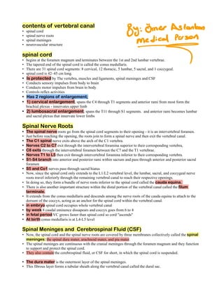
- 1. contents of vertebral canal spinal cord • spinal nerve roots • spinal meninges • neurovascular structure • spinal cord begins at the foramen magnum and terminates between the 1st and 2nd lumbar vertebrae. • The tapered end of the spinal cord is called the conus medullaris. • There are 31 spinal cord segments: 8 cervical, 12 thoracic, 5 lumbar, 5 sacral, and 1 coccygeal. • spinal cord is 42–45 cm long. • Is protected by The vertebra, muscles and ligaments, spinal meninges and CSF • Conducts sensory impulses from body to brain • Conducts motor impulses from brain to body • Controls reflex activities • Has 2 regions of enlargement; • 1) cervical enlargement; spans the C4 through T1 segments and anterior rami from most form the • brachial plexus - innervates upper limb 2) lumbosacral enlargement; spans the T11 through S1 segments. and anterior rami becomes lumbar • and sacral plexus that innervate lower limbs Spinal Nerve Roots The spinal nerve roots go from the spinal cord segments to their opening - it is an intervertebral foramen. • Just before reaching the opening, the roots join to form a spinal nerve and then exit the vertebral canal. • The C1 spinal nerve exits above the arch of the C1 vertebra. • Nerves C2 to C7 exit through the intervertebral foramina superior to their corresponding vertebra, • C8 exits through the intervertebral foramen between the C7 and the T1 vertebrae. • Nerves T1 to L5 then exit through intervertebral foramina inferior to their corresponding vertebra. • S1-S4 branch into anterior and posterior rami within sacrum and pass through anterior and posterior sacral • foramen S5 and Co1 nerves pass through sacral hiatus • Now, since the spinal cord only extends to the L1/L2 vertebral level, the lumbar, sacral, and coccygeal nerve • roots travel inferiorly through the remaining vertebral canal to reach their respective openings. In doing so, they form a bundle of nerve roots inferior to the spinal cord called the cauda equina. • There is also another important structure within the distal portion of the vertebral canal called the filum • terminale. It extends from the conus medullaris and descends among the nerve roots of the cauda equina to attach to the • dorsum of the coccyx, acting as an anchor for the spinal cord within the vertebral canal. in embryo spinal cord occupies whole vertebral canal • by week 8 caudal eminence dissapears and coccyx goes from 6 to 4 • in fetal period VC grows faster than spinal cord so cord "ascends" • At birth conus medullaris is at L4-L5 level • Spinal Meninges and Cerebrospinal Fluid (CSF) Now, the spinal cord and the spinal nerve roots are covered by three membranes collectively called the spinal • meninges: the spinal dura mater, arachnoid mater, and pia mater. The spinal meninges are continuous with the cranial meninges through the foramen magnum and they function • to support and protect the spinal cord. They also contain the cerebrospinal fluid, or CSF for short, in which the spinal cord is suspended. • The dura mater is the outermost layer of the spinal meninges. • This fibrous layer forms a tubular sheath along the vertebral canal called the dural sac. •
- 2. The space that separates the dural sac from the bony walls of the vertebral canal is called the epidural space • it contains the internal vertebral venous plexus, surrounded by epidural fat. • Superiorly, both the dural sac and epidural space extend to the foramen magnum • Inferiorly, the dural sac and epidural space terminate at the level of the S2 vertebra. • The spinal dura mater is innervated by branches of the spinal nerves called the recurrent meningeal nerves. • Next is the arachnoid mater which lines the spinal dura mater and the dural root sheaths internally. • It encloses the subarachnoid space, which is filled with CSF. • Inferiorly, the subarachnoid space extends beyond the conus medullaris of the spinal cord. • The space inferior to the conus medullaris, which extends from the L2 vertebra to the S2 vertebra is called the • lumbar cistern. It contains cerebrospinal fluid and the cauda equina. • The arachnoid mater is separated from the pia mater by CSF, but connective tissue strands called arachnoid • trabeculae make connections between the arachnoid and pia mater through the subarachnoid space. The pia mater is the innermost meningeal layer which covers the spinal cord, spinal nerve roots, and spinal • blood vessels. A fine thread of the spinal pia mater forms the filum terminale extending inferiorly from the conus medullaris. • Laterally, the pia mater forms extensions along the spinal cord called the denticulate ligaments. • These ligaments arise between the anterior and posterior nerve roots on both sides of the spinal cord. • The denticulate ligaments have triangular processes that extend laterally and pass through the arachnoid to • attach to the dura mater. Superiorly the denticulate ligaments attach to the cranial dura mater. • The denticulate ligaments, along with the filum terminale, help suspend the spinal cord in the cerebrospinal fluid • of the subarachnoid space. Arteries The blood supply for the spinal cord comes from a few sources. • First, there are three longitudinal arteries: one anterior spinal artery and two posterior spinal arteries. • The anterior spinal artery originates within the cranium from branches of the vertebral arteries. • It extends inferiorly along the anterior median fissure. Its branches, called the sulcal arteries, enter the spinal • cord through the anterior median fissure. The two posterior spinal arteries also originate in the cranium either as branches of the vertebral arteries • or the posterior inferior cerebellar arteries. The three longitudinal arteries can only supply the superior part of the spinal cord by themselves. • The circulation to the majority of the spinal cord depends on the posterior and anterior segmental medullary • arteries and radicular arteries that run along the spinal nerve roots. The posterior and anterior segmental medullary arteries arise at various vertebral levels from the • spinal branches of the following arteries: ascending cervical, deep cervical, vertebral, posterior intercostal, and lumbar arteries. These arteries supply blood mainly to the cervical and lumbosacral enlargements. Additionally, a branch of an inferior posterior intercostal or upper lumbar artery called the great anterior • segmental medullary artery also reinforces the blood supply to the lower two-thirds of the spinal cord. Lastly, spinal branches also give rise to the radicular arteries, which are small arteries that supply blood to the • spinal nerve roots and their coverings. Unlike the segmental arteries, they do not anastomose with the anterior and posterior spinal arteries. Finally, let's discuss the veins of the spinal cord. The veins of the spinal cord usually follow the arteries. There • are typically three anterior and three posterior spinal veins. They drain into anterior and posterior medullary and radicular veins, which communicate with the • epidural venous plexus. From the epidural venous plexus, blood flows to the dural sinuses of the cranium, the vertebral veins, and • the external vertebral venous plexuses. Summary The spinal cord has 31 spinal segments and pairs of spinal nerves: 8 cervical, 12 thoracic, 5 lumbar, 5 • sacral, and 1 coccygeal.
- 3. The two enlargements of the spinal cord are the cervical enlargement and lumbosacral enlargement. • The spinal cord and the spinal nerve roots are covered by the spinal meninges, which consist of the • dura mater, arachnoid mater, and pia mater. The dura mater is separated from the bony walls of the vertebral canal by the epidural space. • The spinal arachnoid mater is pressed against the dura by the cerebrospinal fluid of the subarachnoid • space. The spinal pia mater directly covers the spinal cord, the spinal nerve roots, and spinal blood vessels. Inferior • to the spinal cord, it continues as a fine thread called the filum terminale. Laterally the spinal pia mater forms extensions along the spinal cord called the denticulate ligaments. • the spinal cord blood supply comes from the anterior spinal artery, the two posterior spinal arteries, and • the posterior and anterior segmental medullary arteries, including the great anterior segmental medullary artery. Venous blood drains through three anterior spinal veins and three posterior spinal veins. •