
Recommended
More Related Content
What's hot
What's hot (20)
Similar to Radiographic assessment of dental caries
Similar to Radiographic assessment of dental caries (20)
More from DrMohamedEkram
More from DrMohamedEkram (8)
Recently uploaded
Recently uploaded (20)
Radiographic assessment of dental caries
- 2. Rationale For The Use Of Intraoral Radiographs Intra-oral radiographyIntra-oral radiography can reveal cariouscan reveal carious lesions which mightlesions which might not be detectednot be detected during a thoroughduring a thorough clinical examination.clinical examination. Cavitating lesion No-cavitation
- 3. Rationale For The Use Of Intraoral Radiographs Both clinical andBoth clinical and radiographic examinationsradiographic examinations are necessary in theare necessary in the detection of dental cariesdetection of dental caries N.B.N.B. Early carious lesionsEarly carious lesions are difficult to detectare difficult to detect radiographically particularlyradiographically particularly when they are small andwhen they are small and limited to the enamel.limited to the enamel.
- 4. EXAMINATIONS PosteriorPosterior bitewing radiographsbitewing radiographs areare the most useful for detectingthe most useful for detecting caries.caries. Periapical radiographsPeriapical radiographs areare primarily used for detectingprimarily used for detecting changes in the periapical andchanges in the periapical and inter-radicular bone.inter-radicular bone.
- 5. EXAMINATIONS Bitewing examinationBitewing examination For childrenFor children : up to 3 years use: up to 3 years use size 0 film, till 6 years use sizesize 0 film, till 6 years use size 1 and after that use size 2.1 and after that use size 2. For adultsFor adults : the most useful: the most useful adult bitewing examinationadult bitewing examination consists of no. 2 size films forconsists of no. 2 size films for separate premolar and molarseparate premolar and molar projections.projections. 2 00 M.Ekram 2211 Up to 3 years 3-6 years After 6 years and adults
- 6. Frequency when radiographs are indicated ? Radiographs are frequently used for detectingRadiographs are frequently used for detecting caries when the teeth are incaries when the teeth are in tight contacttight contact andand cannot be directly inspected.cannot be directly inspected. Radiographs are especially used when theyRadiographs are especially used when they contribute to the diagnosis of dental cariescontribute to the diagnosis of dental caries e.ge.g in clinically inaccessible areas as the rootsin clinically inaccessible areas as the roots of teeth.of teeth.
- 9. Incipient Occlusal Caries Cannot be seen on a radiograph andCannot be seen on a radiograph and must be detected clinicallymust be detected clinically with anwith an explorer. usually radiographs are notexplorer. usually radiographs are not effective till caries reach the dentine.effective till caries reach the dentine.
- 10. Incipient occlusal caries Incipient occlusal caries Not evident radiographically But they require treatment
- 11. Incipient Occlusal Caries The only detectableThe only detectable evidence of an earlyevidence of an early carious lesion at thecarious lesion at the occlusal surface mayocclusal surface may be abe a fine gray shadowfine gray shadow just beneath the (DEJ).just beneath the (DEJ). this should bethis should be differentiated from thedifferentiated from the optical illusionoptical illusion calledcalled mach bandmach band..
- 12. Moderate Occlusal Caries The classic radiographicThe classic radiographic change is achange is a broad basedbroad based of a thin radiolucent zoneof a thin radiolucent zone in the dentine with little orin the dentine with little or no changes in the enamelno changes in the enamel ..
- 13. Moderate Occlusal Caries a broad based thina broad based thin radiolucent zone inradiolucent zone in the dentinethe dentine
- 14. Moderate Occlusal Caries Differential diagnosis:Differential diagnosis: a band of increased opacitya band of increased opacity exists between the lesionexists between the lesion and the pulp chamber. thisand the pulp chamber. this line is not usually seen withline is not usually seen with buccal caries .buccal caries . N.B. careful clinicalN.B. careful clinical examination should beexamination should be carried out .carried out . OcclusalCaries B&Lingcaries
- 15. Severe Occlusal Caries Readily observedReadily observed bothboth clinically andclinically and radiographically.radiographically. Appears as aAppears as a largelarge radiolucency in the crownradiolucency in the crown of the toothof the tooth. the occlusal. the occlusal surface may appearsurface may appear collapsed.collapsed.
- 16. Pitfalls In The Interpretation Of Occlusal Caries 1.Failure to recognize that1.Failure to recognize that occlusalocclusal caries of enamelcaries of enamel will not ordinarily bewill not ordinarily be detectable on radiographs.detectable on radiographs. 2. The carelessness of not observing the2. The carelessness of not observing the the long thin radiolucency that first appears at the ( DEJ) as a sign of occlusal caries. 3. Confusion between occlusal and buccal caries ( clinical examination is essential).
- 18. PROXIMAL CARIESPROXIMAL CARIES Radiographic detection of carious lesions on theRadiographic detection of carious lesions on the proximal surfaces of teeth depends onproximal surfaces of teeth depends on loss ofloss of enough mineralenough mineral to result in a detectable changeto result in a detectable change in radiographic densityin radiographic density N.B.N.B. Approximately 40 %Approximately 40 % demineralization isdemineralization is required.required.
- 19. Proximal caries susceptible -zone Free gingival margin Contact point This region extends from the contact point down to the height of the free gingival margin
- 20. Classification OfClassification Of Proximal CariesProximal Caries Proximal caries can be classified according toProximal caries can be classified according to thethe depth of penetrationdepth of penetration of the lesion through theof the lesion through the enamel and dentin into:enamel and dentin into: Incipient class IIncipient class I Moderate class IIModerate class II Advanced class IIIAdvanced class III Severe class IV
- 21. Incipient Proximal Caries Extends less than halfwayExtends less than halfway through the thickness of enamelthrough the thickness of enamel ( seen only in enamel ).( seen only in enamel ). Non-cavitating lesion
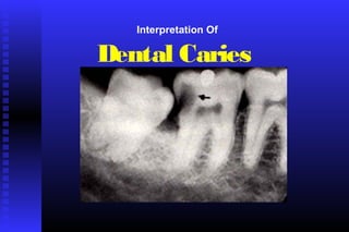
- 22. Incipient proximal caries The general radiographic appearance of anThe general radiographic appearance of an incipient carious lesion is of a radiolucentincipient carious lesion is of a radiolucent “notch”“notch” on the outer surface of the tooth.on the outer surface of the tooth.
- 23. AA magnifying glass is very useful foris very useful for examining incipient carious lesions.examining incipient carious lesions. Incipient proximal caries
- 24. Moderate Proximal Caries Involve more than theInvolve more than the outer half of theouter half of the enamel but are notenamel but are not seen radiographicallyseen radiographically to extend into theto extend into the (DEJ).(DEJ).
- 25. Moderate Proximal Caries These lesions generally haveThese lesions generally have one of three radiographicone of three radiographic appearances :appearances : 1. A1. A triangletriangle with its broad base atwith its broad base at the surface of the tooth.the surface of the tooth. 2.2. DiffuseDiffuse radiolucent image.radiolucent image. 3. A3. A combinationcombination of these twoof these two types.types.
- 26. Advanced Proximal Caries Extends to or throughExtends to or through the (DEJ) and intothe (DEJ) and into dentin but does notdentin but does not extend through theextend through the dentin more than halfdentin more than half the distance towardthe distance toward the pulpthe pulp (( the lesionthe lesion involves both enamelinvolves both enamel and dentinand dentin ).).
- 27. Advanced Proximal Caries The configuration isThe configuration is usuallyusually triangulartriangular butbut may be diffuse or amay be diffuse or a combinationcombination of them.of them.
- 29. Severe Proximal Caries Extends through theExtends through the enamel , through theenamel , through the dentin and more thandentin and more than half the distance towardhalf the distance toward the pulp. the lesionthe pulp. the lesion approaches the pulpapproaches the pulp and may appearand may appear clinically as a cavitationclinically as a cavitation in the tooth.in the tooth.
- 30. Pitfalls In The Interpretation Of Proximal Caries Proximal cariesProximal caries may bemay be confused withconfused with Cervical burnout. CoveredbyboneCoveredbyenamel
- 31. Cervical Burnout
- 32. Pitfalls In The Interpretation Of Proximal Caries Proximal caries mayProximal caries may be confused with:be confused with: Hypoplastic pits Concavities produced by wear.
- 33. Facial , Buccal AndFacial , Buccal And Lingual cariesLingual caries
- 34. Facial , Buccal And Lingual cariesFacial , Buccal And Lingual caries When smallWhen small , appear, appear as small roundas small round radiolucent area.radiolucent area. As they enlargeAs they enlarge theythey become elliptic orbecome elliptic or similunar in shape.similunar in shape.
- 35. Facial , buccal and lingualcaries (Con)Facial , buccal and lingualcaries (Con) They demonstrateThey demonstrate sharpsharp well-defined borderswell-defined borders between the intact andbetween the intact and deminiralized (radiolucent)deminiralized (radiolucent) enamel.enamel. Clinical examinationClinical examination with anwith an explorer is necessary.explorer is necessary. N.B.N.B. It is difficult to differentiate between buccal and lingualIt is difficult to differentiate between buccal and lingual caries on a radiograph.caries on a radiograph.
- 36. Differential Diagnosis Of Buccal And Lingual Caries WITH PROXIMAL CARIESWITH PROXIMAL CARIES A buccal or lingual carious lesion atA buccal or lingual carious lesion at or near the mesial or distal lineor near the mesial or distal line angle of the tooth may project ontoangle of the tooth may project onto a proximal surface and appear asa proximal surface and appear as a proximal lesion.a proximal lesion. Different angulation may be used.Different angulation may be used. N.B. Clinical examination is essential. Proximal caries
- 37. Differential Diagnosis Of Buccal And Lingual Caries WITH OCCLUSAL CARIESWITH OCCLUSAL CARIES It is necessary to examineIt is necessary to examine more than one viewmore than one view ofof the area because a margin of a buccal or lingualthe area because a margin of a buccal or lingual lesion may be superimposed on the (DEJ)lesion may be superimposed on the (DEJ) suggesting occlusal caries.suggesting occlusal caries.
- 38. DIFFERENTIAL DIAGNOSIS OF BUCCAL AND LINGUAL CARIES WITH OCCLUSAL CARIES(con) Also, Occlusal caries will ordinarily be : * more extensive than lingual or buccal caries *it’s outline not as well defined N.B. Clinical examination is is essential.
- 39. Root Surface CariesRoot Surface Caries Involves only the rootsInvolves only the roots of the teeth and is alsoof the teeth and is also calledcalled cemental cariescemental caries..
- 40. Root Surface CariesRoot Surface Caries Radiolucent , illRadiolucent , ill defined saucer-likedefined saucer-like lesions and if small , thelesions and if small , the lesion will be notchedlesion will be notched rather than saucerlike.rather than saucerlike.
- 41. Root Surface CariesRoot Surface Caries
- 42. Pitfalls In the Interpretation Of Root Surface Caries root surface caries may be confused withroot surface caries may be confused with cervical burnoutcervical burnout The true carious lesion may beThe true carious lesion may be distinguished from thedistinguished from the intactintact surfacesurface primarily by :primarily by : Absence of an image of theAbsence of an image of the root edge.root edge. The appearance of aThe appearance of a diffuse roundeddiffuse rounded inner borderinner border where the tooth substancewhere the tooth substance has been lost.has been lost. N.B. clinical examination is essential.
- 43. Recurrent CariesRecurrent Caries OccursOccurs adjacent to a preexisting restoration.adjacent to a preexisting restoration. CariesCaries occur in this region because ofoccur in this region because of :: Inadequate cavity preparationInadequate cavity preparation Defective margins of restorationsDefective margins of restorations incomplete removal of caries.incomplete removal of caries.
- 44. Recurrent cariesRecurrent caries Appears radiographically as a radiolucent area just beneath a restoration. The radiographic appearance depends on : * the amount of decalcification present and * whether a restoration is obscuring the lesion.
- 45. Differential Diagnosis Between Caries And Restorative And Base Materials Radiographic appearance of restorative materialsRadiographic appearance of restorative materials depends on their:depends on their: thickness - density (atomic structure). some can be confused with caries such as :some can be confused with caries such as : old calcium hydroxide , composite , plastic orold calcium hydroxide , composite , plastic or silicate.silicate. They can be differentiated from caries by :They can be differentiated from caries by : 1. their well-defined and classic outline.1. their well-defined and classic outline. 2. dental history and clinical examination.2. dental history and clinical examination.
- 46. Differential Diagnosis Between Caries And Restorative And Base Materials
- 47. Rampant CariesRampant Caries Rampant caries is anRampant caries is an advanced and severeadvanced and severe form of cariesform of caries that affectsthat affects numerous teeth.numerous teeth. Rampant caries isRampant caries is typically seen intypically seen in childrenchildren with poor dietary habits orwith poor dietary habits or inin adultsadults with a decreasedwith a decreased salivary flow.salivary flow.
- 48. Radiation CariesRadiation Caries Occurs in patients who haveOccurs in patients who have received therapeutic radiationreceived therapeutic radiation to the head and neck regionto the head and neck region with a resultantwith a resultant xerostomiaxerostomia .. dark radiolucentdark radiolucent lesionslesions appear at the necks of teethappear at the necks of teeth (cervical caries )(cervical caries ) most obviousmost obvious on the mesial and distalon the mesial and distal aspects.aspects.
- 49. Radiation caries (con)Radiation caries (con) Variation in the depth ofVariation in the depth of destructiondestruction may be present ,may be present , but generally their is uniformitybut generally their is uniformity within a given region of thewithin a given region of the mouth.mouth. Destruction may encircle theDestruction may encircle the tooth causing thetooth causing the entire crownentire crown toto be lost.be lost.