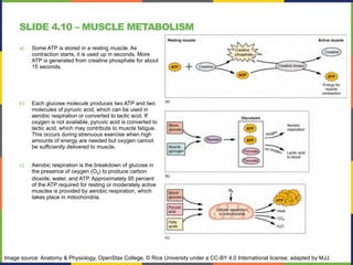
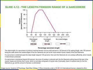
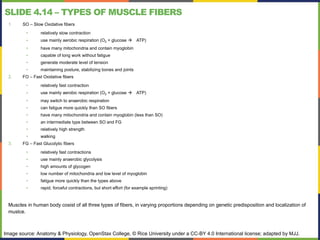
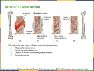
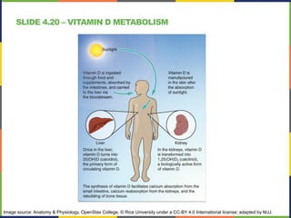
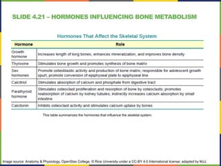
BASICS OF PHYSIOLOGY
The Movement System
SLIDE 4.1 – THREE TYPES OF MUSCLE TISSUE
• Human body contains three types of muscle
tissue:
a) skeletal muscle,
b) cardiac muscle,
c) smooth muscle.
• Despite differences in their appeariance and
played roles, all have in common few things, one
of them is quality called excitability.
• It means that their plasma membranes can
change their electrical states (from polarized to
depolarized) and send an electrical wave called
an action potential along the entire length of the
membrane which leads to contraction.
Image source: Anatomy & Physiology, OpenStax College, © Rice University under a CC-BY 4.0 International license; adapted by MJJ.
SLIDE 4.2 – SKELETAL MUSCLE STRUCTURE
• Each skeletal muscle has three
layers of connective tissue (called
“mysia”) that enclose it and provide
structure to the muscle as a whole,
and also compartmentalize the
muscle fibers within the muscle.
• Inside each skeletal muscle,
muscle fibers are organized into
individual bundles, each called a
fascicle.
• Each fascicle consists of bunch of
muscle fibers.
• Each muscle fiber containts
hundreds to thousands of myfibrils.
myofibrils
ONE MUSCLE CELL = ONE MUSCLE FIBER
Image source: Anatomy & Physiology, OpenStax College, © Rice University under a CC-BY 4.0 International license; adapted by MJJ.
SLIDE 4.3 – THE SARCOMERE
• It is the functional unit of a skeletal muscle fiber, a highly organized arrangement of the contractile myofilaments actin
(thin filament) and myosin (thick filament), along with other support proteins.
• The striated appearance of skeletal muscle fibers is due to the arrangement of the myofilaments of actin and myosin in
sequential order from one end of the muscle fiber to the other.
• The strands of actin (wrapped around with tropomyosin and troponin complex) are much thinner than the myosin
molecules, they are called THIN FILAMENTS, and the myosin strands are called THICK FILAMENTS.
Image source: Anatomy & Physiology, OpenStax College, © Rice University under a CC-BY 4.0 International license; adapted by MJJ.
SLIDE 4.4 – THE MOLECULAR STRUCTURE OF SARCOMERE
This image presents the molecular structure of the thick and thin filaments. Each thick filament is composed of many myosin
molecules, each of which consists of head and tail. The thin filaments are built of double stranded actin subunits chain, each chain
is wrapped with a tropomyosin thread with troponin molecules attached to them. Troponin molecules shields spots, where myosin
heads create cross-bridges with actin chain during the process of muscle contraction.
Image source: Anatomy & Physiology, OpenStax College, © Rice University under a CC-BY 4.0 International license; adapted by MJJ.
SLIDE 4.5 – NEUROMUSCULAR JUNCTION
• NMJ is the site where a motor
neuron’s terminal meets the
muscle. This is where the muscle
fiber first responds to signaling by
the motor neuron.
• At the NMJ, the axon terminal
releases ACh.
• The motor end-plate is the location
of the ACh-receptors in the muscle
fiber sarcolemma.
• When ACh molecules are released,
they diffuse across a minute space
called the synaptic cleft and bind to
the receptors.
Image source: Anatomy & Physiology, OpenStax College, © Rice University under a CC-BY 4.0 International license; adapted by MJJ.
SLIDE 4.6 – THE T-TUBULES
• The sarcolemma has special indentations in its surface that go inside the cell and are in close proximity to the myofibrils.
• These indentations are called T-tubules. Each T-tubule (T like Transverse, but you can also explain it by the T-like shape
of these tubules) is accompanied by two cisterns formed by the reticular channels of the smooth sarcoplasmic reticulum.
• A set of T-tubule and two cisterns form a triad.
• The action potential propagating over the surface of the sarcolemma also includes the T-tubules.
• T-tubules stimulation is transferred to the membranes of the sarcoplasmic reticulum. When activated, the sarcoplasmic
reticulum releases calcium ions into the sarcoplasm by opening calcium ion channels.
Image source: Anatomy & Physiology, OpenStax College, © Rice University under a CC-BY 4.0 International license; adapted by MJJ.
SLIDE 4.7 – CONTRACTION OF A MUSCLE FIBER
• The sequence of events that result in the
contraction of an individual muscle fiber begins
with a signal – the neurotransmitter, Ach – from
the motor neuron innervating that fiber
• A cross-bridge forms between actin and the
myosin heads triggering contraction.
• The local membrane of the fiber will depolarize
as positively charged sodium ions (Na+) enter,
triggering an action potential that spreads to
the rest of the membrane will depolarize,
including the T-tubules, resulting in releasing
the Ca2+ to the sarcoplasm.
• As long as Ca2+ ions remain in the sarcoplasm
to bind to troponin, and as long as ATP is
available, the muscle fiber will continue to
shorten.
This image shows the sequence of events leading to the muscle contraction –
from the nerve impulse arriving to the NMJ to shortening.
Image source: Anatomy & Physiology, OpenStax College, © Rice University under a CC-BY 4.0 International license; adapted by MJJ.
SLIDE 4.8 – THE SLIDING FILAMENT MODEL OF CONTRACTION
• When signaled by a motor neuron, a
skeletal muscle fiber contracts as the
thin filaments are pulled and then slide
past the thick filaments within the fiber’s
sarcomeres.
• This process is known as the sliding
filament model of muscle contraction
• To initiate muscle contraction,
tropomyosin has to expose the myosin-
binding site on an actin filament to allow
cross-bridge formation between the actin
and myosin microfilaments.
• The thin filaments are then pulled by the
myosin heads to slide past the thick
filaments toward the center of the
sarcomere.
• Each head can only pull a very short
distance before it has reached its limit
and must be “re-cocked” before it can
pull again, a step that requires ATP.
Image source: Anatomy & Physiology, OpenStax College, © Rice University under a CC-BY 4.0 International license; adapted by MJJ.
SLIDE 4.9 – CROSS-BRIDGE CYCLE –
STEP BY STEP
a) The active site on actin is exposed as calcium binds
to troponin.
b) The myosin head is attracted to actin, and myosin
binds actin at its actin-binding site, forming the cross-
bridge.
c) During the power stroke, the phosphate generated in
the previous contraction cycle is released. This
results in the myosin head pivoting toward the center
of the sarcomere, after which the attached ADP and
phosphate group are released.
d) A new molecule of ATP attaches to the myosin head,
causing the cross-bridge to detach.
e) The myosin head hydrolyzes ATP to ADP and
phosphate, which returns the myosin to the cocked
position.
Image source: Anatomy & Physiology, OpenStax College, © Rice University under a CC-BY 4.0 International license; adapted by MJJ.
SLIDE 4.10 – MUSCLE METABOLISM
a) Some ATP is stored in a resting muscle. As
contraction starts, it is used up in seconds. More
ATP is generated from creatine phosphate for about
15 seconds.
b) Each glucose molecule produces two ATP and two
molecules of pyruvic acid, which can be used in
aerobic respiration or converted to lactic acid. If
oxygen is not available, pyruvic acid is converted to
lactic acid, which may contribute to muscle fatigue.
This occurs during strenuous exercise when high
amounts of energy are needed but oxygen cannot
be sufficiently delivered to muscle.
c) Aerobic respiration is the breakdown of glucose in
the presence of oxygen (O2) to produce carbon
dioxide, water, and ATP. Approximately 95 percent
of the ATP required for resting or moderately active
muscles is provided by aerobic respiration, which
takes place in mitochondria.
Image source: Anatomy & Physiology, OpenStax College, © Rice University under a CC-BY 4.0 International license; adapted by MJJ.
SLIDE 4.11 – TYPES OF MUSCLE CONTRACTION
• There are two main types of skeletal muscle
contractions:
• isotonic contractions,
• isometric contractions.
• Isotonic contractions - muscle length changes
to move a load. It can be concentric or
eccentric
• Isometric contractions - muscle length does
not change because the load exceeds the
tension the muscle can generate.
Image source: Anatomy & Physiology, OpenStax College, © Rice University under a CC-BY 4.0 International license; adapted by MJJ.
SLIDE 4.11.1 – MOTOR UNITS
• How it happens, that we can modulate the force our muscle is contracting?
When we are trying to lift shopping bag, we are using the precise amount of
force needed to overcome weight of this bag - we don’t grab it with all strength
we got and throw it in the air with excessive power.
• The ability to modulate muscle contraction strength bases on a recruitment
process. Recruitment of motor units is ability to regulate number of motor units
sending impulses to given muscle. The more motor neurons are sending
impulses, the more muscle fibers are contracting and the higher overall force is
generated by muscle.
Image source: Anatomy & Physiology, OpenStax College, © Rice University under a CC-BY 4.0 International license; adapted by MJJ.
SLIDE 4.12 - THE LENGTH-TENSION RANGE OF A SARCOMERE
• The ideal length of a sarcomere to produce maximal tension occurs at 80 percent to 120 percent of its resting length, with 100 percent
being the state where the medial edges of the thin filaments are just at the most-medial myosin heads of the thick filaments.
• If a sarcomere is stretched past this ideal length (beyond 120 percent), thick and thin filaments do not overlap sufficiently, which results
in less tension produced.
• If a sarcomere is shortened beyond 80 percent, the zone of overlap is reduced with the thin filaments jutting beyond the last of the
myosin heads and shrinks the H zone, which is normally composed of myosin tails. Eventually, there is nowhere else for the thin
filaments to go and the amount of tension is diminished.
Image source: Anatomy & Physiology, OpenStax College, © Rice University under a CC-BY 4.0 International license; adapted by MJJ.
SLIDE 4.12.1 - MYOGRAM
• A single action potential from a motor neuron will produce a single contraction in the muscle fibers of its
motor unit. This isolated contraction is called a twitch.
• A single muscle twitch has a latent period, a contraction phase when tension increases, and a relaxation
phase when tension decreases.
Image source: Anatomy & Physiology, OpenStax College, © Rice University under a CC-BY 4.0 International license; adapted by MJJ.
SLIDE 4.13 – WAVE SUMMATION AND TETANUS
a) The excitation-contraction coupling effects of successive motor neuron signaling is
added together which is referred to as wave summation. The bottom of each wave, the
end of the relaxation phase, represents the point of stimulus.
b) When the stimulus frequency is so high that the relaxation phase disappears
completely, the contractions become continuous; this is called tetanus.
Image source: Anatomy & Physiology, OpenStax College, © Rice University under a CC-BY 4.0 International license; adapted by MJJ.
SLIDE 4.14 – TYPES OF MUSCLE FIBERS
1. SO – Slow Oxidative fibers
• relatively slow contraction
• use mainly aerobic respiration (O2 + glucose ! ATP)
• have many mitochondria and contain myoglobin
• capable of long work without fatigue
• generate moderate level of tension
• maintaining posture, stabilizing bones and joints
2. FO – Fast Oxidative fibers
• relatively fast contraction
• use mainly aerobic respiration (O2 + glucose ! ATP)
• may switch to anaerobic respiration
• can fatigue more quickly than SO fibers
• have many mitochondria and contain myoglobin (less than SO)
• an intermediate type between SO and FG
• relatively high strength
• walking
3. FG – Fast Glucolytic fibers
• relatively fast contractions
• use mainly anaerobic glycolysis
• high amounts of glycogen
• low number of mitochondria and low level of myoglobin
• fatigue more quickly than the types above
• rapid, forceful contractions, but short effort (for example sprinting)
Muscles in human body cosist of all three types of fibers, in varying proportions depending on genetic predisposition and localization of
muslce.
Image source: Anatomy & Physiology, OpenStax College, © Rice University under a CC-BY 4.0 International license; adapted by MJJ.
SLIDE 4.15 - EXERCISE AND MUSCLE PERFORMANCE
• Slow fibers are predominantly used in endurance
exercises that require little force but involve numerous
repetitions.
• Long-distance runners have a large number of SO
fibers and relatively few FO and FG fibers.
• Resistance exercises, as opposed to endurance
exercise, require large amounts of FG fibers to
produce short, powerful movements that are not
repeated over long periods
• Body builders have a large number of FG fibers and
relatively few FO and SO fibers..
Image source: Anatomy & Physiology, OpenStax College, © Rice University under a CC-BY 4.0 International license; adapted by MJJ.
SLIDE 4.16 – CARDIAC MUSCLE TISSUE
• Cardiac muscle tissue is only found in the heart.
• Similar to skeletal muscle, cardiac muscle is striated and organized into sarcomeres.
• Cardiac muscle fibers are shorter than skeletal muscle fibers and usually contain only one nucleus, which is
located in the central region of the cell.
• Cardiac muscle fibers cells also are extensively branched and are connected to one another at their ends
by intercalated discs.
• Intercalated discs are part of the sarcolemma and contain two structures important in cardiac muscle
contraction: gap junctions and desmosomes.
Image source: Anatomy & Physiology, OpenStax College, © Rice University under a CC-BY 4.0 International license; adapted by MJJ.
SLIDE 4.17 – SMOOTH MUSCLE TISSUE
Most importat differences of smooth muscles compared to
striated muscles:
1. No striation, thus no sarcomeres.
2. Single nucleus.
3. Dense bodies instead of Z discs.
4. Ca2+ ions come from SR as well as from extracellular
fluid.
5. Calmodulin instead of troponin (Ca2+ binds to calmodulin
enabling development of cross-bridges).
6. The myofibrils are not parallel to each other, they create a
web of filaments like on the pictures.
7. There are no T-tubules.
8. The calcium ions are not completely excreted from the
cells, enabling cells to maintain partially contracted (it is
very important function – it allows to keep diameter of
vessels or respiratory tract walls at strictly defined level).
9. Smooth muscles are not under voluntary control; thus,
they are called involuntary muscles.
10. The triggers for smooth muscle contraction include
hormones, neural stimulation by the ANS, and local
factors. In certain locations, such as the walls of visceral
organs, stretching the muscle can trigger its contraction
(the stretch-relaxation response).
This image shows two different types of the smooth muscles: The single-unit
muscles on the left (typical for internal organs, like stomach) and multi-unit
muscles on the right (can be found for example in the walls of vessels,
respiratory tract or in the eye). In the lower part there is a microscopic view
on sample of smooth muscle with its mononucleated cells.
Image source: Anatomy & Physiology, OpenStax College, © Rice University under a CC-BY 4.0 International license; adapted by MJJ.
SLIDE 4.18 – BONE REPAIR
• The healing of a bone fracture follows a series of progressive steps:
a) A fracture hematoma forms.
b) Internal and external calli form.
c) Cartilage of the calli is replaced by trabecular bone.
d) Remodeling occurs.
Image source: Anatomy & Physiology, OpenStax College, © Rice University under a CC-BY 4.0 International license; adapted by MJJ.
SLIDE 4.20 – VITAMIN D METABOLISM
Image source: Anatomy & Physiology, OpenStax College, © Rice University under a CC-BY 4.0 International license; adapted by MJJ.
SLIDE 4.20 – NUTRIENTS AND BONE HEALTH
This tabel summarizes the role of given nutrients in the bone metabolism.
Image source: Anatomy & Physiology, OpenStax College, © Rice University under a CC-BY 4.0 International license; adapted by MJJ.
SLIDE 4.21 – HORMONES INFLUENCING BONE METABOLISM
This table summarizes the hormones that influence the skeletal system.
Image source: Anatomy & Physiology, OpenStax College, © Rice University under a CC-BY 4.0 International license; adapted by MJJ.
SLIDE 4.22 – CALCIUM HOMEOSTASIS
• The body regulates calcium homeostasis with two pathways; one is signaled to turn on when blood calcium
levels drop below normal and one is the pathway that is signaled to turn on when blood calcium levels are
elevated.
• When all these processes return blood calcium levels to normal, there is enough calcium to bind with the
receptors on the surface of the cells of the parathyroid glands, and this cycle of events is turned off
Image source: Anatomy & Physiology, OpenStax College, © Rice University under a CC-BY 4.0 International license; adapted by MJJ.

04. The Movement System - presentation.pdf

  • 1.
    BASICS OF PHYSIOLOGY TheMovement System
  • 2.
    SLIDE 4.1 –THREE TYPES OF MUSCLE TISSUE • Human body contains three types of muscle tissue: a) skeletal muscle, b) cardiac muscle, c) smooth muscle. • Despite differences in their appeariance and played roles, all have in common few things, one of them is quality called excitability. • It means that their plasma membranes can change their electrical states (from polarized to depolarized) and send an electrical wave called an action potential along the entire length of the membrane which leads to contraction. Image source: Anatomy & Physiology, OpenStax College, © Rice University under a CC-BY 4.0 International license; adapted by MJJ.
  • 3.
    SLIDE 4.2 –SKELETAL MUSCLE STRUCTURE • Each skeletal muscle has three layers of connective tissue (called “mysia”) that enclose it and provide structure to the muscle as a whole, and also compartmentalize the muscle fibers within the muscle. • Inside each skeletal muscle, muscle fibers are organized into individual bundles, each called a fascicle. • Each fascicle consists of bunch of muscle fibers. • Each muscle fiber containts hundreds to thousands of myfibrils. myofibrils ONE MUSCLE CELL = ONE MUSCLE FIBER Image source: Anatomy & Physiology, OpenStax College, © Rice University under a CC-BY 4.0 International license; adapted by MJJ.
  • 4.
    SLIDE 4.3 –THE SARCOMERE • It is the functional unit of a skeletal muscle fiber, a highly organized arrangement of the contractile myofilaments actin (thin filament) and myosin (thick filament), along with other support proteins. • The striated appearance of skeletal muscle fibers is due to the arrangement of the myofilaments of actin and myosin in sequential order from one end of the muscle fiber to the other. • The strands of actin (wrapped around with tropomyosin and troponin complex) are much thinner than the myosin molecules, they are called THIN FILAMENTS, and the myosin strands are called THICK FILAMENTS. Image source: Anatomy & Physiology, OpenStax College, © Rice University under a CC-BY 4.0 International license; adapted by MJJ.
  • 5.
    SLIDE 4.4 –THE MOLECULAR STRUCTURE OF SARCOMERE This image presents the molecular structure of the thick and thin filaments. Each thick filament is composed of many myosin molecules, each of which consists of head and tail. The thin filaments are built of double stranded actin subunits chain, each chain is wrapped with a tropomyosin thread with troponin molecules attached to them. Troponin molecules shields spots, where myosin heads create cross-bridges with actin chain during the process of muscle contraction. Image source: Anatomy & Physiology, OpenStax College, © Rice University under a CC-BY 4.0 International license; adapted by MJJ.
  • 6.
    SLIDE 4.5 –NEUROMUSCULAR JUNCTION • NMJ is the site where a motor neuron’s terminal meets the muscle. This is where the muscle fiber first responds to signaling by the motor neuron. • At the NMJ, the axon terminal releases ACh. • The motor end-plate is the location of the ACh-receptors in the muscle fiber sarcolemma. • When ACh molecules are released, they diffuse across a minute space called the synaptic cleft and bind to the receptors. Image source: Anatomy & Physiology, OpenStax College, © Rice University under a CC-BY 4.0 International license; adapted by MJJ.
  • 7.
    SLIDE 4.6 –THE T-TUBULES • The sarcolemma has special indentations in its surface that go inside the cell and are in close proximity to the myofibrils. • These indentations are called T-tubules. Each T-tubule (T like Transverse, but you can also explain it by the T-like shape of these tubules) is accompanied by two cisterns formed by the reticular channels of the smooth sarcoplasmic reticulum. • A set of T-tubule and two cisterns form a triad. • The action potential propagating over the surface of the sarcolemma also includes the T-tubules. • T-tubules stimulation is transferred to the membranes of the sarcoplasmic reticulum. When activated, the sarcoplasmic reticulum releases calcium ions into the sarcoplasm by opening calcium ion channels. Image source: Anatomy & Physiology, OpenStax College, © Rice University under a CC-BY 4.0 International license; adapted by MJJ.
  • 8.
    SLIDE 4.7 –CONTRACTION OF A MUSCLE FIBER • The sequence of events that result in the contraction of an individual muscle fiber begins with a signal – the neurotransmitter, Ach – from the motor neuron innervating that fiber • A cross-bridge forms between actin and the myosin heads triggering contraction. • The local membrane of the fiber will depolarize as positively charged sodium ions (Na+) enter, triggering an action potential that spreads to the rest of the membrane will depolarize, including the T-tubules, resulting in releasing the Ca2+ to the sarcoplasm. • As long as Ca2+ ions remain in the sarcoplasm to bind to troponin, and as long as ATP is available, the muscle fiber will continue to shorten. This image shows the sequence of events leading to the muscle contraction – from the nerve impulse arriving to the NMJ to shortening. Image source: Anatomy & Physiology, OpenStax College, © Rice University under a CC-BY 4.0 International license; adapted by MJJ.
  • 9.
    SLIDE 4.8 –THE SLIDING FILAMENT MODEL OF CONTRACTION • When signaled by a motor neuron, a skeletal muscle fiber contracts as the thin filaments are pulled and then slide past the thick filaments within the fiber’s sarcomeres. • This process is known as the sliding filament model of muscle contraction • To initiate muscle contraction, tropomyosin has to expose the myosin- binding site on an actin filament to allow cross-bridge formation between the actin and myosin microfilaments. • The thin filaments are then pulled by the myosin heads to slide past the thick filaments toward the center of the sarcomere. • Each head can only pull a very short distance before it has reached its limit and must be “re-cocked” before it can pull again, a step that requires ATP. Image source: Anatomy & Physiology, OpenStax College, © Rice University under a CC-BY 4.0 International license; adapted by MJJ.
  • 10.
    SLIDE 4.9 –CROSS-BRIDGE CYCLE – STEP BY STEP a) The active site on actin is exposed as calcium binds to troponin. b) The myosin head is attracted to actin, and myosin binds actin at its actin-binding site, forming the cross- bridge. c) During the power stroke, the phosphate generated in the previous contraction cycle is released. This results in the myosin head pivoting toward the center of the sarcomere, after which the attached ADP and phosphate group are released. d) A new molecule of ATP attaches to the myosin head, causing the cross-bridge to detach. e) The myosin head hydrolyzes ATP to ADP and phosphate, which returns the myosin to the cocked position. Image source: Anatomy & Physiology, OpenStax College, © Rice University under a CC-BY 4.0 International license; adapted by MJJ.
  • 11.
    SLIDE 4.10 –MUSCLE METABOLISM a) Some ATP is stored in a resting muscle. As contraction starts, it is used up in seconds. More ATP is generated from creatine phosphate for about 15 seconds. b) Each glucose molecule produces two ATP and two molecules of pyruvic acid, which can be used in aerobic respiration or converted to lactic acid. If oxygen is not available, pyruvic acid is converted to lactic acid, which may contribute to muscle fatigue. This occurs during strenuous exercise when high amounts of energy are needed but oxygen cannot be sufficiently delivered to muscle. c) Aerobic respiration is the breakdown of glucose in the presence of oxygen (O2) to produce carbon dioxide, water, and ATP. Approximately 95 percent of the ATP required for resting or moderately active muscles is provided by aerobic respiration, which takes place in mitochondria. Image source: Anatomy & Physiology, OpenStax College, © Rice University under a CC-BY 4.0 International license; adapted by MJJ.
  • 12.
    SLIDE 4.11 –TYPES OF MUSCLE CONTRACTION • There are two main types of skeletal muscle contractions: • isotonic contractions, • isometric contractions. • Isotonic contractions - muscle length changes to move a load. It can be concentric or eccentric • Isometric contractions - muscle length does not change because the load exceeds the tension the muscle can generate. Image source: Anatomy & Physiology, OpenStax College, © Rice University under a CC-BY 4.0 International license; adapted by MJJ.
  • 13.
    SLIDE 4.11.1 –MOTOR UNITS • How it happens, that we can modulate the force our muscle is contracting? When we are trying to lift shopping bag, we are using the precise amount of force needed to overcome weight of this bag - we don’t grab it with all strength we got and throw it in the air with excessive power. • The ability to modulate muscle contraction strength bases on a recruitment process. Recruitment of motor units is ability to regulate number of motor units sending impulses to given muscle. The more motor neurons are sending impulses, the more muscle fibers are contracting and the higher overall force is generated by muscle. Image source: Anatomy & Physiology, OpenStax College, © Rice University under a CC-BY 4.0 International license; adapted by MJJ.
  • 14.
    SLIDE 4.12 -THE LENGTH-TENSION RANGE OF A SARCOMERE • The ideal length of a sarcomere to produce maximal tension occurs at 80 percent to 120 percent of its resting length, with 100 percent being the state where the medial edges of the thin filaments are just at the most-medial myosin heads of the thick filaments. • If a sarcomere is stretched past this ideal length (beyond 120 percent), thick and thin filaments do not overlap sufficiently, which results in less tension produced. • If a sarcomere is shortened beyond 80 percent, the zone of overlap is reduced with the thin filaments jutting beyond the last of the myosin heads and shrinks the H zone, which is normally composed of myosin tails. Eventually, there is nowhere else for the thin filaments to go and the amount of tension is diminished. Image source: Anatomy & Physiology, OpenStax College, © Rice University under a CC-BY 4.0 International license; adapted by MJJ.
  • 15.
    SLIDE 4.12.1 -MYOGRAM • A single action potential from a motor neuron will produce a single contraction in the muscle fibers of its motor unit. This isolated contraction is called a twitch. • A single muscle twitch has a latent period, a contraction phase when tension increases, and a relaxation phase when tension decreases. Image source: Anatomy & Physiology, OpenStax College, © Rice University under a CC-BY 4.0 International license; adapted by MJJ.
  • 16.
    SLIDE 4.13 –WAVE SUMMATION AND TETANUS a) The excitation-contraction coupling effects of successive motor neuron signaling is added together which is referred to as wave summation. The bottom of each wave, the end of the relaxation phase, represents the point of stimulus. b) When the stimulus frequency is so high that the relaxation phase disappears completely, the contractions become continuous; this is called tetanus. Image source: Anatomy & Physiology, OpenStax College, © Rice University under a CC-BY 4.0 International license; adapted by MJJ.
  • 17.
    SLIDE 4.14 –TYPES OF MUSCLE FIBERS 1. SO – Slow Oxidative fibers • relatively slow contraction • use mainly aerobic respiration (O2 + glucose ! ATP) • have many mitochondria and contain myoglobin • capable of long work without fatigue • generate moderate level of tension • maintaining posture, stabilizing bones and joints 2. FO – Fast Oxidative fibers • relatively fast contraction • use mainly aerobic respiration (O2 + glucose ! ATP) • may switch to anaerobic respiration • can fatigue more quickly than SO fibers • have many mitochondria and contain myoglobin (less than SO) • an intermediate type between SO and FG • relatively high strength • walking 3. FG – Fast Glucolytic fibers • relatively fast contractions • use mainly anaerobic glycolysis • high amounts of glycogen • low number of mitochondria and low level of myoglobin • fatigue more quickly than the types above • rapid, forceful contractions, but short effort (for example sprinting) Muscles in human body cosist of all three types of fibers, in varying proportions depending on genetic predisposition and localization of muslce. Image source: Anatomy & Physiology, OpenStax College, © Rice University under a CC-BY 4.0 International license; adapted by MJJ.
  • 18.
    SLIDE 4.15 -EXERCISE AND MUSCLE PERFORMANCE • Slow fibers are predominantly used in endurance exercises that require little force but involve numerous repetitions. • Long-distance runners have a large number of SO fibers and relatively few FO and FG fibers. • Resistance exercises, as opposed to endurance exercise, require large amounts of FG fibers to produce short, powerful movements that are not repeated over long periods • Body builders have a large number of FG fibers and relatively few FO and SO fibers.. Image source: Anatomy & Physiology, OpenStax College, © Rice University under a CC-BY 4.0 International license; adapted by MJJ.
  • 19.
    SLIDE 4.16 –CARDIAC MUSCLE TISSUE • Cardiac muscle tissue is only found in the heart. • Similar to skeletal muscle, cardiac muscle is striated and organized into sarcomeres. • Cardiac muscle fibers are shorter than skeletal muscle fibers and usually contain only one nucleus, which is located in the central region of the cell. • Cardiac muscle fibers cells also are extensively branched and are connected to one another at their ends by intercalated discs. • Intercalated discs are part of the sarcolemma and contain two structures important in cardiac muscle contraction: gap junctions and desmosomes. Image source: Anatomy & Physiology, OpenStax College, © Rice University under a CC-BY 4.0 International license; adapted by MJJ.
  • 20.
    SLIDE 4.17 –SMOOTH MUSCLE TISSUE Most importat differences of smooth muscles compared to striated muscles: 1. No striation, thus no sarcomeres. 2. Single nucleus. 3. Dense bodies instead of Z discs. 4. Ca2+ ions come from SR as well as from extracellular fluid. 5. Calmodulin instead of troponin (Ca2+ binds to calmodulin enabling development of cross-bridges). 6. The myofibrils are not parallel to each other, they create a web of filaments like on the pictures. 7. There are no T-tubules. 8. The calcium ions are not completely excreted from the cells, enabling cells to maintain partially contracted (it is very important function – it allows to keep diameter of vessels or respiratory tract walls at strictly defined level). 9. Smooth muscles are not under voluntary control; thus, they are called involuntary muscles. 10. The triggers for smooth muscle contraction include hormones, neural stimulation by the ANS, and local factors. In certain locations, such as the walls of visceral organs, stretching the muscle can trigger its contraction (the stretch-relaxation response). This image shows two different types of the smooth muscles: The single-unit muscles on the left (typical for internal organs, like stomach) and multi-unit muscles on the right (can be found for example in the walls of vessels, respiratory tract or in the eye). In the lower part there is a microscopic view on sample of smooth muscle with its mononucleated cells. Image source: Anatomy & Physiology, OpenStax College, © Rice University under a CC-BY 4.0 International license; adapted by MJJ.
  • 21.
    SLIDE 4.18 –BONE REPAIR • The healing of a bone fracture follows a series of progressive steps: a) A fracture hematoma forms. b) Internal and external calli form. c) Cartilage of the calli is replaced by trabecular bone. d) Remodeling occurs. Image source: Anatomy & Physiology, OpenStax College, © Rice University under a CC-BY 4.0 International license; adapted by MJJ.
  • 22.
    SLIDE 4.20 –VITAMIN D METABOLISM Image source: Anatomy & Physiology, OpenStax College, © Rice University under a CC-BY 4.0 International license; adapted by MJJ.
  • 23.
    SLIDE 4.20 –NUTRIENTS AND BONE HEALTH This tabel summarizes the role of given nutrients in the bone metabolism. Image source: Anatomy & Physiology, OpenStax College, © Rice University under a CC-BY 4.0 International license; adapted by MJJ.
  • 24.
    SLIDE 4.21 –HORMONES INFLUENCING BONE METABOLISM This table summarizes the hormones that influence the skeletal system. Image source: Anatomy & Physiology, OpenStax College, © Rice University under a CC-BY 4.0 International license; adapted by MJJ.
  • 25.
    SLIDE 4.22 –CALCIUM HOMEOSTASIS • The body regulates calcium homeostasis with two pathways; one is signaled to turn on when blood calcium levels drop below normal and one is the pathway that is signaled to turn on when blood calcium levels are elevated. • When all these processes return blood calcium levels to normal, there is enough calcium to bind with the receptors on the surface of the cells of the parathyroid glands, and this cycle of events is turned off Image source: Anatomy & Physiology, OpenStax College, © Rice University under a CC-BY 4.0 International license; adapted by MJJ.