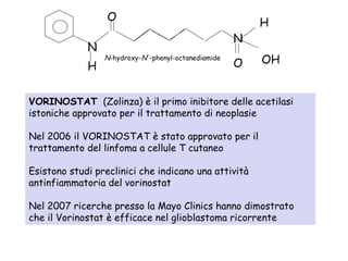
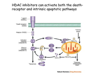
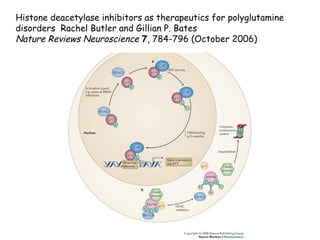
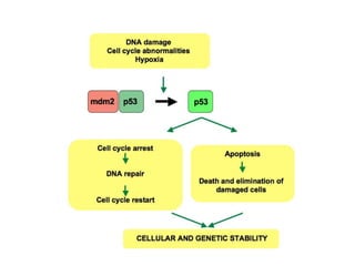
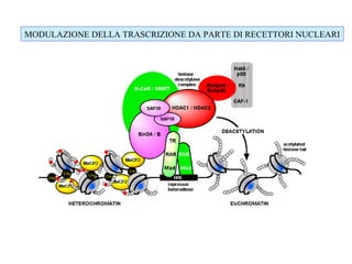
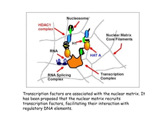
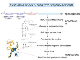
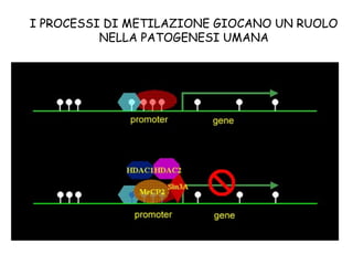
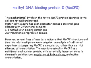
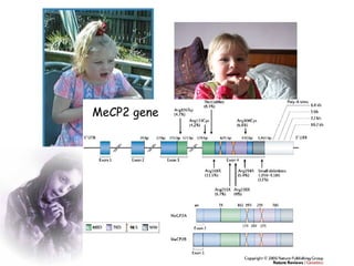
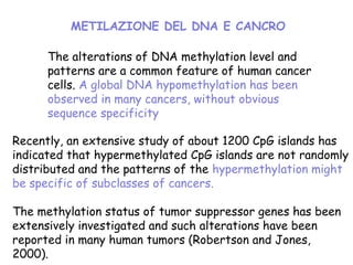
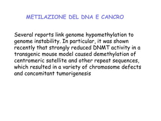
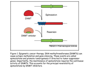
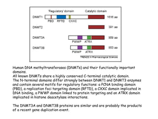
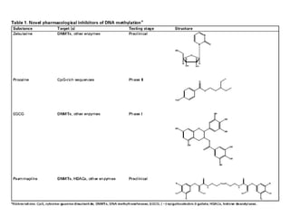
BIOTECNOLOGIE FARMACOLOGICHE
2008/09
LEZIONE 2
CORSO DI LAUREA SPECIALISTICA IN BIOTECNOLOGIE DEL
FARMACO
Adriana Maggi
I PROCESSI DI METILAZIONE GIOCANO UN RUOLO
NELLA PATOGENESI UMANA
The mechanism(s) by which the native MeCP2 protein operates in the
cell are not well understood.
Historically, MeCP2 has been characterized as a proximal gene
silencer with 2 functional domains:
1. a methyl DNA binding domain and
2.a transcription repression domain.
However, several lines of new data indicate that MeCP2 structure and
function relationships are more complex: an analysis of cell-based
experiments suggesting MeCP2 is a regulator, rather than a strict
silencer, of transcription. The new data establish MeCP2 as a
multifunctional nuclear protein, with potentially important roles in
chromatin architecture, regulation of RNA splicing, and active
transcription..
methyl DNA binding protein 2 (MeCP2)
Andreas Rett, che aveva osservato un comportamento insolito, tragico e affascinante insieme,
in alcune bambine sedute nella sala d'aspetto del suo studio. Correva l'anno 1966.
Nel 1999, il team di Huda Zoghbi del Baylor College of Medicine di Houston scoprì che la
sindrome di Rett è associata a una mutazione del gene MeCP2, localizzato sul cromosoma X. Il
gene MeCP2 è un repressore della trascrizione genica.
La proteina MeCP2 è espressa in modo predominante nei neuroni maturi. MeCP2 modula le
connessioni sinaptiche e quindi la comunicazione cellulare attraverso la regolazione di alcuni
geni, tra cui il brain-derived neurotrophic factor (BDNF).
SINDROME DI RETT
Guy J, Gan J, Selfridge J, Cobb S, Bird A. Reversal of neurological defects in
a mouse model of Rett syndrome. Science 2007; 315:1143-1147
Guy et al., con un sistema murino molto sofisticato hanno silenziato la sintesi
della proteina MeCP2 causando la patologia, poi ne hanno reinstaurato la
sintesi con una remissione della sintomatologia
Più recentemente, si è visto che le MeCP2 interagisce con la proteina YB-1
che è coinvolta nella matutazione degli RNA e in particolare della subunità
NR1 dei recettori per il glutammato NMDA
MeCP2 gene
The alterations of DNA methylation level and
patterns are a common feature of human cancer
cells. A global DNA hypomethylation has been
observed in many cancers, without obvious
sequence specificity
Recently, an extensive study of about 1200 CpG islands has
indicated that hypermethylated CpG islands are not randomly
distributed and the patterns of the hypermethylation might
be specific of subclasses of cancers.
The methylation status of tumor suppressor genes has been
extensively investigated and such alterations have been
reported in many human tumors (Robertson and Jones,
2000).
METILAZIONE DEL DNA E CANCRO
Several reports link genome hypomethylation to
genome instability. In particular, it was shown
recently that strongly reduced DNMT activity in a
transgenic mouse model caused demethylation of
centromeric satellite and other repeat sequences,
which resulted in a variety of chromosome defects
and concomitant tumorigenesis
METILAZIONE DEL DNA E CANCRO
Figure 1. Epigenetic cancer therapy. DNA methyltransferases (DNMTs) can
cause ectopic methylation and gene silencing. These events are called
epimutations and promote tumorigenesis if directed to tumor suppressor
genes. Importantly, the maintenance of epimutations requires the continuous
activity of DNMTs. This accounts for the principal reversibility of
epimutations by DNMT inhibitors.
Il mantenimento della metilazione del
DNA (da parte di DNMT1) è
indispensabile nella duplicazione del DNA
per mantenere metilati specifiche porzioni
del DNA.
Nel topo la mutazione di DNMT1 è letale
Human DNA methyltransferases (DNMTs) and their functionally important
domains.
All known DNMTs share a highly conserved C-terminal catalytic domain.
The N-terminal domains differ strongly between DNMT1 and DNMT3 enzymes
and contain several motifs for regulatory functions: a PCNA binding domain
(PBD), a replication foci targeting domain (RFTD), a CXXC domain implicated in
DNA binding, a PWWP domain linked to protein targeting and an ATRX domain
implicated in histone deacetylase interactions.
The DNMT3A and DNMT3B proteins are similar and are probably the products
of a recent gene duplication event.
5-Azacytidine (Vidazae) has been approved
for the treatment of myelodysplastic
syndrome.
This has been a major milestone in the
field of cancer epigenetics and provides an
important validation for the concept of an
epigenetic cancer therapy
Deacetilasi istoniche: una nuova classe di farmaci
nella terapia antitumorale
Generalmente agiscono causando apoptosi di
cellule tumorali,
ma non di cellule non tumorali; le HDAC sono
generalmente associate a fattori di trascrizione
oncogeni.
The effects of HDAC inhibitors on gene expression are highly selective,
leading to transcriptional activation of certain genes such as the cyclin-
dependent kinase inhibitor p21WAF1/CIP1 but repression of others.
HDAC inhibition not only results in acetylation of histones but also
transcription factors such as p53, GATA-1 and estrogen receptor-alpha.
The functional significance of acetylation of non-histone proteins and the
precise mechanisms whereby HDAC inhibitors induce tumor cell growth
arrest, differentiation and/or apoptosis are currently the focus of
intensive research.
Several HDAC inhibitors have shown impressive antitumor activity in vivo
with remarkably little toxicity in preclinical studies and are currently in
phase I clinical trial.
HDACs are classified in four groups based on their homology to
yeast histone deacetylases:
Class I which includes HDAC1, -2, -3 and -8 are related to yeast
RPD3 gene;
Class II which includes HDAC4, -5, -6, -7, -9 and -10 are related
to yeast Hda1 gene;
Class III, also known as the sirtuins are related to the Sir2 gene
and include SIRT1-7, and
Class IV which contains only HDAC11 has features of both Class I
and II.
Classificazione delle deacetilasi istoniche
The “classical” HDIs act exclusively on Class I and Class II HDACs by
binding to the zinc containing catalytic domain of the HDACs. These
classical HDIs fall into several groupings, in order of decreasing
potency:
(i) hyroxamic acids, such as Tricostatine A,
(ii) cyclic tetrapeptides (such as trapoxin B), and the depsipeptides,
(iii) benzamides,
(iv) electrophilic ketones, and
(v) the alophatic acid compounds such as phenylbutyrate and valproic
acid.
"Second generation" HDIs include SAHA/Vorinostat,
Belinostat/PXD101, MS275, LAQ824/LBH589, CI994, and
MGCD0103.
The sirtuin Class III HDACs are NAD+ dependent and are therefore
inhibited by nicotinamides, as well derivatives of NAD,
dihydrocoumarin, naphthopyranone, and 2-hydroxynaphaldehydes.[4]
Classificazione degli inibitori delle istone deacetilasi
VORINOSTAT (Zolinza) è il primo inibitore delle acetilasi
istoniche approvato per il trattamento di neoplasie
Nel 2006 il VORINOSTAT è stato approvato per il
trattamento del linfoma a cellule T cutaneo
Esistono studi preclinici che indicano una attività
antinfiammatoria del vorinostat
Nel 2007 ricerche presso la Mayo Clinics hanno dimostrato
che il Vorinostat è efficace nel glioblastoma ricorrente
N
H
O
OHO
H
N
N-hydroxy-N'-phenyl-octanediamide
HDAC inhibitors can activate both the death-
receptor and intrinsic apoptotic pathways
Histone deacetylase inhibitors as therapeutics for polyglutamine
disorders Rachel Butler and Gillian P. Bates
Nature Reviews Neuroscience 7, 784-796 (October 2006)
P53 puo’ agire a diversi livelli:
Puo’ attivare proteine per il riparo
del DNA
Puo’ regolar e la duplicazione
cellulare a livello G1/S
Puo’ iniziare processi apoptotici
P53
MODULAZIONE DELLA TRASCRIZIONE DA PARTE DI RECETTORI NUCLEARI
Il ruolo degli INSULATORS nella regolazione della
espressione genica
Es. i geni globinici e LCR HS4,
scs di Drosophila che isolano hsp70)
In genere sono sequenze ricche di isole CpG non metilate
Ci sono proteine che legano gli insulators
Transcription factors are associated with the nuclear matrix. It
has been proposed that the nuclear matrix recruits
transcription factors, facilitating their interaction with
regulatory DNA elements.
ESPRESSIONE GENICA IN EUCARIOTE: SEQUENZA DI EVENTI
3’ TRASCRIZIONE
MODIFICAZ.
TP
TRADUZIONE
RNA, trascritto primario
Capping e poliadenilazione
Splicing o maturazione
Fuoriuscita dal nucleo
riconoscimento da parte dei ribosomi
traduzione
Modificazioni post-traduzionali

Sindrome di rett

  • 1.
    BIOTECNOLOGIE FARMACOLOGICHE 2008/09 LEZIONE 2 CORSODI LAUREA SPECIALISTICA IN BIOTECNOLOGIE DEL FARMACO Adriana Maggi
  • 2.
    I PROCESSI DIMETILAZIONE GIOCANO UN RUOLO NELLA PATOGENESI UMANA
  • 3.
    The mechanism(s) bywhich the native MeCP2 protein operates in the cell are not well understood. Historically, MeCP2 has been characterized as a proximal gene silencer with 2 functional domains: 1. a methyl DNA binding domain and 2.a transcription repression domain. However, several lines of new data indicate that MeCP2 structure and function relationships are more complex: an analysis of cell-based experiments suggesting MeCP2 is a regulator, rather than a strict silencer, of transcription. The new data establish MeCP2 as a multifunctional nuclear protein, with potentially important roles in chromatin architecture, regulation of RNA splicing, and active transcription.. methyl DNA binding protein 2 (MeCP2)
  • 4.
    Andreas Rett, cheaveva osservato un comportamento insolito, tragico e affascinante insieme, in alcune bambine sedute nella sala d'aspetto del suo studio. Correva l'anno 1966. Nel 1999, il team di Huda Zoghbi del Baylor College of Medicine di Houston scoprì che la sindrome di Rett è associata a una mutazione del gene MeCP2, localizzato sul cromosoma X. Il gene MeCP2 è un repressore della trascrizione genica. La proteina MeCP2 è espressa in modo predominante nei neuroni maturi. MeCP2 modula le connessioni sinaptiche e quindi la comunicazione cellulare attraverso la regolazione di alcuni geni, tra cui il brain-derived neurotrophic factor (BDNF). SINDROME DI RETT Guy J, Gan J, Selfridge J, Cobb S, Bird A. Reversal of neurological defects in a mouse model of Rett syndrome. Science 2007; 315:1143-1147 Guy et al., con un sistema murino molto sofisticato hanno silenziato la sintesi della proteina MeCP2 causando la patologia, poi ne hanno reinstaurato la sintesi con una remissione della sintomatologia Più recentemente, si è visto che le MeCP2 interagisce con la proteina YB-1 che è coinvolta nella matutazione degli RNA e in particolare della subunità NR1 dei recettori per il glutammato NMDA
  • 5.
  • 6.
    The alterations ofDNA methylation level and patterns are a common feature of human cancer cells. A global DNA hypomethylation has been observed in many cancers, without obvious sequence specificity Recently, an extensive study of about 1200 CpG islands has indicated that hypermethylated CpG islands are not randomly distributed and the patterns of the hypermethylation might be specific of subclasses of cancers. The methylation status of tumor suppressor genes has been extensively investigated and such alterations have been reported in many human tumors (Robertson and Jones, 2000). METILAZIONE DEL DNA E CANCRO
  • 7.
    Several reports linkgenome hypomethylation to genome instability. In particular, it was shown recently that strongly reduced DNMT activity in a transgenic mouse model caused demethylation of centromeric satellite and other repeat sequences, which resulted in a variety of chromosome defects and concomitant tumorigenesis METILAZIONE DEL DNA E CANCRO
  • 8.
    Figure 1. Epigeneticcancer therapy. DNA methyltransferases (DNMTs) can cause ectopic methylation and gene silencing. These events are called epimutations and promote tumorigenesis if directed to tumor suppressor genes. Importantly, the maintenance of epimutations requires the continuous activity of DNMTs. This accounts for the principal reversibility of epimutations by DNMT inhibitors.
  • 9.
    Il mantenimento dellametilazione del DNA (da parte di DNMT1) è indispensabile nella duplicazione del DNA per mantenere metilati specifiche porzioni del DNA. Nel topo la mutazione di DNMT1 è letale
  • 10.
    Human DNA methyltransferases(DNMTs) and their functionally important domains. All known DNMTs share a highly conserved C-terminal catalytic domain. The N-terminal domains differ strongly between DNMT1 and DNMT3 enzymes and contain several motifs for regulatory functions: a PCNA binding domain (PBD), a replication foci targeting domain (RFTD), a CXXC domain implicated in DNA binding, a PWWP domain linked to protein targeting and an ATRX domain implicated in histone deacetylase interactions. The DNMT3A and DNMT3B proteins are similar and are probably the products of a recent gene duplication event.
  • 11.
    5-Azacytidine (Vidazae) hasbeen approved for the treatment of myelodysplastic syndrome. This has been a major milestone in the field of cancer epigenetics and provides an important validation for the concept of an epigenetic cancer therapy
  • 13.
    Deacetilasi istoniche: unanuova classe di farmaci nella terapia antitumorale Generalmente agiscono causando apoptosi di cellule tumorali, ma non di cellule non tumorali; le HDAC sono generalmente associate a fattori di trascrizione oncogeni.
  • 14.
    The effects ofHDAC inhibitors on gene expression are highly selective, leading to transcriptional activation of certain genes such as the cyclin- dependent kinase inhibitor p21WAF1/CIP1 but repression of others. HDAC inhibition not only results in acetylation of histones but also transcription factors such as p53, GATA-1 and estrogen receptor-alpha. The functional significance of acetylation of non-histone proteins and the precise mechanisms whereby HDAC inhibitors induce tumor cell growth arrest, differentiation and/or apoptosis are currently the focus of intensive research. Several HDAC inhibitors have shown impressive antitumor activity in vivo with remarkably little toxicity in preclinical studies and are currently in phase I clinical trial.
  • 15.
    HDACs are classifiedin four groups based on their homology to yeast histone deacetylases: Class I which includes HDAC1, -2, -3 and -8 are related to yeast RPD3 gene; Class II which includes HDAC4, -5, -6, -7, -9 and -10 are related to yeast Hda1 gene; Class III, also known as the sirtuins are related to the Sir2 gene and include SIRT1-7, and Class IV which contains only HDAC11 has features of both Class I and II. Classificazione delle deacetilasi istoniche
  • 16.
    The “classical” HDIsact exclusively on Class I and Class II HDACs by binding to the zinc containing catalytic domain of the HDACs. These classical HDIs fall into several groupings, in order of decreasing potency: (i) hyroxamic acids, such as Tricostatine A, (ii) cyclic tetrapeptides (such as trapoxin B), and the depsipeptides, (iii) benzamides, (iv) electrophilic ketones, and (v) the alophatic acid compounds such as phenylbutyrate and valproic acid. "Second generation" HDIs include SAHA/Vorinostat, Belinostat/PXD101, MS275, LAQ824/LBH589, CI994, and MGCD0103. The sirtuin Class III HDACs are NAD+ dependent and are therefore inhibited by nicotinamides, as well derivatives of NAD, dihydrocoumarin, naphthopyranone, and 2-hydroxynaphaldehydes.[4] Classificazione degli inibitori delle istone deacetilasi
  • 17.
    VORINOSTAT (Zolinza) èil primo inibitore delle acetilasi istoniche approvato per il trattamento di neoplasie Nel 2006 il VORINOSTAT è stato approvato per il trattamento del linfoma a cellule T cutaneo Esistono studi preclinici che indicano una attività antinfiammatoria del vorinostat Nel 2007 ricerche presso la Mayo Clinics hanno dimostrato che il Vorinostat è efficace nel glioblastoma ricorrente N H O OHO H N N-hydroxy-N'-phenyl-octanediamide
  • 18.
    HDAC inhibitors canactivate both the death- receptor and intrinsic apoptotic pathways
  • 19.
    Histone deacetylase inhibitorsas therapeutics for polyglutamine disorders Rachel Butler and Gillian P. Bates Nature Reviews Neuroscience 7, 784-796 (October 2006)
  • 20.
    P53 puo’ agirea diversi livelli: Puo’ attivare proteine per il riparo del DNA Puo’ regolar e la duplicazione cellulare a livello G1/S Puo’ iniziare processi apoptotici P53
  • 22.
    MODULAZIONE DELLA TRASCRIZIONEDA PARTE DI RECETTORI NUCLEARI
  • 23.
    Il ruolo degliINSULATORS nella regolazione della espressione genica Es. i geni globinici e LCR HS4, scs di Drosophila che isolano hsp70) In genere sono sequenze ricche di isole CpG non metilate Ci sono proteine che legano gli insulators
  • 24.
    Transcription factors areassociated with the nuclear matrix. It has been proposed that the nuclear matrix recruits transcription factors, facilitating their interaction with regulatory DNA elements.
  • 25.
    ESPRESSIONE GENICA INEUCARIOTE: SEQUENZA DI EVENTI 3’ TRASCRIZIONE MODIFICAZ. TP TRADUZIONE RNA, trascritto primario Capping e poliadenilazione Splicing o maturazione Fuoriuscita dal nucleo riconoscimento da parte dei ribosomi traduzione Modificazioni post-traduzionali

Editor's Notes

  • #6 a | MECP2 (methyl-CpG-binding protein 2) gene structure. The positions and frequencies of the most common mutations that are associated with Rett syndrome in females are indicated. b | The two main protein isoforms, MeCP2A (486 amino acids) and MeCP2B (498 amino acids), are produced by alternative splicing of the MECP2 transcript and differ in their N-terminal regions, which are encoded by exon 2 of the gene in the case of MeCP2A and exon 1 in MeCP2B. MBD, methyl-CpG binding domain; NLS, nuclear localization signal; poly(A), polyadenylation; TRD, transcriptional repression domain; X, stop codon.
  • #19 HDAC inhibitors can activate both the death-receptor and intrinsic apoptotic pathways. The death-receptor pathway is triggered by the ligation of death receptors, which results in binding of adaptor proteins (FADD) and the recruitment and activation of membrane-proximal activator caspases (caspase-8). These caspases in turn activate downstream effector caspases (caspase-3). The intrinsic apoptotic pathway is activated by internal stresses that induce mitochondrial membrane disruption mediated by pro-apoptotic BCL2 proteins (BAX/BAK). HDAC inhibitors can induce the activation of the intrinsic apoptotic pathway, and anti-apoptotic BCL2 proteins inhibit mitochondrial membrane damage. HDAC inhibitors might still induce cell death in the absence of caspase activation after the production of reactive oxygen species (ROS). Asterix's denote components of the death pathways that are affected by HDAC inhibitors. Those components that inhibit cell death are shown in yellow, and those that promote death are in purple. BAK, BCL2 antagonist/killer protein; BAX, B-cell-associated X protein; BCL2, B-cell lymphoma 2; Cyto c , cytochrome c ; FADD, FAS-associated via death domain; HTRA2, high temperature requirement 2.
  • #20 The function of non-histone proteins is also affected by acetylation (Ac)/deacetylation at specific lysine residues (K). Therefore, histone deacetylase (HDAC) inhibitors could have far reaching effects on multiple cellular processes. Two examples of non-histone acetylated proteins that might be affected by HDAC inhibition and involved in Huntington's disease and other polyglutamine repeat diseases are p53 and heat shock protein 90 (HSP90). a | p53 is acetylated under conditions of stress or HDAC inhibition by its cofactor CREB binding protein (CBP). Acetylation increases p53 activity by enhancing its stability and DNA binding activity. The end result is upregulated expression of p53-controlled genes, such as HTT. b | HSP90 is a chaperone that complexes with other chaperones, such as p23, to maintain correct conformational folding of is client proteins. HDAC6 deacetylates HSP90; inhibition of HDAC6 would result in hyperacetylated HSP90, which would be unable to interact with its co-chaperones and properly fold its client proteins. Inhibition of HDACs in this pathway would therefore lead to misfolded client proteins being targeted for degradation via the ubiquitin–proteasome system. HAT, histone acetyltransferase.