Download to read offline
![International Journal of Biomolecules and Biomedicine
ISSN: 2221-1063 (Print), 2222-503X (Online)
Vol. 20, Issue: 3, p. 1-12, 2025
Website: https://innspub.net
Maurya et al. 7 Int. J. Biomol. Biomed.
Int. J. Biomol. Biomed.
Int. J. Biomol. Biomed.
Int. J. Biomol. Biomed.
inhibitors of both Gram-negative and Gram-positive
bacteria (Bharti et al., 2016; Meher et al., 2024).
Antiviral activity
AgNPs have gained attention for their exceptional
antibacterial properties. Despite their well-
documented antibacterial efficiency, the interaction of
AgNPs with viruses was overlooked until recent
scientific research revealed their intriguing antiviral
activity (Ghosh et al., 2022).
These expanding investigations have demonstrated
AgNPs' potential as powerful antiviral medicines,
particularly against enveloped viruses (Galdiero et al.,
2011). The ease with which enveloped viruses are
spread, their high rates of reproduction and mutation,
and the current absence of comprehensive broad-
spectrum antiviral medicines all contribute to the
necessity for research into AgNP-virus interactions
(Ghosh et al., 2022). The rising threat of enveloped
viruses contributing to imminent pandemics and
biosafety concerns heightens the significance of this
work (Mosidze et al., 2025).
Catheters
Artificial catheters implanted in patients are highly
inclined to contamination which leads to complications.
Catheters made up of polyurethane are coated with
silver nanoparticles for preventing biofilm formation.
The silver nanoparticle-coated catheter is nontoxic and
reduces bacterial growth and helps avoid Catheter-
Associated Ventriculitis (Ahuja et al., 2024).
Orthopaedic implants
The greatest challenge in orthopaedic surgery has
always been bacterial contamination, so to reduce
bacterial resistance, silver nanoparticles (AgNPs)
were used to make the prosthesis. Silver
nanoparticles also began to be used in orthodontic
adhesive for increasing the shear bond strength and
expanding resistance to bacteria(Ahuja et al., 2024).
CONCLUSION
The review covers many synthetic approaches,
including physical, chemical, and environmentally
friendly biological processes. The biological synthesis
is the simplest, quickest, and most cost-effective,
commercial, ecologically friendly, and energy-
efficient method for synthesizing silver nanoparticles.
Chemical methods of producing silver nanoparticles
offer advantages, but they are also dangerous and
environmentally unfriendly.
This review examines the possibilities for mass-
producing silver nanoparticles by a biological
technique. Nanoparticles are believed to have several
biomedical applications. It has a wide range of
medicinal uses, including anti-cancer, antiviral,
antibacterial, and antifungal properties, wound
healing, and anticancer activity. Researchers are now
developing green production methods for silver
nanoparticles, which will be useful for biological
applications. The limitations of standard medical
therapy, as well as the challenges associated with nano-
silver-based technologies, underscore the potential of
silver nanoparticles in biology. Nanostructured
biomaterials and technologies used in modern
biomedicine may come into close contact with AgNPs.
REFERENCES
Adur AJ, Nandini N, Shilpashree Mayachar K,
Ramya R, Srinatha N. 2018. Bio-synthesis and
antimicrobial activity of silver nanoparticles using
anaerobically digested parthenium slurry. Journal of
Photochemistry and Photobiology B: Biology 183, 30-
34. https://doi.org/10.1016/j.jphotobiol.2018.04.020
Ahmad A, Mukherjee P, Senapati S, Mandal D,
Khan MI, Kumar R, Sastry M. 2003.
Extracellular biosynthesis of silver nanoparticles
using the fungus Fusarium oxysporum. Colloids and
Surfaces B: Biointerfaces 28(4), 313-318.
Ahuja P, Rami E, Singh A, Pathak D. 2024.
Green synthesis of silver nanoparticles and their
potential biological applications. In: Nanotechnology
and in silico tools. Elsevier, pp. 97-115. Available from
https://www.sciencedirect.com/science/article/pii/B
9780443154577000228 [accessed 29 November
2024].](https://image.slidesharecdn.com/ijbb-v20-no3-p1-12-251007055010-227373b2/85/Silver-nanoparticles-in-the-biomedical-field-7-320.jpg)


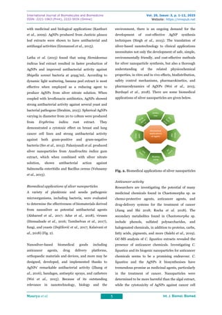
Silver nanoparticles are gaining popularity due to their potential uses in bioengineering and medical diagnosis. Nanoparticles possess specific characteristics, including an enhanced surface-to-volume ratio and improved magnetic properties, making them suitable for various biological applications. They feature several functionalities, a high surface plasmon resonance, a huge surface area, a stable nature, and are simple to produce. Silver nanoparticles have promising uses in biomedical fields such as biomaterials, detection and diagnostics, formulations, medication transport, and medical device coatings. This review covers current research on silver nanoparticles in biomedical applications, including their creation methods, antimicrobial properties, and potential biological uses.


