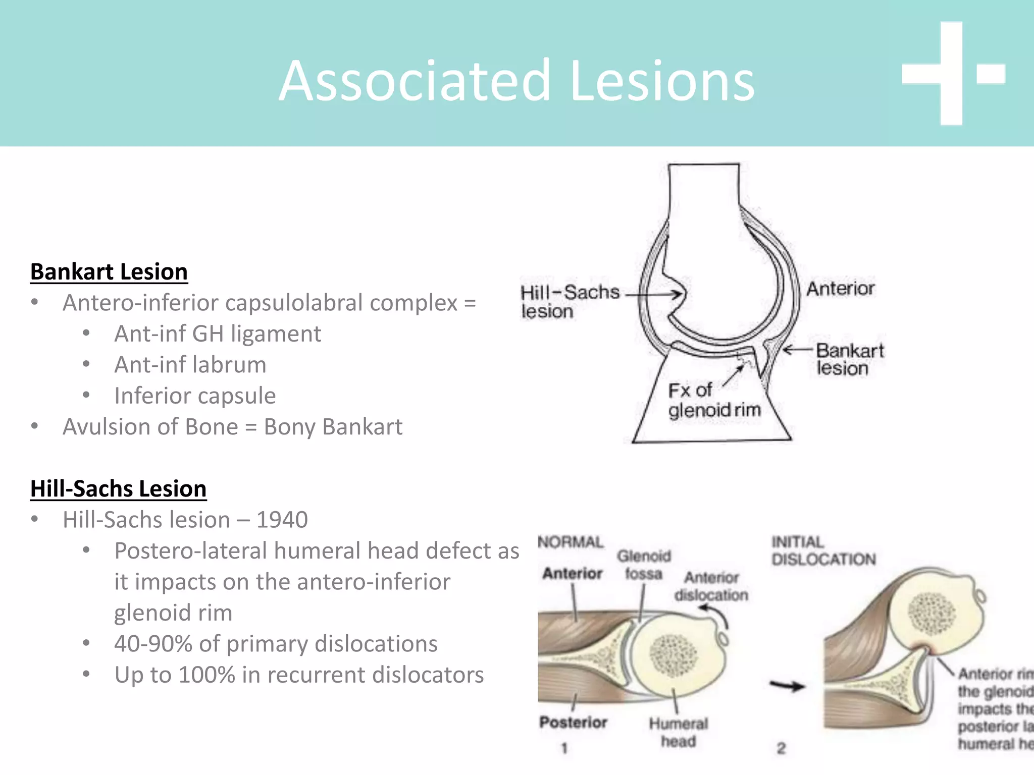
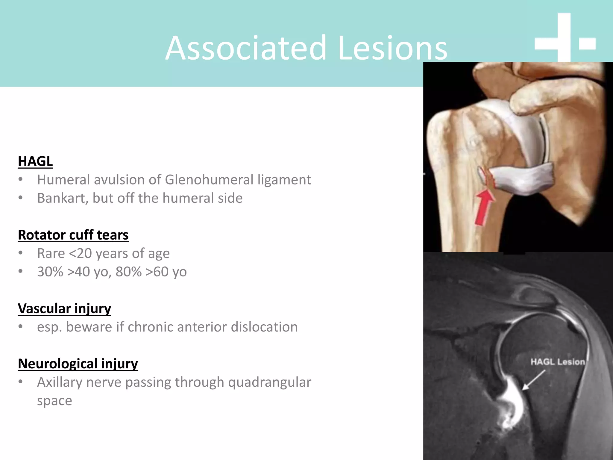
Jarrad Stevens
Orthopaedic Surgeon
drstevens@vbjs.com.aiu
Shoulder Instability
and Ankle Injuries
Dave Slattery
Orthopaedic Surgeon
drslattery@vbjs.com.au
Shoulder Stabilisers
• Shoulder ROM due to 4 articulations
- GH, ST, AC and SC
• Movement mainly from GH and ST
- Abduction approx 2:1 = GH:ST motion
• Humerus: Retroverted 30°
• Glenoid: Retroverted 7°
STABILITY:
• Static and Dynamic stabilisers
• “Concavity - Compression”
• “Suction Cup”
ANATOMY:
Shoulder Stabilisers
STABILITY:
• Glenoid shape
• Cartilage thicker on periphery
• Glenoid deepened by labrum
• GH ligaments and capsule
• These are “check reins” at extremes of
movement
DYNAMIC STABILISERS (Compressors):
• Rotator cuff muscles
• Deltoid
• LHBT – esp against ER in abduction (eg.
throwing)
STATIC STABILISERS
Instability Classification
Chronicity
• Acute, chronic, acute on chronic
Direction
• Anterior, posterior inferior, MDI
Voluntary vs Involuntary
Instability Classification
TUBS
• Traumatic
• Unilateral
• Bankart Lesion
• = Surgery
AMBRI
• Atraumatic
• Multidirection
• Bilateral
• Rehab
• Inferior Capsular Shift
Associated Lesions
Bankart Lesion
• Antero-inferior capsulolabral complex =
• Ant-inf GH ligament
• Ant-inf labrum
• Inferior capsule
• Avulsion of Bone = Bony Bankart
Hill-Sachs Lesion
• Hill-Sachs lesion – 1940
• Postero-lateral humeral head defect as
it impacts on the antero-inferior
glenoid rim
• 40-90% of primary dislocations
• Up to 100% in recurrent dislocators
Associated Lesions
Bankart Lesion
Associated Lesions
HAGL
• Humeral avulsion of Glenohumeral ligament
• Bankart, but off the humeral side
Rotator cuff tears
• Rare <20 years of age
• 30% >40 yo, 80% >60 yo
Vascular injury
• esp. beware if chronic anterior dislocation
Neurological injury
• Axillary nerve passing through quadrangular
space
Typical Presentations
Anterior
• ER, abducted and extended, then force
displaces head anteriorly
Posterior
• axial loading of the IR, Adducted shoulder
OR
• violent muscle contraction (eg.
seizure/electric shock) due to powerful IRs
overcoming weak ERs
Inferior
• Hyperabduction = luxatio erecta
David@melbourneboneandjoint.com
Clinical Examination
Anterior Dislocation
Clinical Examination
Inferior Dislocation
Clinical Examination
Inferior Dislocation
Clinical Examination
Neurovasculature
Clinical Examination
Ligamentous Laxity
• Wynne Davies or Beighton
X Rays
X Rays
Axillary Lateral
X Rays
Anterior Dislocation
X Rays
Anterior Dislocation
X Rays
Anterior Dislocation
X Rays
Posterior Dislocation
X Rays
Posterior Dislocation
X Rays
Posterior Dislocation
X Rays
Posterior Dislocation
X Rays
Inferior Dislocation
Reduction Moves
>200 Described
• Whatever works, without causing more
damage!
Reduction Moves
>200 Described
• Whatever works, without causing more
damage!
• Milch/Stimson/Traction-countertraction
Reduction Moves
Contra-indications to Reduction
• Humeral neck fractures
• Suspected major vessel injury
• Major nerve injury
• Chronic dislocation >48hrs
Post Reduction Treatment
• Confirmation of Reduction
• Immobilisation 1-2/52 – ideally in ER to
tighten S/Scap and reduce bankart lesion
• Aim:
• Restoring function
• Decreasing risk of recurrence
• After immobilisation period, begin RC and
scapular stability exercises to increase
concavity-compression
• Further treatment depends upon risk
profile and imaging – CT/MRI
1st Time Dislocator -
Controversies
• Given reported high risk of recurrence (up
to 92% in active patients) should primary
surgical stabilisation be taking place?
• No clear answer at present, consider if
other factors
• Large HS lesion
• Significant bony bankart ?%
• Persistent displacement of GT #
• Subsequent participation in
contact sports
• Job requires absolute stability
1st Time Dislocator -
Controversies
Ankle Fractures and
Dislocations
WARNING: CONTAINS SURGICAL
IMAGES THAT SOME VIEWERS
MAY FIND
WARNING: CONTAINS SURGICAL
IMAGES THAT SOME VIEWERS
MAY FIND
• 25% of all musculoskeletal
injuries
• 20,000 patients seen each
day in the US
• ATFL injured in isolation 50%
cases
• ATFL + CFL 25% cases
• Isolated AITFL in <5% cases
Ankle Sprains
Ankle Sprains
Ankle Sprains
Ankle Sprains
History
• Female/breastfeeding/pregnant
• Generalised ligamentous laxity
• Cavovarus deformity
• Neurological disorders
• Mechanism of injury
Ankle Sprains
Exam
• Alignment of hindfoot
• Swelling/bruising pattern
• Point tenderness
• Range of motion
• Subtalar joint irritability
• Generalised ligamentous laxity
• Anterior drawer testing
• Syndesmosis stress testing
• Neurovascular status
Ankle Sprains
Imaging
• X-rays – AP, lateral, oblique
weight bearing
• MRI
• Ultrasound unhelpful
Ankle Sprains
Management
• Early mobilisation and
rehabilitation with an
experienced
physiotherapist
• Avoid prolonged non
weight bearing
Ankle Sprains
When to REFER:
• Symptomatic after 3 months
of non-operative treatment
• Intensive rehabilitation
• Compliance
• Functional instability
• Recurrence
• Ligamentous laxity
• Peroneal tendon instability
• Subtle cavovarus foot
Ankle Sprains
When to REFER:
• Symptomatic after 3 months
of non-operative treatment
• Intensive rehabilitation
• Compliance
• Functional instability
• Recurrence
• Ligamentous laxity
• Peroneal tendon instability
• Subtle cavovarus foot
Ankle Sprains
Why not Settling:
• Chronic instability:
– Microinstability
– Gross instability
• Cavovarus foot
• Misdiagnosis
• Concomitant injuries
Ankle Sprains
Surgical Mx:
• Gold standard: anatomic
reconstruction:
– Restore ligaments
– Restore biomechanics
• Brostrom-type
reconstruction – ATFL
imbrication and re-
attachment
• Reinforce with internal brace
device in some patients
• Isolated Medial Malleolar
• Isolated Lateral Malleolar
• Isolated Posterior Malleolar
• Bimalleolar
• Trimalleolar
• Syndesmotic Injuries
• Dislocations
• Plafond/pilon
Ankle Fractures
• Unimalleoli: 70%
• Bilmalleolar: 20%
• Trimalleolar: 7%
• Open: 3%
Incidence
• Cast if undisplaced with no talar shift
• Fix if displaced
• Check the knee x-ray
Isolated Medial
Lateral
Type A: Fracture of the fibula below the level
of the syndesmosis
Type B: Fracture of the fibula at the level of the
syndesmosis
Type C: Fracture of the fibula above the level
of the syndesmosis
Affects joint in all planes
Interosseous disruption
Often associated with posterior fracture
Lateral
• Below the level of syndesmosis
• Generally treat in plaster or CAM 6/52 FWB
Weber A
• At the level of syndesmosis
• ORIF if any talar shift or displacement >2mm
• ORIF is medial structures not intact (functional
bimalleolar fracture)
• Lag screw fixation with neutralising plate
Weber B
Weber B
• Randomised patients to closed contact casting
and ORIF
• PTs >60yrs
• 10% wound complication with ORIF
• 15% malunion rate with CCC
• Have highest incidence of complete
syndesmotic injury
• Plate if fracture in distal third of fibula
• If higher may only require screws if
maisonneuve type injury
Weber C
Syndesmosis
• >25% joint involvement
• >2mm step
• Must use CT to determine as radiographs
inaccurate
• CRIF with AP or PA cannulated screw
• Posterior plate
Posterior Malleolus
Posterior Malleolus
• Any displacement in Bimalleolar and trimalleolar
fractures ORIF
• What about ‘functional bimalleolar’ fracture that
is Weber B and Deltoid rupture:
– examination has been shown to be largely unreliable
in predicting medial injury
– not necessary to repair medial deltoid ligament
– only need to explore medially if you are unable to
reduce the mortise
Bimalleolar
• Assessing the syndesmosis radiographically
Syndesmosis
Radiographic Findings
<6mm
>1mm on Mortise
<4mm
85o
Fracture Dislocation
• Principles in Management
– Early reduction and stabilisation
– Cast / ex-fix or ORIF if swelling appropriate
(usually <6hours or >6 days)
David@melbourneboneandjoint.com
Diabetic Ankle Injuries
• Decision making algorithm the same
• But 6% amputation risk due to infection and
none union
• Rigid fixation
• Sutures in twice as long
• NWB twice as long
Intra-Articular Injuries
• Pilon = Plafond
• High energy axial load
• 75% have associated fibula fractures
Classical Fragments
• Pilon = Plafond
• High energy axial load
• 75% have associated fibula
fractures
• 3 Fragments:
– Volkmann – posterolateral
– Chaput – anterolateral
– Medial
Treatment
• Safe to apply temporising
spanning ex-fix
• Leave until swelling
resolved approx 14 days
• Investigate with CT
Ankle Dislocations
Ankle Dislocations
Treatment
• Safe to apply temporising
spanning ex-fix
• Leave until swelling
resolved approx 14 days
• Investigate with CT
Treatment
Treatment
Treatment
Outcomes
• Want to fix fracture to allow:
– Early ROM
– Early weight bearing
– Restoration of muscle
– Prevention of secondary complications
• High rates of metal problems
• High rates of post traumatic OA
Outcomes
• Want to fix fracture to allow:
– Early ROM
– Early weight bearing
– Restoration of muscle
– Prevention of secondary complications
• High rates of metal problems
• High rates of post traumatic OA

Shoulder and ankle instability