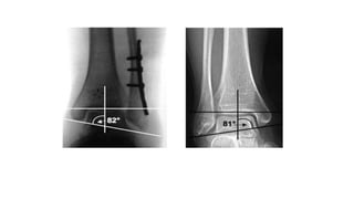
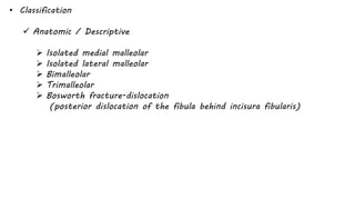
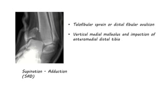
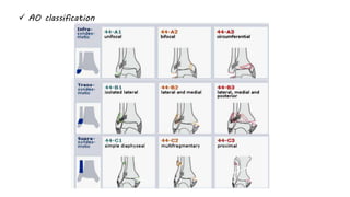
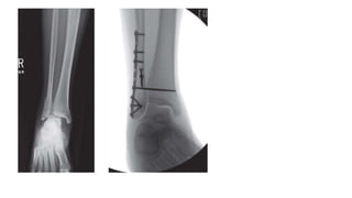
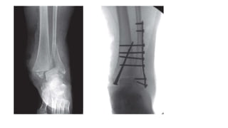
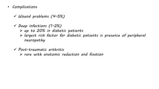
• Injury patterns
 Isolated medial malleolus fracture
 Isolated lateral malleolus fracture
 Bimalleolar fracture
 Posterior malleolus fractures
 Bosworth fracture-dislocations
 Open ankle fractures
 Associated/Isolated syndesmosis injuries
• Radiographs
 AP, Lateral and Obliques
 External rotation stress radiograph
 Most appropriate stress radiograph to assess competency of
deltoid ligament
 A medial clear space of >5mm with external rotation stress
applied to a dorsiflexed ankle is predictive of deep deltoid
disruption
 More sensitive to injury than medial tenderness, ecchymosis, or
edema
• Radiographic measurements
 Talocrural angle
 measured by bisection of line through tibial anatomical axis and
another line through the tips of the malleoli
 shortening of lateral malleoli fractures can lead to increased
talocrural angle
 Talocrural angle is not 100% reliable for estimating restoration of
fibular length
• Classification
 Anatomic / Descriptive
 Isolated medial malleolar
 Isolated lateral malleolar
 Bimalleolar
 Trimalleolar
 Bosworth fracture-dislocation
(posterior dislocation of the fibula behind incisura fibularis)
 Lauge-Hansen
 Based on foot position and force of applied stress/force
 Shown to predict the observed (via MRI) ligamentous
injury in less than 50% of operatively treated fractures
Supination - Adduction
(SAD)
• Talofibular sprain or distal fibular avulsion
• Vertical medial malleolus and impaction of
anteromedial distal tibia
Supination external rotation
(SER)
• Anterior tibiofibular ligament sprain
• Lateral short oblique fibula fracture
(anteroinferior to posterosuperior)
• Posterior tibiofibular ligament rupture
or avulsion of posterior malleolus
• Medial malleolus transverse fracture
or disruption of deltoid ligament
Pronation external rotation
(PER)
• Medial malleolus transverse fracture or
disruption of deltoid ligament
• Anterior tibiofibular ligament disruption
• Lateral short oblique or spiral fracture of
fibula (anterosuperior to posteroinferior)
above the level of the joint
• Posterior tibiofibular ligament rupture or
avulsion of posterior malleolus
Pronation Abduction
(PAB)
• Medial malleolus transverse fracture or disruption
of deltoid ligament
• Anterior tibiofibular ligament sprain
• Transverse comminuted fracture of the fibula
above the level of the syndesmosis
 Denis-Weber (location of fibular fracture)
 A - infrasyndesmotic (generally not associated with ankle instability)
 B - transsyndesmotic
 C – suprasyndesmotic
 AO classification
• General Treatment
 Nonoperative
 Short-leg walking cast/boot
 Isolated nondisplaced medial malleolus fracture or tip avulsions
 Isolated lateral malleolus fracture with < 3mm displacement
and no talar shift
 Posterior malleolar fracture with < 25% joint involvement
or < 2mm step-off
 Operative (Open reduction internal fixation)
 Any talar displacement
 Displaced isolated medial malleolar fracture
 Displaced isolated lateral malleolar fracture
 Bimalleolar fracture and bimalleolar-equivalent fracture
 Posterior malleolar fracture with > 25% or > 2mm step-off
 Bosworth fracture-dislocations
 Open fractures
 Goal of treatment
 stable anatomic reduction of talus in the ankle mortise
 1 mm shift of talus leads to 42% decrease in tibiotalar contact area
 Outcomes
 overall success rate is high
 prolonged recovery expected (2 years to obtain final functional result)
 significant functional impairment often noted
 worse outcomes with: smoking, decreased education, alcohol use,
increased age, presence of medial malleolar fracture
 ORIF superior to closed treatment of bimalleolar fractures
 In Lauge-Hansen supination-adduction fractures, restoration of marginal
impaction of the anteromedial tibial plafond leads to optimal functional
results after surgery
• ISOLATED MEDIAL MALLEOLUS FRACTURE
 Nonoperative (short leg walking cast or cast boot)
 Nondisplaced fracture and tip avulsions
 Operative (ORIF)
 Any displacement or talar shift
 Lag screw fixation (stronger if placed perpendicular to fr line)
 Antiglide plate with lag screw (Best for vertical shear fr)
 Tension band fixation
• ISOLATED LATERAL MALLEOLUS FRACTURE
 Nonoperative (short leg walking cast vs cast boot)
 intact mortise
 no talar shift
 < 3mm displacement
 Classically fractures with more than 4-5 mm of medial clear space
widening on stress radiographs have been considered unstable and
need to be treated surgically
 Recent studies have shown the deep deltoid may be intact with up
to 8-10 mm of widening on stress radiographs
 if the mortise is well reduced, results from operative and non-
operative treatment are similar
 Operative (ORIF)
 if talar shift or > 3 mm of displacement
 can be treated operatively if also treating an ipsilateral syndesmosis
injury
 Open reduction and plating
 plate placement
 lateral
 lag screw fixation with neutralization plating
 bridge plate technique
 posterior
 antiglide technique
 lag screw fixation with neutralization plating
o most common disadvantage of using posterior antiglide plating
is peroneal irritation if the plate is placed too distally
o posterior antiglide plating is biomechanically superior to lateral
plate placement
 intramedullary retrograde screw placement
 Post-operative care
 period of immobilization usually 4-6 weeks after ORIF
 duration of immobilization should be doubled in Diabetic patients
• MEDIAL AND LATERAL (BIMALLEOLAR) FRACTURE
 Operative (ORIF)
 any lateral talar shift
 Technique:
 Fibula (fixed as previously)
 medial malleolus
 cancellous lag screws
 bicortical screws
 tension band wiring
 antiglide plate to treat a vertical medial malleolus
fracture (screws parallel to joint)
• FUNCTIONAL BIMALLEOLAR FRACTURE
(deltoid ligament tear with fibular fracture)
 Operative (ORIF of lateral malleolus)
 can see significant lateral translation of the talus in this pattern
 not necessary to repair medial deltoid ligament
 only need to explore medially if you are unable to reduce the
mortise
• POSTERIOR MALLEOLAR FRACTURE
 Nonoperative (short leg walking cast vs boot)
 < 25% of articular surface involved
(CT rather than Xrays “unreliable”)
 < 2 mm articular stepoff
 syndesmotic stability
 Operative (ORIF)
 > 25% of articular surface involved
 > 2 mm articular stepoff
 syndesmosis injury
 Approach
 posterolateral approach
 posteromedial approach
 Fixation
 anterior to posterior lag screws to capture fragment (if
nondisplaced)
 posterior to anterior lag screw and buttress plate
 antiglide plate
 Syndesmosis injury
 stress examination of syndesmosis still required after posterior
malleolar fixation
 posteroinferior tibiofibular ligament may remain attached to
posterior malleolus and syndesmotic stability may be restored
with isolated posterior malleolar fixation
• BOSWORTH FRACTURE-DISLOCATION
 rare fracture-dislocation of the ankle where the fibula becomes
entrapped behind the tibia and becomes irreducible
 posterolateral ridge of the distal tibia hinders reduction of the fibula
 Operative (ORIF of the fibula in the incisura fibularis)
 indicated in most cases
• OPEN ANKLE FRACTURE
 Operative
 Emergent operative debridement and ORIF
 indicated if soft tissue conditions allow
 External fixation
 According to soft tissue and overall patient conditions.
• ASSOCIATED SYNDESMOTIC INJURY
 suspect injury in all ankle fractures
 most common in Weber C fracture patterns
 up to 25% of tibial shaft fractures will have ankle injury
 Evaluation:
 measure clear space 1 cm above joint
 it has also been reported that there is no actual correlation
between syndesmotic injury and tibiofibular clear space or
overlap measurements
 best option is to assess stability intraoperatively with
abduction/external rotation stress of dorsiflexed foot
 instability of the syndesmosis is greatest in the anterior-
posterior direction
 lateral stress radiograph has more interobserver reliability than
an AP/mortise stress film
 Treatment (operative: syndesmotic screw fixation)
 widening of medial clear space
 tibiofibular clear space (AP) greater than 5 mm
 tibiofibular overlap (mortise) narrowed
 any postreduction malalignment or widening should be treated with
open debridement, reduction, and fixation
 Technique
 length and rotation of fibula must be accurately restored.
 one or two cortical screw(s) 2-4 cm above joint, angled posterior
to anterior 20-30 degrees
 lag technique not desired
 maximum dorsiflexion of ankle not required during screw placement
(can't overtighten a properly reduced syndesmosis)
 Postoperative
 screws should be maintained in place for at least 8-12 weeks
 must remain non-weight bearing, as screws are not biomechanically
strong enough to withstand forces of ambulation
 Controversies
 number of screws
o 1 or 2 no difference
 number of cortices
o 3 or 4 no difference
 size of screws
o 3.5 mm or 4.5 mm screws
 implant material
o (stainless steel screws, titanium screws, suture, bioabsorbable
materials)
 need for hardware removal
o no difference in outcomes seen with hardware maintenance
(breakage or loosening) or removal at 1 year
• Complications
 Wound problems (4-5%)
 Deep infections (1-2%)
 up to 20% in diabetic patients
 largest risk factor for diabetic patients is presence of peripheral
neuropathy
 Post-traumatic arthritis
 rare with anatomic reduction and fixation
Bony-ankle-injuries.pptx

Bony-ankle-injuries.pptx

  • 3.
    • Injury patterns Isolated medial malleolus fracture  Isolated lateral malleolus fracture  Bimalleolar fracture  Posterior malleolus fractures  Bosworth fracture-dislocations  Open ankle fractures  Associated/Isolated syndesmosis injuries
  • 4.
    • Radiographs  AP,Lateral and Obliques  External rotation stress radiograph  Most appropriate stress radiograph to assess competency of deltoid ligament  A medial clear space of >5mm with external rotation stress applied to a dorsiflexed ankle is predictive of deep deltoid disruption  More sensitive to injury than medial tenderness, ecchymosis, or edema
  • 5.
    • Radiographic measurements Talocrural angle  measured by bisection of line through tibial anatomical axis and another line through the tips of the malleoli  shortening of lateral malleoli fractures can lead to increased talocrural angle  Talocrural angle is not 100% reliable for estimating restoration of fibular length
  • 7.
    • Classification  Anatomic/ Descriptive  Isolated medial malleolar  Isolated lateral malleolar  Bimalleolar  Trimalleolar  Bosworth fracture-dislocation (posterior dislocation of the fibula behind incisura fibularis)
  • 8.
     Lauge-Hansen  Basedon foot position and force of applied stress/force  Shown to predict the observed (via MRI) ligamentous injury in less than 50% of operatively treated fractures
  • 9.
    Supination - Adduction (SAD) •Talofibular sprain or distal fibular avulsion • Vertical medial malleolus and impaction of anteromedial distal tibia
  • 10.
    Supination external rotation (SER) •Anterior tibiofibular ligament sprain • Lateral short oblique fibula fracture (anteroinferior to posterosuperior) • Posterior tibiofibular ligament rupture or avulsion of posterior malleolus • Medial malleolus transverse fracture or disruption of deltoid ligament
  • 11.
    Pronation external rotation (PER) •Medial malleolus transverse fracture or disruption of deltoid ligament • Anterior tibiofibular ligament disruption • Lateral short oblique or spiral fracture of fibula (anterosuperior to posteroinferior) above the level of the joint • Posterior tibiofibular ligament rupture or avulsion of posterior malleolus
  • 12.
    Pronation Abduction (PAB) • Medialmalleolus transverse fracture or disruption of deltoid ligament • Anterior tibiofibular ligament sprain • Transverse comminuted fracture of the fibula above the level of the syndesmosis
  • 13.
     Denis-Weber (locationof fibular fracture)  A - infrasyndesmotic (generally not associated with ankle instability)  B - transsyndesmotic  C – suprasyndesmotic
  • 14.
  • 15.
    • General Treatment Nonoperative  Short-leg walking cast/boot  Isolated nondisplaced medial malleolus fracture or tip avulsions  Isolated lateral malleolus fracture with < 3mm displacement and no talar shift  Posterior malleolar fracture with < 25% joint involvement or < 2mm step-off
  • 16.
     Operative (Openreduction internal fixation)  Any talar displacement  Displaced isolated medial malleolar fracture  Displaced isolated lateral malleolar fracture  Bimalleolar fracture and bimalleolar-equivalent fracture  Posterior malleolar fracture with > 25% or > 2mm step-off  Bosworth fracture-dislocations  Open fractures  Goal of treatment  stable anatomic reduction of talus in the ankle mortise  1 mm shift of talus leads to 42% decrease in tibiotalar contact area
  • 17.
     Outcomes  overallsuccess rate is high  prolonged recovery expected (2 years to obtain final functional result)  significant functional impairment often noted  worse outcomes with: smoking, decreased education, alcohol use, increased age, presence of medial malleolar fracture  ORIF superior to closed treatment of bimalleolar fractures  In Lauge-Hansen supination-adduction fractures, restoration of marginal impaction of the anteromedial tibial plafond leads to optimal functional results after surgery
  • 18.
    • ISOLATED MEDIALMALLEOLUS FRACTURE  Nonoperative (short leg walking cast or cast boot)  Nondisplaced fracture and tip avulsions  Operative (ORIF)  Any displacement or talar shift  Lag screw fixation (stronger if placed perpendicular to fr line)  Antiglide plate with lag screw (Best for vertical shear fr)  Tension band fixation
  • 20.
    • ISOLATED LATERALMALLEOLUS FRACTURE  Nonoperative (short leg walking cast vs cast boot)  intact mortise  no talar shift  < 3mm displacement  Classically fractures with more than 4-5 mm of medial clear space widening on stress radiographs have been considered unstable and need to be treated surgically  Recent studies have shown the deep deltoid may be intact with up to 8-10 mm of widening on stress radiographs  if the mortise is well reduced, results from operative and non- operative treatment are similar
  • 21.
     Operative (ORIF) if talar shift or > 3 mm of displacement  can be treated operatively if also treating an ipsilateral syndesmosis injury  Open reduction and plating  plate placement  lateral  lag screw fixation with neutralization plating  bridge plate technique
  • 22.
     posterior  antiglidetechnique  lag screw fixation with neutralization plating o most common disadvantage of using posterior antiglide plating is peroneal irritation if the plate is placed too distally o posterior antiglide plating is biomechanically superior to lateral plate placement  intramedullary retrograde screw placement
  • 24.
     Post-operative care period of immobilization usually 4-6 weeks after ORIF  duration of immobilization should be doubled in Diabetic patients
  • 25.
    • MEDIAL ANDLATERAL (BIMALLEOLAR) FRACTURE  Operative (ORIF)  any lateral talar shift  Technique:  Fibula (fixed as previously)  medial malleolus  cancellous lag screws  bicortical screws  tension band wiring  antiglide plate to treat a vertical medial malleolus fracture (screws parallel to joint)
  • 26.
    • FUNCTIONAL BIMALLEOLARFRACTURE (deltoid ligament tear with fibular fracture)  Operative (ORIF of lateral malleolus)  can see significant lateral translation of the talus in this pattern  not necessary to repair medial deltoid ligament  only need to explore medially if you are unable to reduce the mortise
  • 28.
    • POSTERIOR MALLEOLARFRACTURE  Nonoperative (short leg walking cast vs boot)  < 25% of articular surface involved (CT rather than Xrays “unreliable”)  < 2 mm articular stepoff  syndesmotic stability  Operative (ORIF)  > 25% of articular surface involved  > 2 mm articular stepoff  syndesmosis injury
  • 29.
     Approach  posterolateralapproach  posteromedial approach  Fixation  anterior to posterior lag screws to capture fragment (if nondisplaced)  posterior to anterior lag screw and buttress plate  antiglide plate
  • 30.
     Syndesmosis injury stress examination of syndesmosis still required after posterior malleolar fixation  posteroinferior tibiofibular ligament may remain attached to posterior malleolus and syndesmotic stability may be restored with isolated posterior malleolar fixation
  • 31.
    • BOSWORTH FRACTURE-DISLOCATION rare fracture-dislocation of the ankle where the fibula becomes entrapped behind the tibia and becomes irreducible  posterolateral ridge of the distal tibia hinders reduction of the fibula  Operative (ORIF of the fibula in the incisura fibularis)  indicated in most cases
  • 33.
    • OPEN ANKLEFRACTURE  Operative  Emergent operative debridement and ORIF  indicated if soft tissue conditions allow  External fixation  According to soft tissue and overall patient conditions.
  • 34.
    • ASSOCIATED SYNDESMOTICINJURY  suspect injury in all ankle fractures  most common in Weber C fracture patterns  up to 25% of tibial shaft fractures will have ankle injury
  • 35.
     Evaluation:  measureclear space 1 cm above joint  it has also been reported that there is no actual correlation between syndesmotic injury and tibiofibular clear space or overlap measurements  best option is to assess stability intraoperatively with abduction/external rotation stress of dorsiflexed foot  instability of the syndesmosis is greatest in the anterior- posterior direction  lateral stress radiograph has more interobserver reliability than an AP/mortise stress film
  • 36.
     Treatment (operative:syndesmotic screw fixation)  widening of medial clear space  tibiofibular clear space (AP) greater than 5 mm  tibiofibular overlap (mortise) narrowed  any postreduction malalignment or widening should be treated with open debridement, reduction, and fixation
  • 37.
     Technique  lengthand rotation of fibula must be accurately restored.  one or two cortical screw(s) 2-4 cm above joint, angled posterior to anterior 20-30 degrees  lag technique not desired  maximum dorsiflexion of ankle not required during screw placement (can't overtighten a properly reduced syndesmosis)
  • 40.
     Postoperative  screwsshould be maintained in place for at least 8-12 weeks  must remain non-weight bearing, as screws are not biomechanically strong enough to withstand forces of ambulation  Controversies  number of screws o 1 or 2 no difference  number of cortices o 3 or 4 no difference  size of screws o 3.5 mm or 4.5 mm screws  implant material o (stainless steel screws, titanium screws, suture, bioabsorbable materials)  need for hardware removal o no difference in outcomes seen with hardware maintenance (breakage or loosening) or removal at 1 year
  • 41.
    • Complications  Woundproblems (4-5%)  Deep infections (1-2%)  up to 20% in diabetic patients  largest risk factor for diabetic patients is presence of peripheral neuropathy  Post-traumatic arthritis  rare with anatomic reduction and fixation