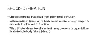
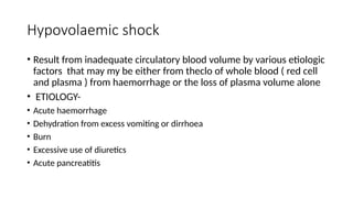
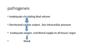
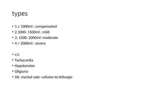
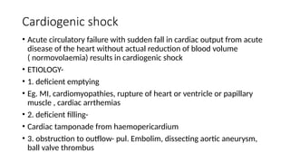
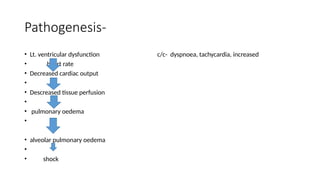
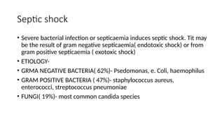
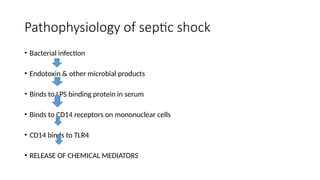
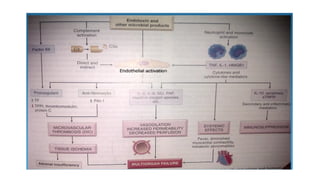
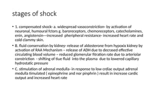
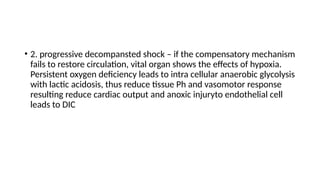
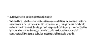
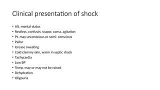
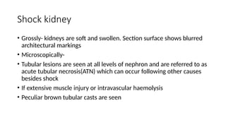
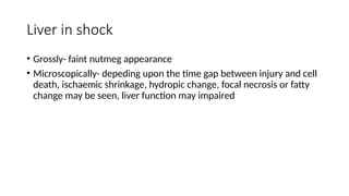
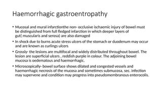
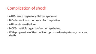
shock
DR. ARCHANA NIKAM
DEPT. OF PATHOLOGY AND MICROBIOLOGY
SHOCK- DEFINATION
• Clinical syndrome that result from poor tissue perfusion
• In this condition tissue in the body do not receive enough oxygen &
nutrients to allow cell to function.
• This ultimately leads to cellular death may progress to organ failure
finally to hole body failure ( death)
classification
• 1. hypovaemic shock
• 2. cardiogenic shock
• 3.septic ( toxaemic ) nshock
• 4. other type-
• a.traumatic hock
• B.neurogenic shock
• C. hypoadrenal shock
Hypovolaemic shock
• Result from inadequate circulatory blood volume by various etiologic
factors that may my be either from theclo of whole blood ( red cell
and plasma ) from haemorrhage or the loss of plasma volume alone
• ETIOLOGY-
• Acute haemorrhage
• Dehydration from excess vomiting or dirrhoea
• Burn
• Excessive use of diuretics
• Acute pancreatitis
pathogeneis
• Inadequate circulating blod volume
• Decrteased cardiac output , low intracardiac pressure
• inadequate oxygen, nutritional supply to all tissue/ organ
• Shock
types
• 1.≤ 1000ml : compensated
• 2.1000- 1500ml : mild
• 3. 1500- 2000ml: moderate
• 4.> 2000ml : severe
• c/c
• Tachycardia
• Hypotension
• Oligiuria
• Alt. mental sate- cofuion to lethargic
Cardiogenic shock
• Acute circulatory failure with sudden fall in cardiac output from acute
disease of the heart without actual reduction of blood volume
( normovolaemia) results in cardiogenic shock
• ETIOLOGY-
• 1. deficient emptying
• Eg. MI, cardiomyopathies, rupture of heart or ventricle or papillary
muscle , cardiac arrthemias
• 2. deficient filling-
• Cardiac tamponade from haemopericardium
• 3. obstruction to outflow- pul. Embolim, dissecting aortic aneurysm,
ball valve thrombus
Pathogenesis-
• Lt. ventricular dysfunction c/c- dyspnoea, tachycardia, increased
• heart rate
• Decreased cardiac output
•
• Descreased tissue perfusion
•
• pulmonary oedema
•
• alveolar pulmonary oedema
•
• shock
Septic shock
• Severe bacterial infection or septicaemia induces septic shock. Tit may
be the result of gram negative septicaemia( endotoxic shock) or from
gram positive septicaemia ( exotoxic shock)
• ETIOLOGY-
• GRMA NEGATIVE BACTERIA( 62%)- Psedomonas, e. Coli, haemophilus
• GRAM POSITIVE BACTERIA ( 47%)- staphylococcus aureus,
enterococci, streptococcus pneumoniae
• FUNGI( 19%)- most common candida species
Pathophysiology of septic shock
• Bacterial infection
• Endotoxin & other microbial products
• Binds to LPS binding protein in serum
• Binds to CD14 receptors on mononuclear cells
• CD14 binds to TLR4
• RELEASE OF CHEMICAL MEDIATORS
Pathophysiology of septic shock
• In septic shock systemic vasodilatation and pooling of blood in the
periphery leads to tissue hypoperfusion and though the cardiac
output is preserved this is accompanying widespread endothelial cell
actvation and often leads to hypercoagulable state that can manifest
DIC .
• Septic shock is associated with change in metabolism that can
directly suppress cellular function. The net is hypo- perfusion and
dysfunction of multi- organ.
• Major factors contributing to the pathophysiology of septic shock are following
• Inflammatory mediators:
• Various microbe cell wall contains LPS( - tive bact.)AND LTG( +tive bact.
( lipotheichoic acid)) these are released bind to free LPS& LTG
• this complex then to cell surface receptor CD14 on macrophage, mononuclear
phagocytic cell. Endothelial cell
• CD14 binds to the signal transducing protein TLR-4 UPON ACTIVATION
CASCADE OF CYTOKINES MEDIATORS TNF, TL-1 , IL-8, INF.
• The compliment cascade are actvated directly or through proteolytic activity of
plasmin resulting in production of anaphylotoxins c3a & c5a, chemotactic
factor c5a causes vasodilatation& increased vascular permeability. Activated
endothelial cell and macrophages release NO contribute to pro- inflammatory
state
• Endothelial cell activation and injury:
• Endothelial cell activation by microbial products or inflammatory
mediators produced by leukocytes. Has 3 major sequel- thrombosis,
increase vascular permeability, vasodilatation.
• Pro- inflammatory cytokines result in increased tissue factor
production by endothelial cell and monocyte the production of
endothelial anti- coagulant factor –tissue factor pathway inhibitor,
thrombomodulin and protein c reduced. Reduced blood flow at the
level of small vesssels produce stasis that reduce the wash out
activated thrombi in small vessels, that lead to tissue hypoperfusion.
• In DIC increase consumption of coagulation factor & platelet leads to
bleeding
• Increase vascular permeability leads exudation of fluid in to
interstitium causing oedema
• Endothelium increase the release of NO
• Metabolic abnormalities: septis pt. exhibit insulin resistance and
hypoglycemia. Cytokines such as TNF & IL-1, stress induced hormone
(glucagon, catecholamine) all drive gluconeogenesis. At the same
time the pro inflammatory cytokines suppress insulin release while
simultenously promoting insulin resistance in the liver and other
tissue, by surface expression of glucose transporter .
• Hyperglycemia reduced neutrophil function & bactericidal activity.
Sepsis also related with adrenal insufficiency & functional deficiencyof
glucacordicoid.
• Immunosupression:
• Hyperinflammatory state activate immunosuppression mechanism
which involves both innate & adaptive immunity by production of
anti- inflammatory mediators ( soluble TNF receptors, IL-receptor
antagonist & IL -10)
• Organ dysfunction
• Systemic hypotension , interstitial oedema, small vessel thrombosis
reduce the delivery of oxygen & to the tisue
• High level of cytokines , sec. mediators reduces myocardial
contractility & CO. increase vascular permeability & endothelial injury
acute respitory distress syndrome ultimately cause multiple organ
failure.
Traumatic shock
• Resulting from trauma initially due to hypovolaemia, but afterwads
suffer from plasma volume loss
ETIOLOGY-
severe inury, surgery with marked blood loss, obstretrical trauma
Neurogenic shock
• Results from causes of interruption of ympathetic vasomotor supply.
• ETIOLOGY-
• High cervical spinal cord injury, accidental high spinal anaesthesia,
severe head injury
Hypoadrenal shock
• Unknown adrenal insufficiency in which pt. fails respond to normally
stress of vtrauma, surgery , illness
• ETIOLOGY- administration of high doses of glucocorticoids
• sec,. Adrenal insufficiency( in tb)
stages of shock
• 1. compensated shock- a. widespread vasoconstriction- by activation of
neuronal, humoural fctors g. baroreceptors, chemoreceptors, catecholamines,
enin, angiotensin---increased pheripheral resistance- increased heart rate and
cold clammy skin.
• B. fluid conservation by kidney- release of aldosterone from hypoxix kidney by
actvation of RAA Mechanism – release of ADH due to decrased effective
circulating blood volume – reduced glomerular fitration rate due to arteriolar
constriction - shifting of tiue fluid into the plasma due to lowered capillary
hydrostatic pressure
• C. stimulation of adrenal medulla- in response to low crdiac output adrenal
medulla timulated ( epinephrine and nor pinphrin ) result in increase cardic
output and increased heart rate
• 2. progressive decompansted shock – if the compensatory mechanism
fails to restore circulation, vital organ shows the effects of hypoxia.
Persistent oxygen deficiency leads to intra cellular anaerobic glycolysis
with lactic acidosis, thus reduce tissue Ph and vasomotor response
resulting reduce cardiac output and anoxic injuryto endothelial cell
leads to DIC
• 3.irreversible decompensated shock –
• When thre is failure to restoration o circulation by compensatory
mechanism or by therapeutic intervention, the process of shock
enters the irreversible stage. Widespread cell injury is reflected in
lyssomal enzyme leakage , nitric oxide reduced myocardial
contractability, acute tubular necrosis ultimately death.
Clinical presentation of shock
• Alt. mental status
• Restless, confusin, stupor, coma, agitation
• Pt. may unconscious or semi- conscious
• Pallor
• Icrease sweating
• Cold clammy skin, warm in septic shock
• Tachycardia
• Low BP
• Temp. may or may not be raised
• Dehydration
• Oligouria
Morphology of shock
Characteristic feature is multisystem failure
Due to prolonged hypoxia resulting in degeneration and necrosis in
various organs.
Basicaly major organ affected are the brain, heart, lungs, and kidney.
Morphologic changes also noticed in the adrenal gland, git, liver, and
other organ.
Hypoxic encephalopathy
• Occurs Due to cerebral ischaemia followedby alt. state of
consciousness.
• BP falls 50mmhg causes Hypotension in prolonged shock and cardiac arrest,
brain get suffers from ischaemic damage with loss of cortical functions, coma
and a vegetative state
• Gross- severe ischaemic necrosis esp. at border zone between the ant. And
middle cerebral arteries
• Microscopically- noticeable changes occurs if ischaemia prolonged for 12 to
24 hrs. neurons, particularly purkinje cells, are more prone to develop the
effects of ischaemia. The cytoplasmof the affected neurons is intensely
eosinophilic and nucleus small pyknotic. Dead and dying nerve cells are
replaced by gliosis
Heart in shock
• More vulnerable to the effects of hypoxia
• Heart is affected in cardiogenic as well as in other forms of shock.
There are 2 types of morphologic changes in heart in all types of
shock
• 1) haemorrhage and necrosis- there may be small or large ischaemic
areas or infarct, particularly located in the subepicardial and
subendocardial region.
• 2) zonal lesion- these are opaque transverse contraction bands in the
myocytes near the intercalated disc
Shock lung
• Lungs due to dual blood supply are generally not affected by
hypovolaemic shock but septic shock the morphologic changes in
lungs are quite prominent termed shock lung
• Gross- lungs are heavy and wet
• Microscopically- ards brifly changes include congestion , interstitial
and alveolar oedema, interstitial lymphocytic infiltrate, alveolar
hyaline membranes, thickening and fibrosis of alveolar septa, and
fibrin and platelet thrombi in the pulmonary microvasculature.
Shock kidney
• Grossly- kidneys are soft and swollen. Section surface shows blurred
architectural markings
• Microscopically-
• Tubular lesions are seen at all levels of nephron and are referred to as
acute tubular necrosis(ATN) which can occur following other causes
besides shock
• If extensive muscle injury or intravascular haemolysis
• Peculiar brown tubular casts are seen
Liver in shock
• Grossly- faint nutmeg appearance
• Microscopically- depeding upon the time gap between injury and cell
death, ischaemic shrinkage, hydropic change, focal necrosis or fatty
change may be seen, liver function may impaired
Haemorrhagic gastroentropathy
• Mucosal and mural infarctionthe non- occlusive ischaemic injury of bowel must
be distinguished from full fledged infarction in which deeper layers of
gut( muscularis and serosa) are also damaged
• In shock due to burns acute stress ulcers of the stomach or duodenum may occur
and are known as curlings ulcers
• Grossly- the lesions are multifocal and widely distributed throughout bowel. The
lesion are superficial ulcers , reddish purple in colour. The adjoining bowel
mucosa is oedematous and haemorrhagic.
• Microscopically- bowel surface shows dilated and congested vessels and
haemorrhagic necrosis of the mucosa and sometimes submucosa, sec. infection
may supervene and condition may progress into pseudomembranous enterocolis.
Complication of shock
• ARDS- acute respiratory distress syndrome
• DIC- desseminated intravascular coagulation
• ARF- acute renal failure
• MODS- multiple organ dysfunction syndrome.
• With progression of the condition , pt. may develop stupor, coma, and
death.
Adrenals in shock
• The adrenals show stress response in shock. This includes release of
aldosterone in response to hypoxic kidney, relase of glucocorticoids
from adrenal cortex and catecholamines like adrenaline from adrenal
medulla. In severe shock , acute adrenal haemorrhagic necrosis may
occur.

shock & types ,pathogenesis of shock.pptx

  • 1.
    shock DR. ARCHANA NIKAM DEPT.OF PATHOLOGY AND MICROBIOLOGY
  • 2.
    SHOCK- DEFINATION • Clinicalsyndrome that result from poor tissue perfusion • In this condition tissue in the body do not receive enough oxygen & nutrients to allow cell to function. • This ultimately leads to cellular death may progress to organ failure finally to hole body failure ( death)
  • 3.
    classification • 1. hypovaemicshock • 2. cardiogenic shock • 3.septic ( toxaemic ) nshock • 4. other type- • a.traumatic hock • B.neurogenic shock • C. hypoadrenal shock
  • 4.
    Hypovolaemic shock • Resultfrom inadequate circulatory blood volume by various etiologic factors that may my be either from theclo of whole blood ( red cell and plasma ) from haemorrhage or the loss of plasma volume alone • ETIOLOGY- • Acute haemorrhage • Dehydration from excess vomiting or dirrhoea • Burn • Excessive use of diuretics • Acute pancreatitis
  • 5.
    pathogeneis • Inadequate circulatingblod volume • Decrteased cardiac output , low intracardiac pressure • inadequate oxygen, nutritional supply to all tissue/ organ • Shock
  • 6.
    types • 1.≤ 1000ml: compensated • 2.1000- 1500ml : mild • 3. 1500- 2000ml: moderate • 4.> 2000ml : severe • c/c • Tachycardia • Hypotension • Oligiuria • Alt. mental sate- cofuion to lethargic
  • 7.
    Cardiogenic shock • Acutecirculatory failure with sudden fall in cardiac output from acute disease of the heart without actual reduction of blood volume ( normovolaemia) results in cardiogenic shock • ETIOLOGY- • 1. deficient emptying • Eg. MI, cardiomyopathies, rupture of heart or ventricle or papillary muscle , cardiac arrthemias • 2. deficient filling- • Cardiac tamponade from haemopericardium • 3. obstruction to outflow- pul. Embolim, dissecting aortic aneurysm, ball valve thrombus
  • 8.
    Pathogenesis- • Lt. ventriculardysfunction c/c- dyspnoea, tachycardia, increased • heart rate • Decreased cardiac output • • Descreased tissue perfusion • • pulmonary oedema • • alveolar pulmonary oedema • • shock
  • 9.
    Septic shock • Severebacterial infection or septicaemia induces septic shock. Tit may be the result of gram negative septicaemia( endotoxic shock) or from gram positive septicaemia ( exotoxic shock) • ETIOLOGY- • GRMA NEGATIVE BACTERIA( 62%)- Psedomonas, e. Coli, haemophilus • GRAM POSITIVE BACTERIA ( 47%)- staphylococcus aureus, enterococci, streptococcus pneumoniae • FUNGI( 19%)- most common candida species
  • 10.
    Pathophysiology of septicshock • Bacterial infection • Endotoxin & other microbial products • Binds to LPS binding protein in serum • Binds to CD14 receptors on mononuclear cells • CD14 binds to TLR4 • RELEASE OF CHEMICAL MEDIATORS
  • 12.
    Pathophysiology of septicshock • In septic shock systemic vasodilatation and pooling of blood in the periphery leads to tissue hypoperfusion and though the cardiac output is preserved this is accompanying widespread endothelial cell actvation and often leads to hypercoagulable state that can manifest DIC . • Septic shock is associated with change in metabolism that can directly suppress cellular function. The net is hypo- perfusion and dysfunction of multi- organ.
  • 13.
    • Major factorscontributing to the pathophysiology of septic shock are following • Inflammatory mediators: • Various microbe cell wall contains LPS( - tive bact.)AND LTG( +tive bact. ( lipotheichoic acid)) these are released bind to free LPS& LTG • this complex then to cell surface receptor CD14 on macrophage, mononuclear phagocytic cell. Endothelial cell • CD14 binds to the signal transducing protein TLR-4 UPON ACTIVATION CASCADE OF CYTOKINES MEDIATORS TNF, TL-1 , IL-8, INF. • The compliment cascade are actvated directly or through proteolytic activity of plasmin resulting in production of anaphylotoxins c3a & c5a, chemotactic factor c5a causes vasodilatation& increased vascular permeability. Activated endothelial cell and macrophages release NO contribute to pro- inflammatory state
  • 14.
    • Endothelial cellactivation and injury: • Endothelial cell activation by microbial products or inflammatory mediators produced by leukocytes. Has 3 major sequel- thrombosis, increase vascular permeability, vasodilatation. • Pro- inflammatory cytokines result in increased tissue factor production by endothelial cell and monocyte the production of endothelial anti- coagulant factor –tissue factor pathway inhibitor, thrombomodulin and protein c reduced. Reduced blood flow at the level of small vesssels produce stasis that reduce the wash out activated thrombi in small vessels, that lead to tissue hypoperfusion.
  • 15.
    • In DICincrease consumption of coagulation factor & platelet leads to bleeding • Increase vascular permeability leads exudation of fluid in to interstitium causing oedema • Endothelium increase the release of NO
  • 16.
    • Metabolic abnormalities:septis pt. exhibit insulin resistance and hypoglycemia. Cytokines such as TNF & IL-1, stress induced hormone (glucagon, catecholamine) all drive gluconeogenesis. At the same time the pro inflammatory cytokines suppress insulin release while simultenously promoting insulin resistance in the liver and other tissue, by surface expression of glucose transporter . • Hyperglycemia reduced neutrophil function & bactericidal activity. Sepsis also related with adrenal insufficiency & functional deficiencyof glucacordicoid.
  • 17.
    • Immunosupression: • Hyperinflammatorystate activate immunosuppression mechanism which involves both innate & adaptive immunity by production of anti- inflammatory mediators ( soluble TNF receptors, IL-receptor antagonist & IL -10) • Organ dysfunction • Systemic hypotension , interstitial oedema, small vessel thrombosis reduce the delivery of oxygen & to the tisue
  • 18.
    • High levelof cytokines , sec. mediators reduces myocardial contractility & CO. increase vascular permeability & endothelial injury acute respitory distress syndrome ultimately cause multiple organ failure.
  • 19.
    Traumatic shock • Resultingfrom trauma initially due to hypovolaemia, but afterwads suffer from plasma volume loss ETIOLOGY- severe inury, surgery with marked blood loss, obstretrical trauma
  • 20.
    Neurogenic shock • Resultsfrom causes of interruption of ympathetic vasomotor supply. • ETIOLOGY- • High cervical spinal cord injury, accidental high spinal anaesthesia, severe head injury
  • 21.
    Hypoadrenal shock • Unknownadrenal insufficiency in which pt. fails respond to normally stress of vtrauma, surgery , illness • ETIOLOGY- administration of high doses of glucocorticoids • sec,. Adrenal insufficiency( in tb)
  • 22.
    stages of shock •1. compensated shock- a. widespread vasoconstriction- by activation of neuronal, humoural fctors g. baroreceptors, chemoreceptors, catecholamines, enin, angiotensin---increased pheripheral resistance- increased heart rate and cold clammy skin. • B. fluid conservation by kidney- release of aldosterone from hypoxix kidney by actvation of RAA Mechanism – release of ADH due to decrased effective circulating blood volume – reduced glomerular fitration rate due to arteriolar constriction - shifting of tiue fluid into the plasma due to lowered capillary hydrostatic pressure • C. stimulation of adrenal medulla- in response to low crdiac output adrenal medulla timulated ( epinephrine and nor pinphrin ) result in increase cardic output and increased heart rate
  • 23.
    • 2. progressivedecompansted shock – if the compensatory mechanism fails to restore circulation, vital organ shows the effects of hypoxia. Persistent oxygen deficiency leads to intra cellular anaerobic glycolysis with lactic acidosis, thus reduce tissue Ph and vasomotor response resulting reduce cardiac output and anoxic injuryto endothelial cell leads to DIC
  • 24.
    • 3.irreversible decompensatedshock – • When thre is failure to restoration o circulation by compensatory mechanism or by therapeutic intervention, the process of shock enters the irreversible stage. Widespread cell injury is reflected in lyssomal enzyme leakage , nitric oxide reduced myocardial contractability, acute tubular necrosis ultimately death.
  • 25.
    Clinical presentation ofshock • Alt. mental status • Restless, confusin, stupor, coma, agitation • Pt. may unconscious or semi- conscious • Pallor • Icrease sweating • Cold clammy skin, warm in septic shock • Tachycardia • Low BP • Temp. may or may not be raised • Dehydration • Oligouria
  • 26.
    Morphology of shock Characteristicfeature is multisystem failure Due to prolonged hypoxia resulting in degeneration and necrosis in various organs. Basicaly major organ affected are the brain, heart, lungs, and kidney. Morphologic changes also noticed in the adrenal gland, git, liver, and other organ.
  • 27.
    Hypoxic encephalopathy • OccursDue to cerebral ischaemia followedby alt. state of consciousness. • BP falls 50mmhg causes Hypotension in prolonged shock and cardiac arrest, brain get suffers from ischaemic damage with loss of cortical functions, coma and a vegetative state • Gross- severe ischaemic necrosis esp. at border zone between the ant. And middle cerebral arteries • Microscopically- noticeable changes occurs if ischaemia prolonged for 12 to 24 hrs. neurons, particularly purkinje cells, are more prone to develop the effects of ischaemia. The cytoplasmof the affected neurons is intensely eosinophilic and nucleus small pyknotic. Dead and dying nerve cells are replaced by gliosis
  • 28.
    Heart in shock •More vulnerable to the effects of hypoxia • Heart is affected in cardiogenic as well as in other forms of shock. There are 2 types of morphologic changes in heart in all types of shock • 1) haemorrhage and necrosis- there may be small or large ischaemic areas or infarct, particularly located in the subepicardial and subendocardial region. • 2) zonal lesion- these are opaque transverse contraction bands in the myocytes near the intercalated disc
  • 29.
    Shock lung • Lungsdue to dual blood supply are generally not affected by hypovolaemic shock but septic shock the morphologic changes in lungs are quite prominent termed shock lung • Gross- lungs are heavy and wet • Microscopically- ards brifly changes include congestion , interstitial and alveolar oedema, interstitial lymphocytic infiltrate, alveolar hyaline membranes, thickening and fibrosis of alveolar septa, and fibrin and platelet thrombi in the pulmonary microvasculature.
  • 30.
    Shock kidney • Grossly-kidneys are soft and swollen. Section surface shows blurred architectural markings • Microscopically- • Tubular lesions are seen at all levels of nephron and are referred to as acute tubular necrosis(ATN) which can occur following other causes besides shock • If extensive muscle injury or intravascular haemolysis • Peculiar brown tubular casts are seen
  • 31.
    Liver in shock •Grossly- faint nutmeg appearance • Microscopically- depeding upon the time gap between injury and cell death, ischaemic shrinkage, hydropic change, focal necrosis or fatty change may be seen, liver function may impaired
  • 32.
    Haemorrhagic gastroentropathy • Mucosaland mural infarctionthe non- occlusive ischaemic injury of bowel must be distinguished from full fledged infarction in which deeper layers of gut( muscularis and serosa) are also damaged • In shock due to burns acute stress ulcers of the stomach or duodenum may occur and are known as curlings ulcers • Grossly- the lesions are multifocal and widely distributed throughout bowel. The lesion are superficial ulcers , reddish purple in colour. The adjoining bowel mucosa is oedematous and haemorrhagic. • Microscopically- bowel surface shows dilated and congested vessels and haemorrhagic necrosis of the mucosa and sometimes submucosa, sec. infection may supervene and condition may progress into pseudomembranous enterocolis.
  • 33.
    Complication of shock •ARDS- acute respiratory distress syndrome • DIC- desseminated intravascular coagulation • ARF- acute renal failure • MODS- multiple organ dysfunction syndrome. • With progression of the condition , pt. may develop stupor, coma, and death.
  • 34.
    Adrenals in shock •The adrenals show stress response in shock. This includes release of aldosterone in response to hypoxic kidney, relase of glucocorticoids from adrenal cortex and catecholamines like adrenaline from adrenal medulla. In severe shock , acute adrenal haemorrhagic necrosis may occur.