
Recommended
More Related Content
Similar to SERIES ‘‘ATSERS TASK FORCE STANDARDISATION OF LUNGFUNCTION.docx
Similar to SERIES ‘‘ATSERS TASK FORCE STANDARDISATION OF LUNGFUNCTION.docx (20)
More from klinda1
More from klinda1 (20)
Recently uploaded
Recently uploaded (20)
SERIES ‘‘ATSERS TASK FORCE STANDARDISATION OF LUNGFUNCTION.docx
- 1. SERIES ‘‘ATS/ERS TASK FORCE: STANDARDISATION OF LUNG FUNCTION TESTING’’ Edited by V. Brusasco, R. Crapo and G. Viegi Number 2 in this Series Standardisation of spirometry M.R. Miller, J. Hankinson, V. Brusasco, F. Burgos, R. Casaburi, A. Coates, R. Crapo, P. Enright, C.P.M. van der Grinten, P. Gustafsson, R. Jensen, D.C. Johnson, N. MacIntyre, R. McKay, D. Navajas, O.F. Pedersen, R. Pellegrino, G. Viegi and J. Wanger CONTENTS Background . . . . . . . . . . . . . . . . . . . . . . . . . . . . . . . . . . . . . . . . . . . . . . . . . . . . . . . . . . . . . . . 320 FEV1 and FVC manoeuvre . . . . . . . . . . . . . . . . . . . . . . . . . . . . . . . . . . . . . . . . . . . . . . . . . . . . 321 Definitions . . . . . . . . . . . . . . . . . . . . . . . . . . . . . . . . . . . . . . . . . . . . . . . . . . . . . . . . . . . . . . . 321 Equipment . . . . . . . . . . . . . . . . . . . . . . . . . . . . . . . . . . . . . . . . . . . . . . . . . . . . . . . . . . . . . . . 321 Requirements . . . . . . . . . . . . . . . . . . . . . . . . . . . . . . . . . . . . . . . . . . . . . . . . . . . . . . . . . . . 321
- 2. Display . . . . . . . . . . . . . . . . . . . . . . . . . . . . . . . . . . . . . . . . . . . . . . . . . . . . . . . . . . . . . . . . 321 Validation . . . . . . . . . . . . . . . . . . . . . . . . . . . . . . . . . . . . . . . . . . . . . . . . . . . . . . . . . . . . . . 322 Quality control . . . . . . . . . . . . . . . . . . . . . . . . . . . . . . . . . . . . . . . . . . . . . . . . . . . . . . . . . . . 322 Quality control for volume-measuring devices . . . . . . . . . . . . . . . . . . . . . . . . . . . . . . . . . . . 322 Quality control for flow-measuring devices . . . . . . . . . . . . . . . . . . . . . . . . . . . . . . . . . . . . . 323 Test procedure . . . . . . . . . . . . . . . . . . . . . . . . . . . . . . . . . . . . . . . . . . . . . . . . . . . . . . . . . . . . 323 Within-manoeuvre evaluation . . . . . . . . . . . . . . . . . . . . . . . . . . . . . . . . . . . . . . . . . . . . . . . . . . 324 Start of test criteria. . . . . . . . . . . . . . . . . . . . . . . . . . . . . . . . . . . . . . . . . . . . . . . . . . . . . . . . 324 End of test criteria . . . . . . . . . . . . . . . . . . . . . . . . . . . . . . . . . . . . . . . . . . . . . . . . . . . . . . . . 324 Additional criteria . . . . . . . . . . . . . . . . . . . . . . . . . . . . . . . . . . . . . . . . . . . . . . . . . . . . . . . . . 324 Summary of acceptable blow criteria . . . . . . . . . . . . . . . . . . . . . . . . . . . . . . . . . . . . . . . . . . . 325 Between-manoeuvre evaluation . . . . . . . . . . . . . . . . . . . . . . . . . . . . . . . . . . . . . . . . . . . . . . . . 325
- 3. Manoeuvre repeatability . . . . . . . . . . . . . . . . . . . . . . . . . . . . . . . . . . . . . . . . . . . . . . . . . . . . 325 Maximum number of manoeuvres . . . . . . . . . . . . . . . . . . . . . . . . . . . . . . . . . . . . . . . . . . . . . 326 Test result selection . . . . . . . . . . . . . . . . . . . . . . . . . . . . . . . . . . . . . . . . . . . . . . . . . . . . . . . . 326 Other derived indices . . . . . . . . . . . . . . . . . . . . . . . . . . . . . . . . . . . . . . . . . . . . . . . . . . . . . . . 326 FEVt . . . . . . . . . . . . . . . . . . . . . . . . . . . . . . . . . . . . . . . . . . . . . . . . . . . . . . . . . . . . . . . . . . 326 Standardisation of FEV1 for expired volume, FEV1/FVC and FEV1/VC . . . . . . . . . . . . . . . . . . . . 326 FEF25–75% . . . . . . . . . . . . . . . . . . . . . . . . . . . . . . . . . . . . . . . . . . . . . . . . . . . . . . . . . . . . . . 326 PEF . . . . . . . . . . . . . . . . . . . . . . . . . . . . . . . . . . . . . . . . . . . . . . . . . . . . . . . . . . . . . . . . . . 326 Maximal expiratory flow–volume loops . . . . . . . . . . . . . . . . . . . . . . . . . . . . . . . . . . . . . . . . . . . 326 Definitions . . . . . . . . . . . . . . . . . . . . . . . . . . . . . . . . . . . . . . . . . . . . . . . . . . . . . . . . . . . . . . 326 Equipment . . . . . . . . . . . . . . . . . . . . . . . . . . . . . . . . . . . . . . . . . . . . . . . . . . . . . . . . . . . . . 327 Test procedure . . . . . . . . . . . . . . . . . . . . . . . . . . . . . . . . . . . . . . . . . . . . . . . . . . . . . . . . . . 327
- 4. Within- and between-manoeuvre evaluation . . . . . . . . . . . . . . . . . . . . . . . . . . . . . . . . . . . . . . 327 Flow–volume loop examples . . . . . . . . . . . . . . . . . . . . . . . . . . . . . . . . . . . . . . . . . . . . . . . . . 327 Reversibility testing . . . . . . . . . . . . . . . . . . . . . . . . . . . . . . . . . . . . . . . . . . . . . . . . . . . . . . . . . 327 Method . . . . . . . . . . . . . . . . . . . . . . . . . . . . . . . . . . . . . . . . . . . . . . . . . . . . . . . . . . . . . . . . 327 Comment on dose and delivery method . . . . . . . . . . . . . . . . . . . . . . . . . . . . . . . . . . . . . . . . 328 Determination of reversibility . . . . . . . . . . . . . . . . . . . . . . . . . . . . . . . . . . . . . . . . . . . . . . . . . 328 VC and IC manoeuvre . . . . . . . . . . . . . . . . . . . . . . . . . . . . . . . . . . . . . . . . . . . . . . . . . . . . . . . 329 Definitions . . . . . . . . . . . . . . . . . . . . . . . . . . . . . . . . . . . . . . . . . . . . . . . . . . . . . . . . . . . . . . . 329 AFFILIATIONS For affiliations, please see Acknowledgements section CORRESPONDENCE V. Brusasco Internal Medicine
- 5. University of Genoa V.le Benedetto XV, 6 I-16132 Genova Italy Fax: 39 103537690 E-mail: [email protected] Received: March 23 2005 Accepted after revision: April 05 2005 European Respiratory Journal Print ISSN 0903-1936 Online ISSN 1399-3003 Previous articles in this series: No. 1: Miller MR, Crapo R, Hankinson J, et al. General considerations for lung function testing. Eur Respir J 2005; 26: 153–161. EUROPEAN RESPIRATORY JOURNAL VOLUME 26 NUMBER 2 319 Eur Respir J 2005; 26: 319–338
- 6. DOI: 10.1183/09031936.05.00034805 Copyright�ERS Journals Ltd 2005 c VC and IVC . . . . . . . . . . . . . . . . . . . . . . . . . . . . . . . .329 IC . . . . . . . . . . . . . . . . . . . . . . . . . . . . . . . . . . . . . . .329 Equipment . . . . . . . . . . . . . . . . . . . . . . . . . . . . . . . . . .329 Test procedure . . . . . . . . . . . . . . . . . . . . . . . . . . . . . . .329 VC . . . . . . . . . . . . . . . . . . . . . . . . . . . . . . . . . . . . . .329 IC . . . . . . . . . . . . . . . . . . . . . . . . . . . . . . . . . . . . . . .330 Use of a nose clip . . . . . . . . . . . . . . . . . . . . . . . . . . .330 Within-manoeuvre evaluation . . . . . . . . . . . . . . . . . . . . .330 Between-manoeuvre evaluation . . . . . . . . . . . . . . . . . . .330 Test result selection . . . . . . . . . . . . . . . . . . . . . . . . . . .330 Peak expiratory flow . . . . . . . . . . . . . . . . . . . . . . . . . . .330 Definition . . . . . . . . . . . . . . . . . . . . . . . . . . . . . . . . . . .330 Equipment . . . . . . . . . . . . . . . . . . . . . . . . . . . . . . . . . .330 Test procedure . . . . . . . . . . . . . . . . . . . . . . . . . . . . . . .330
- 7. Within-manoeuvre evaluation . . . . . . . . . . . . . . . . . . . . .331 Between-manoeuvre evaluation . . . . . . . . . . . . . . . . . . .331 Test result selection . . . . . . . . . . . . . . . . . . . . . . . . . . .331 Maximum voluntary ventilation . . . . . . . . . . . . . . . . . . .331 Definition . . . . . . . . . . . . . . . . . . . . . . . . . . . . . . . . . . .331 Equipment . . . . . . . . . . . . . . . . . . . . . . . . . . . . . . . . . .331 Test procedure . . . . . . . . . . . . . . . . . . . . . . . . . . . . . . .331 Within-manoeuvre evaluation . . . . . . . . . . . . . . . . . . . . .331 Between-manoeuvre evaluation . . . . . . . . . . . . . . . . . . .331 Test result selection . . . . . . . . . . . . . . . . . . . . . . . . . . .331 Technical considerations . . . . . . . . . . . . . . . . . . . . . . . .331 Minimal recommendations for spirometry systems . . . . . .331 BTPS correction . . . . . . . . . . . . . . . . . . . . . . . . . . . . . .332 Comments . . . . . . . . . . . . . . . . . . . . . . . . . . . . . . . .332 Test signals for spirometer testing . . . . . . . . . . . . . . . . .333 Method . . . . . . . . . . . . . . . . . . . . . . . . . . . . . . . . . . .333 Accuracy test . . . . . . . . . . . . . . . . . . . . . . . . . . . . . .333 Repeatability test . . . . . . . . . . . . . . . . . . . . . . . . . . . .333
- 8. Test signals for PEF meter testing . . . . . . . . . . . . . . . . .333 Method . . . . . . . . . . . . . . . . . . . . . . . . . . . . . . . . . . .333 Accuracy test . . . . . . . . . . . . . . . . . . . . . . . . . . . . . .333 Repeatability test . . . . . . . . . . . . . . . . . . . . . . . . . . . .334 Test signals for MVV testing . . . . . . . . . . . . . . . . . . . . . .334 Abbreviations. . . . . . . . . . . . . . . . . . . . . . . . . . . . . . . . .334 Appendix . . . . . . . . . . . . . . . . . . . . . . . . . . . . . . . . . . . .335 KEYWORDS: Peak expiratory flow, spirometry, spirometry standardisation, spirometry technique, spirometry traning, ventilation BACKGROUND Spirometry is a physiological test that measures how an individual inhales or exhales volumes of air as a function of time. The primary signal measured in spirometry may be volume or flow. Spirometry is invaluable as a screening test of general respiratory health in the same way that blood pressure provides important information about general cardiovascular health. However, on its own, spirometry does not lead clinicians directly to an aetiological diagnosis. Some indica- tions for spirometry are given in table 1. In this document, the most important aspects of spirometry are the forced vital capacity (FVC), which is the volume delivered during an expiration made as forcefully and completely as possible starting from full inspiration, and the forced expira-
- 9. tory volume (FEV) in one second, which is the volume delivered in the first second of an FVC manoeuvre. Other spirometric variables derived from the FVC manoeuvre are also addressed. Spirometry can be undertaken with many different types of equipment, and requires cooperation between the subject and the examiner, and the results obtained will depend on technical as well as personal factors (fig. 1). If the variability of the results can be diminished and the measurement accuracy can be improved, the range of normal values for populations can be narrowed and abnormalities more easily detected. The Snowbird workshop held in 1979 resulted in the first American Thoracic Society (ATS) statement on the standardisation of spirometry [1]. This was updated in 1987 and again in 1994 [2, 3]. A similar initiative was undertaken by the European Community for Steel and Coal, resulting in the first European standardisation document in 1983 [4]. This was then updated in 1993 as the official statement of the European Respiratory Society (ERS) [5]. There are generally only minor differences between the two most recent ATS and ERS statements, except that the ERS statement includes absolute lung volumes and the ATS does not. This document brings the views of the ATS and ERS together in an attempt to publish standards that can be applied more TABLE 1 Indications for spirometry Diagnostic To evaluate symptoms, signs or abnormal laboratory tests To measure the effect of disease on pulmonary function
- 10. To screen individuals at risk of having pulmonary disease To assess pre-operative risk To assess prognosis To assess health status before beginning strenuous physical activity programmes Monitoring To assess therapeutic intervention To describe the course of diseases that affect lung function To monitor people exposed to injurious agents To monitor for adverse reactions to drugs with known pulmonary toxicity Disability/impairment evaluations To assess patients as part of a rehabilitation programme To assess risks as part of an insurance evaluation To assess individuals for legal reasons Public health Epidemiological surveys Derivation of reference equations
- 11. Clinical research STANDARDISATION OF SPIROMETRY M.R. MILLER ET AL. 320 VOLUME 26 NUMBER 2 EUROPEAN RESPIRATORY JOURNAL widely. The statement is structured to cover definitions, equipment and patient-related procedures. All recording devices covered by this statement must meet the relevant requirements, regardless of whether they are for monitoring or diagnostic purposes. There is no separate category for ‘‘monitoring’’ devices. Although manufacturers have the responsibility for producing pulmonary function testing systems that satisfy all the recommendations presented here, it is possible that, for some equipment, meeting all of them may not always be achievable. In these circumstances, manufacturers should clearly identify which equipment requirements have not been met. While manufacturers are responsible for demonstrating the accuracy and reliability of the systems that they sell, it is the user who is responsible for ensuring that the equipment’s measurements remain accurate. The user is also responsible for following local law, which may have additional requirements. Finally, these guidelines are minimum guidelines, which may not be sufficient for all settings, such as when conducting research, epidemiological studies, longitudinal evaluations and occupa- tional surveillance. FEV1 AND FVC MANOEUVRE Definitions FVC is the maximal volume of air exhaled with maximally
- 12. forced effort from a maximal inspiration, i.e. vital capacity performed with a maximally forced expiratory effort, expressed in litres at body temperature and ambient pressure saturated with water vapour (BTPS; see BTPS correction section). FEV1 is the maximal volume of air exhaled in the first second of a forced expiration from a position of full inspiration, expressed in litres at BTPS. Equipment Requirements The spirometer must be capable of accumulating volume for o15 s (longer times are recommended) and measuring volumes of o8 L (BTPS) with an accuracy of at least ¡3% of reading or ¡0.050 L, whichever is greater, with flows between 0 and 14 L?s-1. The total resistance to airflow at 14.0 L?s-1 must be ,1.5 cmH2O?L -1 ?s-1 (0.15 kPa?L-1?s-1; see Minimal recommen- dations for spirometry systems section). The total resistance must be measured with any tubing, valves, pre-filter, etc. included that may be inserted between the subject and the spirometer. Some devices may exhibit changes in resistance due to water vapour condensation, and accuracy requirements must be met under BTPS conditions for up to eight successive FVC manoeuvres performed in a 10-min period without inspiration from the instrument. Display For optimal quality control, both flow–volume and volume–
- 13. time displays are useful, and test operators should visually inspect the performance of each manoeuvre for quality assurance before proceeding with another manoeuvre. This inspection requires tracings to meet the minimum size and resolution requirements set forth in this standard. Displays of flow versus volume provide more detail for the initial portion (first 1 s) of the FVC manoeuvre. Since this portion of the manoeuvre, particularly the peak expiratory flow (PEF), is correlated with the pleural pressure during the manoeuvre, the flow–volume display is useful to assess the magnitude of effort during the initial portions of the manoeuvre. The ability to overlay a series of flow–volume curves registered at the point of maximal inhalation may be helpful in evaluating repeatability and detecting sub- maximal efforts. However, if the point of maximal inhala- tion varies between blows, then the interpretation of these results is difficult because the flows at identical measured volumes are being achieved at different absolute lung volumes. In contrast, display of the FVC manoeuvre as a volume–time graph provides more detail for the latter part of the manoeuvre. A volume–time tracing of sufficient size also allows independent measurement and calculation of parameters from the FVC manoeuvres. In a display of multiple trials, the sequencing of the blows should be apparent to the user. For the start of test display, the volume–time display should include o0.25 s, and preferably 1 s, before exhalation starts (zero volume). This time period before there is any change in volume is needed to calculate the back extrapolated volume (EV; see Start of test criteria section) and to evaluate effort during the initial portion of the manoeuvre. Time zero, as defined by EV, must be presented as the zero point on the graphical output.
- 14. The last 2 s of the manoeuvre should be displayed to indicate a satisfactory end of test (see End of test criteria section). When a volume–time curve is plotted as hardcopy, the volume scale must be o10 mm?L-1 (BTPS). For a screen display, 5 mm?L-1 is satisfactory (table 2). The time scale should be o20 mm?s-1, and larger time scales are preferred (o30 mm?s-1) when manual measure- ments are made [1, 6, 7]. When the volume–time plot is used in conjunction with a flow–volume curve (i.e. both display methods are provided for interpretations and no hand �������� ���� ������ ��� ���� �������� ������ � � ����� � � � � � ������ ��� ��� ��� ������ ���������� �� ������� ����� ����� �
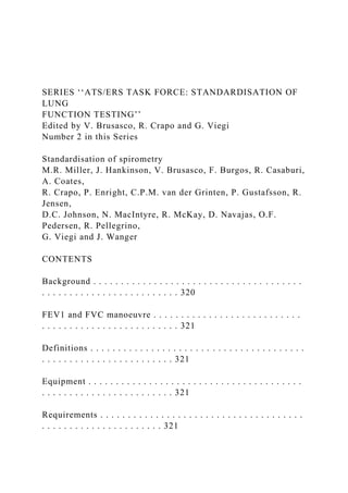
- 15. ����� ����� � ��������� �������� ����� � � � �������� ��������� ����� � ��������� �������� �� ������ FIGURE 1. Spirometry standardisation steps. M.R. MILLER ET AL. STANDARDISATION OF SPIROMETRY c EUROPEAN RESPIRATORY JOURNAL VOLUME 26 NUMBER 2 321 measurements are performed), the time scale requirement is reduced to 10 mm?s-1 from the usually required minimum of 20 mm?s-1 (table 2). The rationale for this exception is that the flow–volume curve can provide the means for quality assessment during the initial portion of the FVC manoeuvre. The volume–time curve can be used to evaluate the latter part of the FVC manoeuvre, making the time scale less critical.
- 16. Validation It is strongly recommended that spirometry systems should be evaluated using a computer-driven mechanical syringe or its equivalent, in order to test the range of exhalations that are likely to be encountered in the test population. Testing the performance of equipment is not part of the usual laboratory procedures (see Test signals for spirometer testing section). Quality control Attention to equipment quality control and calibration is an important part of good laboratory practice. At a minimum, the requirements are as follows: 1) a log of calibration results is maintained; 2) the documentation of repairs or other alterations which return the equipment to acceptable opera- tion; 3) the dates of computer software and hardware updates or changes; and 4) if equipment is changed or relocated (e.g. industrial surveys), calibration checks and quality-control procedures must be repeated before further testing begins. Key aspects of equipment quality control are summarised in table 3. Calibration is the procedure for establishing the relationship between sensor-determined values of flow or volume and the actual flow or volume. A calibration check is different from calibration and is the procedure used to validate that the device is within calibration limits, e.g. ¡3% of true. If a device fails its calibration check, then a new calibration procedure or equipment maintenance is required. Calibration checks must be undertaken daily, or more frequently, if specified by the manufacturer. The syringe used to check the volume calibration of spiro- meters must have an accuracy of ¡15 mL or ¡0.5% of the full
- 17. scale (15 mL for a 3-L syringe), and the manufacturer must provide recommendations concerning appropriate intervals between syringe calibration checks. Users should be aware that a syringe with an adjustable or variable stop may be out of calibration if the stop is reset or accidentally moved. Calibration syringes should be periodically (e.g. monthly) leak tested at more than one volume up to their maximum; this can be done by attempting to empty them with the outlet corked. A dropped or damaged syringe should be considered out of calibration until it is checked. With regard to time, assessing mechanical recorder time scale accuracy with a stopwatch must be performed at least quarterly. An accuracy of within 2% must be achieved. Quality control for volume-measuring devices The volume accuracy of the spirometer must be checked at least daily, with a single discharge of a 3-L calibrated syringe. Daily calibration checking is highly recommended so that the onset of a problem can be determined within 1 day, and also to help define day-to-day laboratory variability. More frequent checks may be required in special circumstances, such as: 1) during industrial surveys or other studies in which a large number of subject manoeuvres are carried out, the equipment’s calibration should be checked more frequently than daily [8]; and 2) when the ambient temperature is changing (e.g. field studies), volume accuracy must be checked more frequently than daily and the BTPS correction factor appropriately updated. The accuracy of the syringe volume must be considered in determining whether the measured volume is within accep- table limits. For example, if the syringe has an accuracy of 0.5%, a reading of ¡3.5% is appropriate. The calibration syringe should be stored and used in such a
- 18. way as to maintain the same temperature and humidity of the testing site. This is best accomplished by keeping the syringe in close proximity to the spirometer, but out of direct sunlight and away from heat sources. Volume-type spirometer systems must be evaluated for leaks every day [9, 10]. The importance of undertaking this daily test cannot be overstressed. Leaks can be detected by applying a constant positive pressure of o3.0 cmH2O (0.3 kPa) with the spirometer outlet occluded (preferably at or including the mouthpiece). Any observed volume loss .30 mL after 1 min indicates a leak [9, 10] and needs to be corrected. TABLE 2 Recommended minimum scale factors for time, volume and flow on graphical output Instrument display Hardcopy graphical outputParameter Resolution required Scale factor Resolution required Scale factor Volume# 0.050 L 5 mm?L-1 0.025 L 10 mm?L-1
- 19. Flow# 0.200 L?s-1 2.5 mm?L-1?s-1 0.100 L?s-1 5 mm?L-1?s-1 Time 0.2 s 10 mm?s-1 0.2 s 20 mm?s-1 #: the correct aspect ratio for a flow versus volume display is two units of flow per one unit of volume. TABLE 3 Summary of equipment quality control Test Minimum interval Action Volume Daily Calibration check with a 3-L syringe Leak Daily 3 cmH2O (0.3 kPa) constant pressure for 1 min Volume linearity Quarterly 1-L increments with a calibrating syringe measured over entire volume range Flow linearity Weekly Test at least three different flow ranges Time Quarterly Mechanical recorder check with stopwatch Software New versions Log installation date and perform test using ‘‘known’’ subject STANDARDISATION OF SPIROMETRY M.R. MILLER ET AL.
- 20. 322 VOLUME 26 NUMBER 2 EUROPEAN RESPIRATORY JOURNAL At least quarterly, volume spirometers must have their calibration checked over their entire volume range using a calibrated syringe [11] or an equivalent volume standard. The measured volume should be within ¡3.5% of the reading or 65 mL, whichever is greater. This limit includes the 0.5% accuracy limit for a 3-L syringe. The linearity check procedure provided by the manufacturer can be used if it is equivalent to one of the following procedures: 1) consecutive injections of 1-L volume increments while comparing observed volume with the corresponding cumulative measured volume, e.g. 0–1, 1–2, 2–3,…6–7 and 7–8 L, for an 8-L spirometer; and 2) injection of a 3-L volume starting at a minimal spirometer volume, then repeating this with a 1-L increment in the start position, e.g. 0–3, 1–4, 2–5, 3–6, 4–7 and 5–8 L, for an 8-L spirometer. The linearity check is considered acceptable if the spirometer meets the volume accuracy requirements for all volumes tested. Quality control for flow-measuring devices With regards to volume accuracy, calibration checks must be undertaken at least daily, using a 3-L syringe discharged at least three times to give a range of flows varying between 0.5 and 12 L?s-1 (with 3-L injection times of ,6 s and ,0.5 s). The volume at each flow should meet the accuracy requirement of ¡3.5%. For devices using disposable flow sensors, a new sensor from the supply used for patient tests should be tested each day. For linearity, a volume calibration check should be per-
- 21. formed weekly with a 3-L syringe to deliver three relatively constant flows at a low flow, then three at a mid-range flow and finally three at a high flow. The volumes achieved at each of these flows should each meet the accuracy requirement of ¡3.5%. Test procedure There are three distinct phases to the FVC manoeuvre, as follows: 1) maximal inspiration; 2) a ‘‘blast’’ of exhalation; and 3) continued complete exhalation to the end of test (EOT). The technician should demonstrate the appropriate technique and follow the procedure described in table 4. The subject should inhale rapidly and completely from functional residual capacity (FRC), the breathing tube should be inserted into the subject’s mouth (if this has not already been done), making sure the lips are sealed around the mouthpiece and that the tongue does not occlude it, and then the FVC manoeuvre should be begun with minimal hesitation. Reductions in PEF and FEV1 have been shown when inspiration is slow and/or there is a 4–6 s pause at total lung capacity (TLC) before beginning exhalation [12]. It is, therefore, important that the preceding inspiration is fast and any pause at full inspiration be minimal (i.e. only for 1–2 s). The test assumes a full inhalation before beginning the forced exhalation, and it is imperative that the subject takes a complete inhalation before beginning the manoeuvre. The subject should be prompted to ‘‘blast,’’ not just ‘‘blow,’’ the air from their lungs, and then he/ she should be encouraged to fully exhale. Throughout the manoeuvre, enthusiastic coaching of the subject using appro- priate body language and phrases, such as ‘‘keep going’’, is required. It is particularly helpful to observe the subject with occasional glances to check for distress, and to observe the tracing or computer display during the test to help ensure maximal effort. If the patient feels ‘‘dizzy’’, the manoeuvre
- 22. should be stopped, since syncope could follow due to prolonged interruption of venous return to the thorax. This is more likely to occur in older subjects and those with airflow limitation. Performing a vital capacity (VC) manoeuvre (see VC and IC manoeuvre section), instead of obtaining FVC, may help to avoid syncope in some subjects. Reducing the effort part- way through the manoeuvre [13] may give a higher expiratory volume in some subjects, but then is no longer a maximally forced expiration. Well-fitting false teeth should not be routinely removed, since they preserve oropharyngeal geome- try and spirometry results are generally better with them in place [14]. With appropriate coaching, children as young as 5 yrs of age are often able to perform acceptable spirometry [15]. The technicians who are involved in the pulmonary function testing of children should be specifically trained to deal with such a situation. A bright, pleasant atmosphere, TABLE 4 Procedures for recording forced vital capacity Check the spirometer calibration Explain the test Prepare the subject Ask about smoking, recent illness, medication use, etc. Measure weight and height without shoes Wash hands Instruct and demonstrate the test to the subject, to include Correct posture with head slightly elevated
- 23. Inhale rapidly and completely Position of the mouthpiece (open circuit) Exhale with maximal force Perform manoeuvre … Reference Ranges for Spirometry Across All Ages A New Approach Sanja Stanojevic1,2, Angie Wade1, Janet Stocks2, John Hankinson3, Allan L. Coates4, Huiqi Pan1, Mark Rosenthal5, Mary Corey4, Patrick Lebecque6, and Tim J. Cole1 1Medical Research Council Centre of Epidemiology for Child Health, and 2Portex Respiratory Unit, University College London Institute of Child Health, London, United Kingdom; 3Hankinson Consulting, Valdosta, Georgia; 4Department of Respiratory Medicine, Hospital for Sick Children, Toronto, Canada; 5Royal Brompton Hospital, London, United Kingdom; and 6Pediatric Pulmonology and Cystic Fibrosis Unit, Cliniques St. Luc, Université Catholique de Louvain, Brussels, Belgium Rationale: The Third National Health and Nutrition Examination Survey (NHANES III) reference is currently recommended for inter- preting spirometry results, but it is limited by the lack of
- 24. subjects younger than 8 years and does not continuously model spirometry across all ages. Objectives: By collating pediatric data from other large- population surveys, we have investigated ways of developing reference ranges that more accurately describe the relationship between spirometric lung function and height and age within the pediatric age range, and allow a seamless transition to adulthood. Methods: Data were obtained from four surveys and included 3,598 subjects aged 4–80 years. The original analyses were sex specific and limited to non-Hispanic white subjects. An extension of the LMS (lambda, mu, sigma) method, widely used to construct growth ref- erence charts, was applied. Measurements and Main Results: The extended models have four important advantages over the original NHANES III analysis as follows: (1) they extend the reference data down to 4 years of age, (2) they incorporate the relationship between height and age in a way that is biologically plausible, (3) they provide smoothly changing curves to describe the transition between childhood and adulthood, and (4) they highlight the fact that the range of normal values is highly de-
- 25. pendent on age. Conclusions: The modeling technique provides an elegant solution to a complex and longstanding problem. Furthermore, it provides a bio- logically plausible and statistically robust means of developing contin- uous reference ranges from early childhood to old age. These dynamic models provide a platform from which future studies can be devel- oped to continue to improve the accuracy of reference data for pulmo- nary function tests. Keywords: spirometry; pulmonary function; reference values Reference data are important for interpreting pulmonary func- tion test results and can aid in the management of respiratory diseases (1). As measurement techniques, equipment, and popula- tion characteristics evolve, there is a concurrent need to keep reference data up-to-date to reflect these changes. Currently in the United States, the American Thoracic Society (ATS) guidelines recommend the use of the Third National Health and Nutrition Examination Survey (NHANES III) reference as the standard for interpreting spirometry results in the U.S. population (2, 3). The NHANES III dataset is one of the few references spanning childhood and adulthood that is also nationally representative and generalizable. However, the NHANES III reference is lim- ited by a lack of subjects younger than 8 years, which often results in reference data being extrapolated to younger ages. Subbarao and coworkers (4) have demonstrated the inaccuracies in inter-
- 26. preting results at younger ages using NHANES III, and the ATS strongly discourages extrapolation of reference data beyond the intended age/height range (3, 4). The alternative, to use pediat- ric reference equations before switching to NHANES III, intro- duces discontinuities between equations at the transition point and can lead to further misinterpretation. In recognizing these limitations, it may be reasonable to col- late other available pediatric data to extend the NHANES III reference to younger ages. Collation of previously collected refer- ence data has been shown to be a reasonable alternative to col- lecting new data provided the data are available for reanalysis and that the datasets are relatively homogeneous (5). Such an extended dataset could also be reanalyzed in an attempt to model the transition between childhood and adulthood continuously, tak- ing into consideration the combined effects of height and age. Additional inconsistencies in pediatric reference data include the selection of explanatory factors used to predict lung function. There is no doubt that spirometric lung function is related to height and, in adults, both FEV1 and FVC are known to decrease with age. However, in children, as a result of the growth process, age and height are highly correlated, thus some references have chosen to omit age from prediction models. The studies that ad- justed for both height and age in children tend to describe either an absolute association (additive) (2, 6, 7) or a proportional one (multiplicative) (8) and, in some cases, develop age-specific equa- tions (6, 9). Adjustment for age using a proportional model may be especially important during periods of rapid growth, such as
- 27. during puberty when lung and somatic growth may not be syn- chronized (8, 10). AT A GLANCE COMMENTARY Scientific Knowledge on the Subject Accurate interpretation of lung function tests relies on ref- erence ranges which distinguish the effects of disease from growth and development. Limited data from young children limit the accuracy with which early lung disease may be identified. What This Study Adds to the Field These extended models provide more accurate reference ranges for spirometry with transition into adulthood and also incorporate age-related differences in between-subject vari- ability, improving the definition of lower limits of normal. (Received in original form August 23, 2007; accepted in final form November 14, 2007) Supported by Asthma UK (S.S.) and UK Medical Research Council grant G9827821 (T.J.C.). Correspondence and requests for reprints should be addressed to Sanja Stanojevic, M.Sc., Portex Respiratory Physiology Unit, UCL Institute of Child Health, 30 Guilford Street, London, WC1N 1EH UK. E-mail: [email protected]
- 28. Am J Respir Crit Care Med Vol 177. pp 253–260, 2008 Originally Published in Press as DOI: 10.1164/rccm.200708- 1248OC on November 15, 2007 Internet address: www.atsjournals.org Spirometric lung function is further complicated because the variability of individual measurements around the median is not uniform across the age/height range and there is skewness in the distributions. This means that conventional multiple regression analysis is not adequate to model the complex relationship be- tween body size and lung function. This study investigated ways to develop more appropriate ref- erence ranges that could describe the relationship between lung function and height and age more accurately within the pediatric age range, while also including the adult age range and the tran- sition between childhood and adulthood. Preliminary results were presented in abstract form at the 2007 ATS conference (11). METHODS Outcome Measures We have chosen to focus on three spirometry outcomes: FEV1, FVC, and forced expiratory flow, midexpiratory phase (FEF25–75), plus the FEV1/FVC ratio. FEF25–75 is referred to as MMEF in some centers, but
- 29. will be referred to here as the FEF25–75. The outcomes were modeled in terms of sex, age, and height. Data Collation To supplement the data from the NHANES III survey, the lead authors of several large-population surveys that had measured children across a wide age range, including those younger than 8 years, were contacted to obtain original data. Positive responses were limited to those from indi- viduals personally known to the authors. Data from four surveys were obtained, as summarized in Table 1. Briefly, the NHANES III is a large, representative, stratified, random survey of the U.S. population aged 8 to 80 years (2). For these analyses, the original data were reanalyzed to be consistent with the 1994 ATS criteria. These data were supplemented with pediatric reference data published by Rosenthal and colleagues, who sampled children aged 4 to 19 from 12 London schools in the early 1990s (7). This reference is cur- rently recommended by the British Thoracic Society for children in the United Kingdom. Additional pediatric data were available from a Belgian study (12), which used spirometry to validate the forced oscillation tech-
- 30. nique in children aged 5 to 18 years. Finally, data from an older Canadian study (13), which measured healthy individuals aged 4 to 40 years to de- velop prediction equations, were also included in the analysis, of which only the pediatric subset younger than 20 years was used to increase the proportion of children within the collated dataset (13). Two-thirds of the original NHANES III population was of African- American or Mexican-American ethnic origin and approximately one- third of the Rosenthal data were nonwhite. Rosenthal and colleagues (7) classified nonwhite subjects as Afro-Caribbean, Oriental, Middle Eastern, Pakistani/Bangladeshi, and other; of these, only 39 subjects were Afro- Caribbean. The other two surveys were limited to non-Hispanic white subjects. Due to known ethnic and racial differences in lung function, models developed for this study were limited to data from non- Hispanic white subjects, but were subsequently compared with data from African- American and Mexican-American subjects. The ethnic and racial data currently available for these analyses were not sufficient to develop eth- nically and racially specific ‘‘all age’’ reference ranges that would be robust enough to apply in multiethnic populations,
- 31. To identify possible transcription errors, each dataset was examined individually for obvious outliers and impossible values. Because these data have been published previously, the datasets were relatively clean and very few data points (,1%) needed to be excluded. Statistical Analysis Conventional multiple regression analysis relies on four assumptions: (1) a linear relationship, (2) constant variability of values around the mean across the range of height and age, (3) a normally distributed outcome variable, and (4) that the combined effect of the covariates is additive. In the case of spirometric measures of lung function, these assumptions are rarely met. The LMS (lambda, mu, sigma) method (14), widely used to con- struct growth reference charts, is an extension of regression analysis that includes three components: (1) the median (mu), which represents how the outcome variable changes with an explanatory variable (e.g., height or age); (2) the coefficient of variation (sigma), which models the spread of values around the mean and adjusts for any nonuniform dispersion; and (3) the skewness (lambda), which models the departure of the vari-
- 32. ables from normality using a Box-Cox transformation. Because height is not the only explanatory variable that needs to be considered, we applied the LMS method using the GAMLSS package (15) in the statistical program R (Version 2.4.1; R Foundation, http://www. r-project.org) to allow for modeling of more than one explanatory variable—in this case, height, age, and potential between-center differ- ences. Separate models were developed for males and females. Smoothly changing curves were applied to remove the effects of sampling and mea- surement variability across the height and age range without distorting the underlying relationship, which allowed the effects of height and age to be TABLE 1. CHARACTERISTICS OF DATA INCLUDED IN ANALYSIS, WHICH WERE RESTRICTED TO NON- HISPANIC WHITE SUBJECTS First Author (Reference) Country No. in Study Non-Hispanic White Subjects Included in Analysis
- 33. Age Range (yr) Population Selection Exclusion Criteria Equipment Hankinson (2) NHANES III U.S. 20,627 2,273 8–80 Random sample of the U.S. population living in households Symptomatic, history of smoking, ,2 acceptable maneuvers Dry rolling seal spirometer Rosenthal (7) U.K. 1,007 761 4–19 12 London schools Acute respiratory illness in the 3 wk before testing, or symptoms suggesting chronic respiratory disease 12-L dry rolling
- 34. seal spirometer (OHIO 840) Lebecque (12) Belgium 377 316 5–18 Children in private schools from upper-middle-class families Prior history of wheezing or use of bronchodilator, chronic cough, exercise intolerance, frequent or severe upper respiratory tract infections, use of tobacco products, major health problems (particularly cardiac or thoracic surgery), asthma of parents or siblings, or a history of respiratory
- 35. infection during the month before the study Automated 8-L water sealed spirometer (Eagle 1) Corey (13) Canada 337 248 5–19 Not specified Lung disease Wedge spirometer 254 AMERICAN JOURNAL OF RESPIRATORY AND CRITICAL CARE MEDICINE VOL 177 2008 modeled as a smooth transition from adolescence into adulthood as a function of age. The goodness of fit was assessed using the Schwartz Bayesian criterion (SBC), which compares consecutive models directly while adjusting for the increased complexity to determine the simplest model with best fit. Any reduction of SBC is considered technically important, but in practice we chose to balance reductions with clinical relevance and biological plausibility. A detailed description of the sta-
- 36. tistical methodology is planned. The fitted model provides height/age/sex-specific values of the three elements of the distribution (median, coefficient of variation (CV), and skewness). The spirometry standard deviation (SD) is in the same units as the outcome (i.e., L or L/s), whereas the CV is defined as 100 3 (SD/ median). The median is the predicted value for the individual, which, together with the CV and skewness, allows the individual’s measurement to be converted to a z score; z scores are normally distributed with a mean of 0 and an SD of 1. The lower limit of normal is officially defined as the fifth percentile of the distribution that corresponds to a z score of 21.64 (1, 3). We also discuss the clinical definition that assumes a CV of 10% and defines the normal range as two CVs on either side of 100% predicted (i.e., a normal range of 80 to 120%). RESULTS The models were based on 3,598 non-Hispanic white subjects aged 4 to 80 years, 2,182 (60.5%) of whom were younger than 20 years and 271 (7.5%) of whom were younger than 8 years. A description of the demographic characteristics of the study popu- lation can be found in Table 2. Children from the NHANES III reference were taller (height-for-age z score) (16) and heavier
- 37. (weight-for-age z score) (data not shown) than children in the other three datasets. On the basis of the original distributions of each of the three outcomes (FEV1, FVC, and FEF25–75) studied, height and age were nonlinear, the spread of values around the mean was nonuniform for both height and age, and there was also evidence that the distributions of all three outcomes were skewed. FEF25–75 results were not available for the British data, so models for these data are based on 700 fewer subjects. Median Trends The models for all three outcomes were dependent on height and age, and logarithmic transformation of both the outcome and explanatory variables was necessary. The resulting models de- scribe a multiplicative and allometric height relationship, where all three spirometric outcomes are proportional to height raised to the power 2.5. For example, a 1% increase in height corre- sponds to a 2.5% increase in spirometry. The median volumes for each of the outcomes, smoothed by age, are presented in Figure 1. Despite age and height being highly correlated, there was a sig- nificant and independent effect of age after adjusting for height (Figure 2). Variability The LMS method also quantifies the spread of values around the median, which is essential information when determining the range of expected lung function values in a normal population. After adjustment for the effects of height and age, the between- subject variability, characterized by the CV, demonstrated impor- tant age-related trends (Figure 3). The between-subject
- 38. variability was highly age dependent, being greatest in children younger than 11 years and increasing steadily with increasing age in adults after the age of 30. The variability of FEF25–75 was noticeably larger than for FEV1 and FVC. The commonly quoted ‘‘normal range’’ of 80 to 120% predicted assumes a CV of 10%; however, as can be seen from Figure 3, even for FVC, this only occurs over a limited age range of 15 to 35 years. By contrast, at 5 to 6 years of age, the CV for FEV1 and FVC is 15%, corresponding to a normal range of 70 to 130% predicted. The CV for FEF25–75 at age 5 to 6 years is 20%, corresponding to 60 to 140% predicted, and by age 50, the CV for FEF25–75 has widened to 30%, a normal range of 40 to 160%. Skewness After adjustment for height and age, there was little evidence of skewness for FEV1 and FVC. By contrast, there was significant skewness in FEF25–75 and the FEV1/FVC ratio for both sexes, which was incorporated into the prediction models. The age-related changes in FEV1 and FVC were accompanied by age-related changes in the ratio (FEV1/FVC) (Figure 4). As can be seen, the frequently quoted predicted FEV1/FVC of 0.7 is not in fact attained until around 50 years of age in males and considerably later in females, being noticeably higher during childhood and lower in the elderly. The range of ‘‘normal
- 39. values’’ for this ratio is age dependent, being wider in both the young and the elderly, and sex differences are apparent, with females having greater predicted values of FEV1/FVC than males at all ages and which are most marked in late puberty (Figure 4). Between-Center Differences The models were further explored by evaluating the extent to which between-center differences affected the expected refer- ence range. After adjustment for height and age, there were small but significant between-center differences in FEV1 for both males and females and in FVC for females. Compared with NHANES III, median values from Lebecque (12) and Corey (13) were 2 to 3% greater after adjustment, whereas those from Rosenthal and colleagues (7) were approximately 4% smaller. Interestingly, no between-center differences were observed for FVC in males or for FEF25–75 in either sex. TABLE 2. SUMMARY OF DATA INCLUDED IN ANALYSIS No. Male % (n) Female % (n) Age (yr) Height (cm) Height-for-Age z Score* Mean (SD) Range Mean (SD) Range Mean (SD) Range Total 3,598 45 (1,621) 55 (1,977) 16† 4.0 to 80 156.7 (17.6) 107.4 to 206.5 0.33 (1.0) –4.1 to 4.1 NHANES (2)
- 40. Adult 1,552 35.2 (546) 64.8 (1,006) 45.0 (19.1) 17 to 80 167.4 (9.8) 144.7 to 206.5 NA NA Pediatric 721 48.6 (350) 51.5 (371) 11.9 (2.5) 8.0 to 16.9 150.2 (15.1) 117.0 to 191.9 0.52 (1.1) 24.1 to 4.1 Rosenthal (7) 761 59.0 (453) 41.0 (315) 11.7 (3.7) 4.4 to 18.8 148.0 (20.0) 107.4 to 191.6 0.18 (0.9) 23.1 to 3.8 Lebecque (12) 316 52.5 (166) 47.5 (150) 12.0 (3.5) 5.0 to 18.9 149.0 (18.1) 112.0 to 186.0 0.21 (0.9) 22.8 to 3.1 Corey (13) 248 44.4 (110) 55.7 (138) 11.1 (3.5) 4.0 to 18.0 144.7 (17.8) 109.0 to 183.0 0.26 (1.1) 22.4 to 3.2 Definition of abbreviations: NA 5 not applicable; NHANES 5 National Health and Nutrition Examination Survey. * z scores calculated using the British 1990 growth reference charts (16). † Median age: Age was not normally distributed, therefore median is presented instead of mean. Stanojevic, Wade, Stocks, et al.: Pediatric Reference Ranges for Spirometry 255 Ethnic Differences The NHANES III African-American subjects had considerably lower FEV1 and FVC, but similar flows and FEV1/FVC compared
- 41. with non-Hispanic white subjects (Figure 5). With the exception of FVC in females, Mexican Americans had similar values to non- Hispanic whites. Ethnic and racial differences varied according to sex, generally being more marked in females. Of significance is that the standard deviations for each of the sex-specific ethnic z scores were approximately 1, which could facilitate develop- ment of race- and sex-specific adjustment factors to account for the shift in values. Comparison with the Original NHANES III Equations Figure 6 compares the current model with the original NHANES III equations in terms of the median and the lower limit of normal. Although the new model is not dramatically different from the original, three major advantages of the current approach can be seen. First, the current models extend the reference down to 4 years of age, thereby improving the accuracy with which normal values can be predicted in very young children; it can be seen that the original NHANES III equations underpredict lung function in healthy children younger than 10 years and therefore fail to identify early lung disease. Second, smoothly changing curves describe the transition between childhood and early adulthood. Third, the age-dependent between-subject variability is quanti- fied, thereby allowing improved precision with which to define the lower limits of normal at all ages. Reference Equations The methods used do not produce equations per se but compre- hensive look-up tables that can be applied in a Microsoft Excel add-in module. The module can be found at www.growinglungs.
- 42. org.uk (Pediatric Reference Ranges for Spirometry). The module can also be easily implemented into current commercial spiro- meters, upon request by manufacturers. The program facilitates prospective interpretation of a single observation or retrospective analysis of an entire dataset to calculate z scores, % predicted, or centiles. DISCUSSION This study presents a new approach to modeling spirometry data, which produces ‘‘all age’’ reference curves using a single, smoothly age-changing model to explain the complex relation- ship between lung function and height and age during puberty and early adulthood. In addition, the dataset extends the NHANES III reference to include children as young as 4 years and uses age- dependent between-subject variability to establish the lower limits of normal. This study also confirms previous observations regarding the rapid decrease in the FEV1/FVC ratio with age (17, 18). In Figure 3, we demonstrate that the ratio is not fixed at 0.70, as recommended by the Global Initiative for Chronic Obstructive Lung Disease (19, 20), but is markedly age dependent. The initial high values reflect the relatively large airways in relation to lung volumes in early life, which are associated with a short expiratory time constant and rapid lung emptying, whereas during adoles-
- 43. cence, the rapid decline in FEV1/FVC probably reflects the differ- ent rates of lung and airway growth (dysanaptic growth) during this period, which may be particularly marked in males, in whom lung growth continues for several years after somatic growth has ceased (8, 17, 18). A key feature of this study is the proportional model that adjusts for measures of body size and age in a way that is biologi- cally plausible, where the nonlinear height relationship approximates Figure 1. The median volumes for each out- come, smoothed by age. Note the relatively higher FEF25–75 compared with FVC and FEV1 in females compared with males. 256 AMERICAN JOURNAL OF RESPIRATORY AND CRITICAL CARE MEDICINE VOL 177 2008 the three-dimensional shape of the chest. Inclusion of an age adjustment in addition to height allows the complex changes during puberty to be accounted for without the need to undertake pubertal staging, which may be impractical in many clinical and research settings. The ethnic/race trends in Figure 5 complement those described
- 44. in the original analysis of the NHANES III dataset and high- light the fact that ethnic/race adjustments are complex and in- consistent, such that using the same adjustment factor for both sexes and for all outcomes, as commonly reported (i.e., 12%), is unlikely to be appropriate (3). For these analyses, we did not have access to sufficient ethnicity- and race-specific data in children younger than 8 years or for other groups not included in the NHANES III dataset. Having now established suitable modeling techniques to allow development of all-age reference ranges, a further initiative will be required to collate more ethnicity- and race-specific spirometric data from healthy children (especially those younger than 8 yr) and adults, so that the exercise can be extended for multiethnic application. Implications for Practice In addition to allowing more accurate predictions of expected val- ues in younger children and a smooth transition between pediatric and adult reference data, the ability to quantify the age/height- adjusted between-subject variability has major implications for defining clinical thresholds of normal. Respiratory clinicians are familiar with expressing lung function results as % predicted (i.e., 100 *[observed/predicted]), where the predicted value comes from a reference equation incorporating sex, age, and height. A value of 100% predicted represents the median reference value, with a range of values around the median indicating between- subject variability. For FEV1 and FVC, this variability has con- ventionally been taken to be a CV of 10% and the normal range is 62 CVs of the median (i.e., 80–120%). This is valid as long as the CV is genuinely 10%. However, when actual variability of the
- 45. three spirometric outcomes is plotted as a function of age (Figure 4), it can be seen that a CV of 10% is only observed over a narrow age range of between 15 and 35 years. In younger children and older adults, the CV approaches 15% for FEV1, which extends the normal range to 70 to 130%. Given this wider range of normal values in younger and older subjects, age-specific cutoffs for the lower limit of normal are essential because failure to account for this increased variability will incorrectly flag individuals as ‘‘abnor- mal.’’ This problem is exacerbated by the differences in between- subject variability between different spirometric outcomes. An alternative approach is to express results in terms of a z score rather than % predicted, because z scores combine the % predicted and CV into a single number: z score 5 (% pre- dicted – 100)/CV. Regardless of the CV, the range of normal values is consistent as the z score changes in relation to the CV. Thus, although 80% predicted represents the lower limit of nor- mal when the CV is 10%, it is well within the normal range if the CV is greater than this. This further emphasizes the need to know the between-subject CV for each outcome if results that have been expressed as % predicted are to be interpreted correctly. Although the ATS statement on interpretation of lung func- tion tests does not explicitly recommend the use of z scores, it does state that the lower limit of normal corresponds to the fifth
- 46. percentile of the frequency distribution (3). When data are nor- mally distributed, z scores correspond directly to percentiles such that a z score of 21.64 is equivalent to the fifth percentile (1). Thus, the lower limit of normal can be defined as % pre- dicted 21.64 3 CV. Regardless of whether z scores, percentiles, or % predicted are used to interpret results, it is imperative to consider the between-subject CV when determining the lower limit of normal. Strengths and Limitations We have demonstrated that it is possible to collate data from more than one center, and have established a foundation on which Figure 2. Height trends in FEV1 at eight specific ages dem- onstrate that, for any given height, age is as important to consider in determining the reference range, especially dur- ing puberty. In contrast to adulthood, where there is a de- cline with age, throughout childhood at any given height an older subject can be expected to have higher values of lung function. This effect is most marked during puberty. Stanojevic, Wade, Stocks, et al.: Pediatric Reference Ranges for Spirometry 257 larger international and more comprehensive datasets can be built. The small differences observed between centers could be due to equipment or software differences, measurement
- 47. technique, and/or true population differences. For instance, the Canadian data are more than 30 years old and there may be differences in population characteristics, such as timing of puberty. It is re- markable that, despite the cohort effects, the differences between centers were minimal and not likely to be clinically important. Ideally, each center should develop its own reference ranges; however, in practice, this is rarely feasible (22, 23). In effect, this combined dataset describes a typical center, trading off a slight reduction in precision, due to the increased between-center vari- ability, against a reduction in bias. Combining data from more than one center provides a possible way forward to address the ongoing practical problem of applying reference data in centers that lack their own reference. Nevertheless, centers should con- tinue to validate reference equations with a sample of healthy con- trol subjects from their own population to test for any systematic Figure 3. Between-subject variability, ex- pressed as the coefficient of variantion (CV) for each of the three spirometric outcomes. A CV of 10% corresponds to a normal range of 80 to 120% predicted. As can be seen, the CV for FVC and FEV1 is near … Disclaimer: This is a machine generated PDF of selected content from our databases. This functionality is provided
- 48. solely for your convenience and is in no way intended to replace original scanned PDF. Neither Cengage Learning nor its licensors make any representations or warranties with respect to the machine generated PDF. The PDF is automatically generated "AS IS" and "AS AVAILABLE" and are not retained in our systems. CENGAGE LEARNING AND ITS LICENSORS SPECIFICALLY DISCLAIM ANY AND ALL EXPRESS OR IMPLIED WARRANTIES, INCLUDING WITHOUT LIMITATION, ANY WARRANTIES FOR AVAILABILITY, ACCURACY, TIMELINESS, COMPLETENESS, NON- INFRINGEMENT, MERCHANTABILITY OR FITNESS FOR A PARTICULAR PURPOSE. Your use of the machine generated PDF is subject to all use restrictions contained in The Cengage Learning Subscription and License Agreement and/or the Gale Academic OneFile Terms and Conditions and by using the machine generated PDF functionality you agree to forgo any and all claims against Cengage Learning or its licensors for your use of the machine generated PDF functionality and any output derived therefrom. Spirometry in Primary Care Practice(*) Authors: Tam Eaton, Steve Withy, Jeffrey E. Garrett, Jill Mercer, Robert M. L. Whitlock and Harry H. Rea Date: Aug. 1999 From: Chest(Vol. 116, Issue 2) Publisher: Elsevier B.V. Document Type: Article
- 49. Length: 4,638 words The Importance of Quality Assurance and the Impact of Spirometry Workshops Objective: To determine the quality of spirometry performed in primary care practice and to assess the impact of formal training. Design: Randomized, controlled prospective interventional study. Setting: Primary care practice, Auckland City, New Zealand. Participants: Thirty randomly selected primary care practices randomized to "trained" or "usual" groups. One doctor and one practice nurse were nominated to participate from each practice. Interventions: "Trained" was defined as participation in an "initial" spirometry workshop at week 0 and a "maintenance of standards" workshop at week 12. "Usual" was defined as no formal training until week 12, when participants they attended the same "initial" workshop provided for the trained group. The study duration was 16 weeks. Each practice was provided with a spirometer to be used at their clinical discretion. Measurements and results: Spirometry data were uploaded weekly and analyzed using American Thoracic Society (ATS) criteria for acceptability and reproducibility. The workshops were assessed objectively with practical and written assessments, confirming a significant training effect. However, analysis of spirometry performed in clinical practice by the trained practitioners revealed three acceptable blows in only 18.9% of patient tests.
- 50. In comparison, 5.1% of patient tests performed by the usual practitioners had three acceptable blows (p [is less than] 0.0001). Only 13.5% of patient tests in the trained group and 3.4% in the usual group (p [is less than] 0.0001) satisfied full acceptability and reproducibility criteria. However, 33.1% and 12.5% of patient tests in the trained and usual groups, respectively (p [is less than] 0.0001), achieved at least two acceptable blows, the minimum requirement. Nonacceptability was largely ascribable to failure to satisfy end-of-test criteria; a blow of at least 6 s. Visual inspection of the results of these blows as registered on the spirometer for the presence of a plateau on the volume-time curve suggests that [is less than] 15% were acceptable. Conclusions: Although a significant training effect was demonstrated, the quality of the spirometry performed in clinical practice did not generally satisfy full ATS criteria for acceptability and reproducibility. Further study would be required to determine the clinical impact. However, the ATS guidelines allow for the use of data from unacceptable or nonreproducible maneuvers at the discretion of the interpreter. Since most of the failures were end-of-test related, the [FEV.sub.1] levels are likely to be valid. Our results serve to emphasize the importance of effective training and quality assurance programs to the provision of successful spirometry in primary care practice. (CHEST 1999; 116:416- 423) Key words: primary care practice; quality assurance; randomized controlled; spirometry; spirometry workshops
- 51. Abbreviations: ATS = American Thoracic Society; PEF = peak expiratory flow Spirometry is pivotal to the screening, diagnosis, and monitoring of respiratory disease and is increasingly advocated in primary care practice. Earlier this year, CHEST published a comprehensive supplement entitled "Strategies in Preserving Lung Health and Preventing COPD and Associated Diseases."[1] This major new initiative is known as the National Lung Health Education Program and is directed at primary care physicians. The campaign is clearly underpinned by spirometry. COPD is a leading cause of both morbidity and mortality, not only in the United States but also in most developed countries, including New Zealand.[2,3] However, airflow obstruction is also a marker of increased risk of death from heart disease, lung cancer, and stroke.[4-6] A recent consensus statement of the European Respiratory Society emphasized the importance of spirometry in allowing the early diagnosis of COPD in asymptomatic patients.[7] Smoking cessation strategies then may be more appropriately directed, potentially yielding an immense public health gain. Although spirometry is often described as a simple screening test, due consideration is essential not only of equipment selection, but, importantly, of test performance and correct interpretation of the results. In 1991, the American Thoracic Society (ATS) Statement on Lung Function Testing stated: "The largest single source of within subject variability is improper performance of the test."[8] This was persuasively addressed in the Lung Health study, particularly with regard to the importance of ongoing maintenance of standards.[9,10] Hence,
- 52. effective training and quality assurance are vital prerequisites for successful spirometry.[11] While well-established criteria for acceptability and reproducibility have been widely disseminated, it is by no means certain that these are adhered to in clinical practice. Excepting research studies and accredited pulmonary function laboratories,[12] there are no formal quality assurance programs in place. Quality assurance is crucial to prevent misleading results and misdiagnoses. If spirometry is to be promoted as a screening tool in primary care practice, it is important that careful attention is paid to ensuring that quality standards are met. No previous study has formally assessed spirometry performance in primary care practice. The development of "smart" spirometers has enabled the quality of spirometry performed in clinical practice to be objectively assessed using the ATS criteria for acceptability and reproducibility.[11] We aimed to determine both the quality and the impact of training on spirometry performed in primary care practice. MATERIALS AND METHODS Participants A randomized, controlled prospective study was performed to examine the quality of spirometry in primary care practice. Practitioners were invited to participate by an introductory letter, sent to 301 primary care practices; 119 of 301 practices (40%) accepted. Thirty of 119 practices (25%), each nominating one doctor and one nurse, were randomly selected to make up the final study group. Local ethics committee approval was obtained.
- 53. Three separate evaluations were performed: 1. Spirometry quality (using ATS criteria) was examined in all patient tests (n = 1,012); 2. Practical and written assessments were used to quantify the training effect of the spirometry workshops; and 3. Indications for (n = 580) and interpretations of (n = 559) spirometry were inspected using a randomly selected subgroup. Study Design As shown in Figure 1, practices were randomly assigned to spirometry training ("trained" group, n = 15) or to the performance of spirometry without prior instruction ("usual" group, n = 15). In the trained group, the doctors and practice nurses attended an "initial" spirometry workshop at week 0 and a further "maintenance of standards" workshop at week 12. In the usual group, the spirometer was delivered to the doctor and nurse with instructions for its operation, but no training in spirometry performance was given. At week 12, they attended the same spirometry workshop provided in week 0 for the trained group. Spirometry Workshops The initial workshop was 2 h in duration and comprised theoretical and practical aspects of spirometry performance, with particular attention paid to acceptability and reproducibility
- 54. criteria and the importance of quality assurance (see "Appendix"). The spirometry workshops were led by the clinical director of our pulmonary function laboratory, two consultant pulmonologists, a clinical respiratory scientific officer, and charge respiratory technician. All had [is greater than] 15 years of experience in the field. This workshop was held at week 0 for the trained group and week 12 for the usual group. The trained group received a 90-rain "maintenance of standards" workshop at week 12. Quality assurance was emphasized. Following the workshops, the results of the written and practical assessments and feedback on the spirometry they had performed in the previous 12 weeks (provisionally analyzed for acceptability and reproducibility) were discussed individually with each practitioner. Spirometers Each practice was provided with a handheld spirometer (2120 U; Vitalograph Limited; Buckingham, UK) certified by Dr. R.O. Crapo, MD (Medical Director of the Pulmonary Laboratory at LDS Hospital, Salt Lake City, UT) as meeting ATS standards.[11] This unit uses a pneumotachograph flow sensor. A perceived advantage of the unit was the provision of "built-in" quality assurance features, based on ATS criteria.[11] Prompts were displayed after each blow for an unacceptable blow (eg, slow start, or abrupt end), for recommending the performance of three or more blows, and giving the variability between the two largest values for [FEV.sub.1] and for FVC. Data Collection
- 55. During the study period, only the nominated doctor and/or practice nurse from each practice used the spirometer. Both groups were provided with the clinical indications for spirometry.[13] They were advised to use the spirometer entirely at their clinical discretion. The spirometer had a 100-test memory, allowing data to be uploaded at weekly intervals; in this way any study effect on the generation of spirometry by the participants was minimized. A unique patient identification code was entered. Standard demographic data (age, sex, height, and ethnicity) were entered with the derivation of normal predicted values.[14,15] For study purposes, only expiratory parameters were measured: [FEV.sub.1], FVC, peak expiratory flow rate, and forced expiratory flow (midexpiratory phase). Primary Outcome Assessments Spirometry Quality Assurance: All the spirograms generated by the practitioners during the study period were analyzed for acceptability and reproducibility as specified by the ATS quality criteria.[11] Acceptability Criteria: Individual spirograms were judged "acceptable" if all of the following were satisfied: good start (as defined by an extrapolated volume [is less than] 5% of FVC or 150 mL, whichever is greater); satisfactory exhalation for [is greater than or equal to] 6 s; free from abrupt end; and free from cough. Reproducibility Criteria: After three acceptable spirograms were identified, the following tests were applied. Were the two largest FVC values within 200 mL of each other? Were the two largest [FEV.sub.1] values within 200 mL of each other? If both these criteria were satisfied, the test was judged reproducible.
- 56. "Good start" has also been defined by time-to-peak expiratory flow (PEF). Time-to-PEF is not currently a standard recommendation in the ATS criteria[11] and, hence, was not used in the overall analysis of acceptability. However, the spirometry unit used in this study incorporated quality prompts with poor start (defined as a time-to PEF [is greater than] 85 ms), as did the Lung Health study.[9] Since blows of [is less than] 6 s may be acceptable if a plateau is reached, a random selection of these blows was scrutinized by two experienced pulmonologists. All blows [is less than] 4 s were automatically judged nonacceptable. A plateau was defined as no visible change in volume for at least 1 s. Practical and Written Spirometry Assessments: Both groups were formally assessed on practical performance of spirometry at week 19. (see "Appendix"). Each practitioner was asked to perform spirometry on a "naive subject" and was scored on a scale of 1 to 10 by one of five trained examiners. Although the examiners were not blinded, scoring bias was minimized by using stringent objective criteria. A score of 8 was judged "acceptable." The trained group completed a written assessment before and immediately after the week 0 workshop. The same assessment was repeated before the week 12 workshops in both groups. The assessment included only material presented at the workshop. We developed a video of "poorly performed" spirometry containing five errors to be identified. The written and practical assessments were fed back to the trained group at week 12 (ie, the formative assessment).
- 57. Indications for and Interpretation of Spirometry: At the end of the study, each practice was provided with 25 patient identifications, randomly selected from their patients who had performed spirometry in the preceding 16 weeks. Patient demographic data were recorded, and the doctor was asked to specify the indication for spirometry (eg, screening of an asymptomatic smoker). For each spirometric record, the doctor was asked to provide an interpretation, choosing from the following possibilities: normal, early small airways disease, obstruction, restriction, mixed obstruction/restriction, inability to interpret due to inadequate spirometry, and other. These records were then reviewed by two experienced pulmonologists. The same information was provided as was available to the primary care physician; patient demographic data, "indication," forced expiratory indexes with normal predicted values, the expiratory curve, and the acceptability and reproducibility prompts. Primary care physician interpretations were then marked as "correct" or "incorrect." Statistical Analysis Data were normally distributed and were presented as mean (SD). Proportions were compared by Fisher's Exact Test. Changes in scores were analyzed using paired Student's t test. Logistic regression was used to explore predictors of "acceptable" spirometry. A p value [is less than] 0.05 was considered significant. All analyses were performed on a personal computer (IBM-compatible) using appropriate software (SAS, version 6.1 for Windows; SAS Institute; Cary, NC). RESULTS A total of 1,012 patient tests (2,928 blows) were performed. The
- 58. mean number (SD) of patient tests per practice per week was 2.3 (2.4). Patient demographic data included the following: mean age, 46.0 years (range, 5 to 90 years); and patients were equally divided by gender (male/female ratio, 1:0.98). The majority of patients were either white (83%) or Maori/Pacific Islander (12.5%), reflecting the ethnic distribution in the Auckland region. Spirometry Quality Assurance Spirometry quality for weeks 0 to 11 is shown in Table 1. Patient tests satisfied ATS criteria for acceptability and reproducibility more frequently in the trained group than in the usual group (p [is less than] 0.0001). Nonacceptability was largely ascribable to failure to satisfy end-of-test criteria (Table 2). From a total of 2,928 blows, 825 (28%) were [is greater than] 6 s, and 1,380 (47%) were [is less than] 4 s and were rejected automatically. The remaining 723 blows (25%) of 4 to [is less than] 6 s duration may have been acceptable on further scrutiny. Visual inspection of 245 blows (33%) demonstrated a plateau in 90 (37%). This finding suggests that [is less than] 15% of blows that were judged unacceptable on the grounds of inadequate duration were acceptable. Individual blows were more often unacceptable in the usual group (p [is less than] 0.0001); however, following the week 12 workshop, a clear training effect was observed (p [is less than] 0.0001) (Fig 2). The proportion of acceptable blows was then consistent with the trained group. Lung function and spirometry quality assurance data were tabulated by age (Table 3). On logistic regression, the major determinant of an acceptable maneuver was "training" (p = 0.0001). Blows were more often unacceptable at the extremes of age ([is less than] 10
- 59. years and [is greater than] 80 years; p = 0.01), in women (p = 0.05), and in non-Europeans (p = 0.006). Data Trained Usual No. of patient tests 275 471 No. of blows 923 1,285 Patient tests 3 blows 242 (88.0) 369 (78.3) [is greater than or equal to] 3 acceptable blows 52 (18.9) 24 (5.1) [is greater than or equal to] reproducible blows 37 (13.5) 16 (3.4) [is greater than or equal to] 2 acceptable blows([dagger]) 91 (33.1) 59 (19.5) Data p Value No. of patient tests No. of blows Patient tests 3 blows 0.0008 [is greater than or equal to] 3 acceptable blows <0.0001 [is greater than or equal to] reproducible blows <0.0001 [is greater than or equal to] 2 acceptable blows([dagger]) <0.0001 Data Trained Usual No. of blows 923 1,285
- 60. Blows with poor start([dagger]) 32 (3) 32 (2) Blows with unsatisfactory exhalation Duration < 6 s 565 (61) 1,078 (84) Abrupt end 263 (28) 674 (52) Blows with cough 16 (2) 26 (2) Blows with poor start([dagger]) 296 (32) 461 (36) Data p Value No. of blows Blows with poor start([dagger]) NS Blows with unsatisfactory exhalation Duration < 6 s <0.0001 Abrupt end <0.001 Blows with cough NS Blows with poor start([dagger]) NS Table 1--Spirometry Quality Assurance Data From Weeks 0 to 11 Comparing Trained With Usual Practitioners(*) (*) Values given as No. (%), unless otherwise indicated. ([dagger]) The ATS statement specifically states that "the only criterion for unacceptable subject performance is fewer than two acceptable curves."(11) Table 2--Spirometry Quality Assurance Data From Weeks 0 to 11 and Reasons for Lack of Acceptability(*) No. of Age, yr Patients [FEV.sub.1], %pred [FVC.sub.1], %pred
- 61. < 10 50 85.8 (16.4) 87.9 (16.8) 10-19 163 87.7 (19.4) 94.3 (20.8) 20-29 240 97.7 (21.5) 97.7 (22.3) 30-39 344 99.2 (22.2) 98.9 (21.6) 40-49 348 97.3 (23.3) 96.4 (24.3) 50-59 291 95.7 (23.2) 93.8 (23.1) 60-69 315 83.4 (21.4) 79.1 (18.2) 70-79 197 85.2 (21.8) 83.7 (20.6) 80-89 35 83.1 (22.5) 82.7 (25.0) Patient Tests With [is greater than or equal to] 3 Acceptable Age, yr [FEV.sub.1]/FVC, %pred Blows, No. (%)([dagger]) < 10 102.3 (10.2) 0 (0) 10-19 95.5 (13.2) 0 (0) 20-29 101.6 (11.8) 2 (2.1) 30-39 101.4 (13.9) 11 (8.6) (*) Values given as No. (%) unless otherwise indicated. NS = not significant. ([dagger]) Poor start defined as the extrapolated volume > 5% FVC or 150 mL, whichever amount is greater. ([double dagger]) Poor start defined as time-to PEF > 85 ms, as in the Lung Health study? Table 3--Lung Function and Spirometry Quality Assurance Data Characterized by Age(*) 40-49 99.9 (14.8) 11 (9.0)
- 62. 50-59 99.3 (17.1) 15 (12.9) 60-69 101.0 (20.5) 22 (20.0) 70-79 98.7 (32.2) 14 (18.2) 80-89 95.5 (20.3) 1 (5.9) (*) Values given as mean [+ or -] SD. %pred = percent predicted. ([dagger]) Weeks 0 to 11. Practical and Written Spirometry Assessments The practical spirometry assessment performed at week 12 demonstrated a higher proportion of practitioners performing "acceptable" spirometry in the trained group (16/24 [67%]) than in the usual group (4/25 [16%]) (p = 0.0004) (Fig 3). Written assessments were performed by the trained group before and after their spirometry workshop at week 0, with mean preworkshop scores of 16 (range, 14.3 to 17.7) for doctors and 6.2 (range, 4.5 to 7.8) for nurses. Immediately after the workshop, scores improved to a mean scores of 26.3 (range, 23.7 to 28.8) and 17.6 (range, 15.3 to 20), respectively (p [is less than] 0.0001) (Fig 4). At week 12, immediately before the maintenance of standards workshop, the scores had fallen for doctors to a mean of 23.5 (range, 21.7 to 25.3; p = 0.02) but had not for nurses (mean, 17.1; range, 13.6 to 20.6). Indications for and Interpretation of Spirometry A random selection of 580 of 1,012 patient tests (57%) were further analyzed. Patient demographics did not differ significantly in this subgroup. The indications given for performing spirometry were the following: management of asthma (41%); investigation of respiratory symptoms (22%); COPD (14%);
- 63. screening of asymptomatic smokers (8%); and insurance/medical exams (3%). Practitioners believed that results of spirometry testing helped in counseling smokers in 13% of patient tests. A total of 559 patient tests were assessed by two experienced pulmonologists. The primary care physician's interpretation was judged correct in 296 of 559 patient tests (53%). The proportion of correct interpretations did not differ significantly between the trained and usual groups of primary care physicians. DISCUSSION This is the first study to formally address the quality of spirometry performed in primary care practice. Spirometry performed in clinical primary care practice did not generally satisfy ATS criteria for acceptability and reproducibility, both before and after formal training, although a significant training effect was observed. Is it possible the ATS criteria are unnecessarily rigorous? Published data would suggest not. The Lung Health Study demonstrated that in only 2.1% of test sessions were participants unable to produce three acceptable FVC maneuvers, with the two best [FEV.sub.1] values matching within 5% or 100 mL.[9] This was more stringent than the ATS criteria of 200 mL that we used.[11] However, a direct comparison with the Lung Health study may not be entirely appropriate due to differences in the study population; patients with COPD may find it easier to comply, especially with end-of-test criteria. The primary aim of our study was to address the quality of spirometry performed in clinical practice. This necessarily required the use of well- defined standard objective criteria. We believe that these
- 64. criteria served the purpose of assessing the effect of training, but this study was not designed to assess the impact of poorly performed spirometry on clinical practice. There is a danger of undue negativity based on an uncritical "interpretation" of our data. The ATS statement allows for the use of data from unacceptable or nonreproducible maneuvers at the discretion of the interpreter. We appreciate that a blow of [is less than] 6 s may be acceptable if a plateau has been achieved. Further analysis suggested that this was only achieved in a small proportion of blows. It is likely that primary care practitioners, practice nurses, and, indeed, the patients have been well schooled in the use of a peak flow meter for asthma. Since asthma was given as the indication for testing in 40% of eases, this may be a possible explanation for the relatively "good starts" but unacceptably "short" blows. However, since the majority of test failures were end-of-test related, the [FEV.sub.1] level is likely to be valid and could be used for serial monitoring. Failure to meet the ATS criteria does not necessarily imply that the test performance was clinically invalid or unusable. Satisfaction of rigid criteria is likely to be crucial only when abnormalities are marginal. In most eases, results are likely to be interpretable if values are within normal limits or are substantially deranged. However, minor abnormalities should be interpreted with great care. Our spirometry workshops were successful, as judged by the traditional performance criteria of written, and practical assessments. These assessments were incorporated into our workshops both to reinforce educational goals and to provide objective measures of the efficacy of our training. The pre-
- 65. workshop written assessment exposed low baseline knowledge. Immediately postworkshop, the scores improved significantly, albeit representing short-term recall only. The results of retesting at week 19, demonstrated the need for continuing education to maintain standards. Our training program was very consistent in the magnitude of its training effect. Following week 12, when the usual groups had received the "initial" spirometry workshop given to the trained group at week 0, both groups were achieving a very similar proportion of acceptable blows. The practical assessments also demonstrated a significant training effect. However, although statistically significant, the improvement associated with training may not have been of clinical value. An acceptable practical assessment did not necessarily translate into acceptable spirometry when performed in clinical practice. Prior to the development of "smart" spirometers, we did not have the ability to objectively assess spirometry performed in clinical practice. It is perhaps not unexpected that the results were appreciably different from those obtained using more traditional assessments. It is well recognized that knowledge may not reliably predict behavior.[16] Although we primarily addressed the quality of spirometry performance, the interpretations of spirometry were incorrect in almost 50% of the eases reviewed. Accurate interpretation is highly dependent not only on a well-performed procedure, but also on an appreciation of physiology, appropriate choice of normal values, and clinical knowledge of the patient. Our results indicate an important gap in knowledge and understanding that dearly requires more training and experience than our
- 66. workshops could provide. It is certainly possible that longer, more intensive workshops may have produced better results. However, if spirometry is to be widely available in primary care practice, the sheer logistics of training and maintaining standards among large numbers of practitioners dictates a condensed and pragmatic training program. Our workshops did pay particular attention to the acceptability and reproducibility criteria and to the importance of quality assurance. Following the workshops, all practitioners at week 12 also received individual feedback on their written and practical assessments. Most importantly, we thought, they were given individual feedback on the quality of the spirometry they had performed in the previous 19, weeks, which had been provisionally analyzed for acceptability and reproducibility. The Lung Health Study demonstrated that, even in a dedicated research setting with meticulous attention to quality, technician performance fell over time.[9] However, the quality of spirometry not only improved dramatically, but was maintained with regular monitoring of test session quality and prompt individual feedback. We selected the Vitalograph 2120 spirometer with its inbuilt quality assurance prompts with the expectation that it would improve the quality of spirometry. This, unfortunately, did not appear to be the ease, although, to our knowledge, a comparison of performance quality between spirometers with and without quality prompts has not been done. Even when a spirometer is available on-site, underuse remains a problem, as demonstrated by a Canadian study.[17] Despite almost 60% of doctors having direct access to spirometry equipment, primary care doctors had a low index of suspicion for
- 67. COPD and markedly underused spirometry. In our study, the mean number of patient tests per week was only 2.3. The reasons for this are likely to be multifactorial and may change with further education and the use of incentives. Acknowledging the recent National Lung Health Education Program publication,[1] it is disappointing that, despite education, spirometer was dearly … Vol. 333 No. 11 INCENTIVE SPIROMETRY FOR PULMONARY COMPLICATIONS IN SICKLE CELL DISEASES 699 INCENTIVE SPIROMETRY TO PREVENT ACUTE PULMONARY COMPLICATIONS IN SICKLE CELL DISEASES P AUL S. B ELLET , M.D., K
- 68. AREN A. K ALINYAK , M.D., R AKESH S HUKLA , P H .D., M ICHAEL J. G
- 69. ELFAND , M.D., AND D ONALD L. R UCKNAGEL , M.D., P H .D. Abstract Background.
- 70. This study was designed to de- termine the incidence of thoracic bone infarction in pa- tients with sickle cell diseases who were hospitalized with acute chest or back pain above the diaphragm and to test the hypothesis that incentive spirometry can decrease the incidence of atelectasis and pulmonary infiltrates. Methods. We conducted a prospective, randomized trial in 29 patients between 8 and 21 years of age with sickle cell diseases who had 38 episodes of acute chest or back pain above the diaphragm and were hospitalized. Each episode of pain was considered to be an independ- ent event. At each hospitalization, patients with normal or unchanged chest radiographs on admission were ran- domly assigned to treatment with spirometry or to a con- trol nonspirometry group. Each patient in the spirometry group took 10 maximal inspirations using an incentive spi- rometer every two hours between 8 a.m. and 10 p.m. and while awake during the night until the chest pain subsid- ed. A second radiograph was obtained three or more days after admission, or sooner if clinically necessary, to de- termine the incidence of pulmonary complications. Bone scanning was performed no sooner than two days after hospital admission to determine the incidence of thoracic bone infarction. Results.
- 71. The incidence of thoracic bone infarction was 39.5 percent (15 of 38 hospitalizations). Pulmonary complications (atelectasis or infiltrates) developed during only 1 of 19 hospitalizations of patients assigned to the spirometry group, as compared with 8 of 19 hospitaliza- tions of patients in the nonspirometry group (P � 0.019). Among patients with thoracic bone infarction, no pulmo- nary complications developed in those assigned to the spirometry group during a total of seven hospitalizations, whereas they developed during five of eight hospitaliza- tions in the nonspirometry group (P � 0.025). Conclusions. Thoracic bone infarction is common in patients with sickle cell diseases who are hospitalized with acute chest pain. Incentive spirometry can prevent the pul- monary complications (atelectasis and infiltrates) associat- ed with the acute chest syndrome in patients with sickle cell diseases who are hospitalized with chest or back pain above the diaphragm. (N Engl J Med 1995;333:699-703.)
- 72. From the Division of General Pediatrics (P.S.B.), the Division of Hematology– Oncology and the Cincinnati Comprehensive Sickle Cell Center (K.A.K., D.L.R.), and the Department of Radiology (M.J.G.), Children’s Hospital Medical Center; and the Department of Environmental Health, Division of Biostatistics and Epidemiology, University of Cincinnati College of Medicine (R.S.) — all in Cincinnati. Address reprint requests to Dr. Bellet at the Department of Pedi- atrics, Children’s Hospital Medical Center, 3333 Burnet Ave., Cincinnati, OH 45229-3039. Supported in part by a grant (5 P60 HL 15996) from the National Heart, Lung, and Blood Institute. P ATIENTS with sickle cell diseases are prone to an acute chest syndrome of chest pain and the pres- ence of pulmonary infiltrates on chest radiography. 1 The cause of most cases of the acute chest syndrome is
- 73. uncertain. 2 Pneumonia is often among the causes con- sidered, but bacterial, viral, or mycoplasma infection is infrequently documented. 3-8 Vaso-occlusion due to in- travascular sickling of red cells in the lung and embo- lism of thrombus or bone marrow 9 have never been shown conclusively to be present in the majority of cas- es. Although the illness is frequently self-limited when the infiltrate is confined to a small area, it can progress rapidly and may be fatal. 10 In some patients with the acute chest syndrome, ra- dionuclide imaging showed focal changes in the bony
- 74. thorax (ribs, sternum, and thoracic vertebrae) indica- tive of bone infarction. 11 In a retrospective analysis of bone scans, we found a high degree of correlation be- tween thoracic bone infarction and the presence of a pulmonary infiltrate. 12 We propose that in many cases the primary event leading to the acute chest syndrome is thoracic bone infarction, which predisposes patients to the development of the acute pulmonary complica- tions of atelectasis or infiltrates. Analgesia to relieve the pain of splinting and the use of the incentive spi- rometer to ensure lung aeration may prevent these complications. The incentive spirometer measures the inspiratory capacity of the lungs and is designed to en- courage deeper inspiratory effort. To test our hypothe- sis, we conducted a prospective, randomized trial of in- centive spirometry in patients with sickle cell diseases who were hospitalized with acute chest or back pain above the diaphragm. The incidence of thoracic bone infarction was also determined. M
- 75. ETHODS Patients and Study Design All patients received health care at the Comprehensive Sickle Cell Center at Children’s Hospital Medical Center, Cincinnati. Twenty-nine patients (14 female and 15 male) between 8 and 21 years of age with sickle cell diseases who had acute chest or back pain above the diaphragm and who were admitted to the hospital were enrolled in the study between October 1, 1990, and August 1, 1994. Twenty-three patients had homozygous sickle cell anemia, three had sickle cell–hemoglobin C disease, two had sickle cell– b � - thalassemia, and one had sickle cell–hemoglobin D disease (Los An- geles). Reasons for hospital admission included acute chest or back pain, usually unrelieved by two doses of intravenous morphine; fe- ver; respiratory distress; a sharp decrease in the hemoglobin concen- tration; and a need for oxygen. These 29 patients were
- 76. hospitalized a total of 38 times — 21 had 1 hospitalization, 7 had 2 , and 1 had 3. Patients less than 7 years old were not enrolled because of their difficulty in learning how to use the incentive spirometer effectively. Patients were admitted to the Hematology– Oncology Service of the Children’s Hospital Medical Center, and the attending hematologist was responsible for their treatment. The investigators supervised the use of the incentive spirometer and the collection of data. The study protocol was approved by the institutional review board, and The New England Journal of Medicine Downloaded from nejm.org at SLIPPERY ROCK UNIV on May 3, 2020. For personal use only. No other uses without permission. Copyright © 1995 Massachusetts Medical Society. All rights reserved. 700 THE NEW ENGL AND JOURNAL OF MEDICINE Sept. 14, 1995 informed consent was obtained from all study patients or their par- ents or legal guardians.
- 77. A complete history was taken and a physical examination per- formed when the patients presented to the Comprehensive Sickle Cell Center or the emergency department. Some had pain elsewhere than in the chest or back, and others did not. The following tests were per- formed: complete blood and differential counts, reticulocyte count, measurement of hemoglobin F and S concentrations, blood culture if the patient was febrile, measurement of oxygen saturation with a pulse oximeter while the patient breathed room air, and chest radi- ography. At each hospitalization, patients with normal chest radio- graphs or radiographs that were unchanged since the previous exam- ination were randomly assigned to one of two groups — a spirometry group that received standard care as well as the use of the incentive spirometer, and a nonspirometry group that received standard care only. Bone scanning was performed during each hospitalization to de- termine the incidence of thoracic bone infarction. Incentive spirometry was used by patients in the spirometry group every two hours between 8 a.m. and 10 p.m. and while they were awake at night until the chest pain had subsided. The stand- ard Volurex volumetric incentive spirometer (Diemolding
- 78. Health- care Division, Canastota, N.Y.) was used to measure inspiratory ca- pacity. The patients were asked to take 10 maximal inspirations, and the inspiratory capacities on the 4th, 5th, and 6th inspirations were measured and recorded. A second chest radiograph was ob- tained at least three days after admission to the hospital, or sooner if clinically necessary, to determine whether pulmonary complica- tions (atelectasis or infiltrates) had developed. We grouped atelec- tasis and infiltrates together because it is often difficult to distin- guish between them radiographically. The chest radiographs were interpreted in a blinded manner; that is, the radiologists did not know whether a patient was assigned to the spirometry group or the nonspirometry group during a given hospitalization. Bone scanning was performed two hours after the intravenous administration of technetium Tc 99m medronate (0.185 mCi per kilogram of body weight; maximum, 12 mCi) no sooner than two days after admis- sion to the hospital to determine the incidence of thoracic bone in- farction. General-purpose or high-resolution parallel-hole collima- tion was used. Intravenous fluid — 5 percent dextrose in 0.45 percent sodium chloride — was routinely given at 1 to 1.5 times the maintenance rate for at least the initial 24 hours of hospitalization. Antibiotics were given for oral temperatures greater than 38
- 79. ° C and for suspected or known bacterial infection. Blood transfusions were given when the hemoglobin concentration fell below 6 g per deci- liter. Different analgesic agents were used to treat pain. The narcotics included morphine, hydromorphone, meperidine, methadone, fenta- nyl, oxycodone, and codeine (with acetaminophen). The dosage was adjusted according to the patient’s comfort or tolerance. Non- narcot- ic analgesic agents included ketorolac, naproxen, ibuprofen, and acetaminophen. The amount of narcotics given during each hospital- ization was recorded in milligrams of morphine equivalents per kilo- gram of body weight 13 and for ketorolac in milligrams of morphine equivalents per kilogram, assuming that 30 mg of intravenous keto- rolac equals 12 mg of intravenous morphine.
- 80. 14 The total narcotic dos- age was compared in the spirometry and nonspirometry groups (Ta- ble 1) as a measure of the amount of pain that was experienced. Statistical Analysis The data were entered in our computer data base and analyzed with SAS software. Patients were randomly assigned to the spirome- try or nonspirometry group at each hospitalization, according to the forced-randomization procedure of Taves. 15 All dichotomized data were analyzed with Fisher’s exact test for two-by-two tables, and all continuous data were analyzed with Student’s t-test. All the tests were two-tailed. Logistic-regression analysis was used to assess the effect of incentive spirometry on decreasing the incidence of pulmo- nary complications (atelectasis or infiltrates), independent of other confounding variables.
- 81. Since 8 of 29 patients had more than one hospitalization, we in- vestigated whether each hospitalization could be treated as an inde- pendent event. To do this, we used the method described by Liang and Zeger. 16 Two sets of analyses were compared to assess the effec- tiveness of incentive spirometry in preventing pulmonary complica- tions. In one analysis, the existence of within-patient correlation was assumed, whereas in the other, a within-patient correlation of zero was assumed. The P values for the regression coefficients for treat- ment effect were almost identical (0.0252 and 0.0261 for the inde- pendence model and the exchangeable-correlation model, respec- tively). These two Liang–Zeger analyses gave P values close to that obtained with Fisher’s exact test (P � 0.019). Therefore, Fisher’s ex- act test, which assumes the independence of events, could be used
- 82. in the analysis to determine whether incentive spirometry prevents pulmonary complications. As a measure of the effectiveness of the patient’s performance in *Plus –minus values are means � SD. †N denotes number of hospitalizations. Patients were assigned to one of the two groups at each admission. ‡N � 14. §N � 16. ¶N �
- 83. 17. Table 1. Selected Clinical Features of Patients Assigned to Spi- rometry or Standard Care without Spirometry during Hospitalization. * C LINICAL F EATURE S PIROMETRY (N �
- 84. 19)† N ONSPIROMETRY (N � 19)† Sex — F/M 8/11 11/8 Age — yr 15.0 � 4.2 16.8 � 3.0 Genotype — no. Homozygous sickle cell anemia 14 16 Sickle cell–hemoglobin C disease 3 1
- 85. Sickle cell–hemoglobin D disease (Los Angeles) 0 2 Sickle cell– b � -thalassemia 2 0 Temperature (oral) on admission — ° C 37.6 � 1.2 37.3 � 0.8 Respiratory rate — per min 24.2