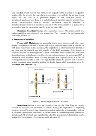
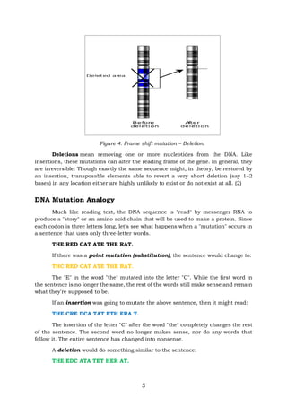
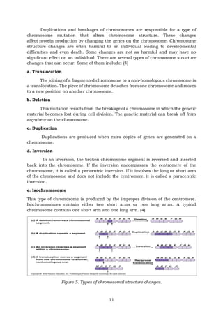
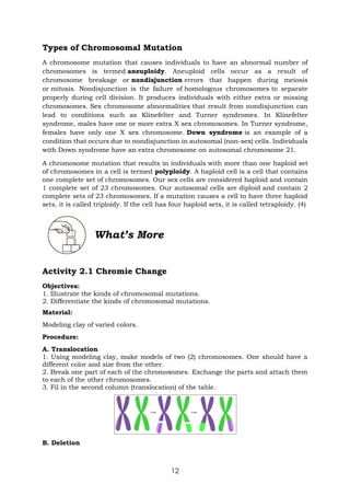
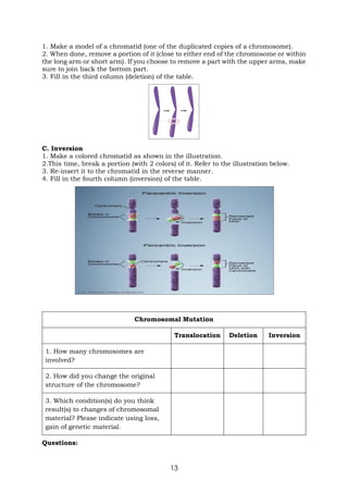
1. The document provides information about gene mutations, including definitions, types (point mutations and frameshift mutations), and examples.
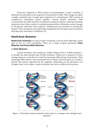
2. Point mutations include substitutions (silent, missense, nonsense), and involve a change to a single nucleotide. Frameshift mutations are generally more serious as they involve insertions or deletions of nucleotides that shift the reading frame.
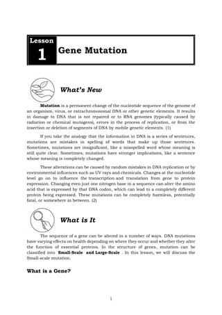
3. The document uses examples and analogies to explain how different mutation types can alter DNA sequences and cause changes to mRNA and resulting protein sequences and functions.


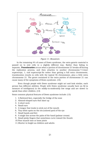


![[Appendix 2] RPMS Tool for MT I-IV SY 2020-2021 in the time of COVID-19.docx](https://cdn.slidesharecdn.com/ss_thumbnails/appendix2rpmstoolformti-ivsy2020-2021inthetimeofcovid-19-220912212152-fe7544c1-thumbnail.jpg?width=640&height=640&fit=bounds)


