Schindler's disease
•Download as PPTX, PDF•
2 likes•1,252 views

Schindler's disease, also known as Kanzaki disease or alpha-N-acetylgalactosaminidase deficiency, is a rare lysosomal storage disorder caused by a deficiency of the enzyme alpha-NAGA. This enzyme deficiency leads to an accumulation of glycosphingolipids in the body's cells and tissues. There are three main types which vary in symptoms and age of onset, from a severe early childhood type 1 to a milder adult-onset type 2. The disease has no cure but researchers are investigating enzyme replacement therapy, gene therapy, and bone marrow transplants.

Recommended
More Related Content
What's hot
What's hot (20)
Similar to Schindler's disease
Similar to Schindler's disease (20)
More from kavanvyas1
Recently uploaded
Recently uploaded (20)
Schindler's disease
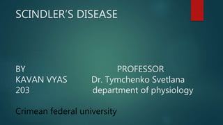
- 1. SCINDLER’S DISEASE BY PROFESSOR KAVAN VYAS Dr. Tymchenko Svetlana 203 department of physiology Crimean federal university
- 2. PLAN OF DESCRIPTION WHAT IS SCHINDLER’S DISEASE SYNONYMS CAUSES & REASON TYPE OF DISEASE SYMPTOMS DIAGNOSIS TRAETMENT
- 3. Lysosomal storage diseases (LSDs) are a group of approximately 50 rare inherited metabolic disorders that result from defects in lysosomal function. Lysosomes are sacs of enzymes within cells that digest large molecules and pass the fragments on to other parts of the cell for recycling. This process requires several critical enzymes. If one of these enzymes is defective, because of a mutation, the large molecules accumulate within the cell, eventually killing it. What are lysosomal storage disorders?
- 4. What is schindler’s disease? Schindler disease, also known as Kanzaki disease and alpha-N- acetylgalactosaminidase deficiency, it is a rare disease found in humans which is caused by a deficiency in the enzyme alpha-NAGA (alpha-N- acetylgalactosaminidase). This is lysosomal storage disorder.
- 5. Continue A deficiency of the alpha-NAGA enzyme leads to an accumulation of glycosphingolipids throughout the body. This accumulation of sugars gives rise to the clinical features
- 6. Continue Location of NAGA gene 22q13.2 Schindler disease, type III 22q13.2 Schindler disease, type I (on 22nd chromosome and q stands for long arm of the chromosome ) ( while 13 is for band and 2 stands for sub band) Schindler disease is an autosomal recessive disorder, so that one an abnormal allele from both parents in order to have the disease.
- 7. Other names of schindler’s disease alpha-galactosidase B deficiency alpha- galNAc deficiency alpha N-acetylgalactosaminidase deficiency alpha-NAGA deficiency angiokeratome corporis diffusum- glycopeptiduria GALB deficiency Kanzaki disease lysosomal glycoaminoacid storage disease - NAGA deficiency neuroaxonal dystrophy, Schindler type 1 neuronal axonal dystrophy, Schindler type
- 8. Cause of Scindler’s disease Schindler disease is caused by loss-of-function mutations in NAGA, a gene encoding a lysosomal exoglycosidase enzyme called α-N- acetylgalactosaminidase (α-NAGAL) that cleaves terminal α-N- acetylgalactosamine (GalNAc) residues from glycopeptides and glycolipids. N- Acetylgalactosamine is necessary for intercellular communication, and is concentrated in sensory nerve structures absent activity of the enzyme leads to the abnormal accumulation of certain complex compounds (glycosphingolipids, glycoproteins, and oligosaccharides) in certain tissues of the body and in urine
- 9. Types of schindler’s disease There are mainly 3 types of schindler’s disease Type 1 Type 2 OR kanzaki disease Type 3 The classification is based on the the age when patient shows symtoms of the disease, apart this… slight difference is also present in symptoms among these 3 types.
- 10. Type 1 disease Schindler disease type I - also called infantile type is the most severe form. Babies with Schindler disease type I appear healthy at birth, but at age 8 to 15 months they stop developing new skills and begin losing skills they have already acquired. These children develop , seizures and eventually lose awareness of their surrounds.
- 11. Continue Hearing and visual impairments occurs , low muscle tone and weakness , even sometimes complete muscle rigidity occurs. involuntary muscle spasms that result in slow, stiff movements (spasticity) misalignment of the eyes (strabismus) involuntary, rapid eye movements (nystagmus) and visual impairment due to the gradual deterioration of the nerves of the eyes (optic atrophy) leads to blindness.
- 12. Continue Children with this form of the disease do not usually live past early childhood.
- 13. Type 2 disease Schindler disease type II or Kanzaki disease occurs In the adult-onset form symptoms may not appear until the second or third decade of life. A distinctive symptom of Schindler disease type II is involvement of small blood vessels (telangiectasia) in the skin that cause reddish small skin lesions, and an increase of its layer stratum corneum (hyperkeratosis) referred to as angiokeratomas.
- 14. Continue Angiokeratomas may first be restricted to a single area (localized), such as the lower torso, and then appear later in additional locations (e.g., from the lower torso to the chest area). These reddish lesions may be flat or raised and vary in colour, and may occur in clusters. Affected individuals may also have these lesions in other areas of the body such as the mucous membranes including the mouth and eyes.
- 16. Continue The dilation of small lymph vessels may lead to swelling (lymphedema) particularly of the lower extremities. Individuals have also mild intellectual impairment, but do not show the serious neurological complications associated with Schindler disease type I.
- 17. Continue Additional symptoms have been reported in the medical literature including vertigo, hearing loss, ringing in the ears (tinnitus), and muscle weakness , experiences pain crises. Complication in development of central and peripheral nerve occurs.
- 18. Individuals may also develop distinctive facial features including mildly coarse features, thick lips, a depressed nasal bridge and an enlarged tip of the nose.
- 19. Type 3 disease Schindler disease type III, is an intermediate form the disorder. Symptoms can range from more serious intellectual impairment, neurological dysfunction. seizures to milder neurological and psychiatric issues such as speech and language delays and mild autism-like symptoms.
- 20. Continue Behaviour problem and mental retardation occurs. Type 3 disease is less severe than type 1 disease but more severe than type 2 disease.
- 22. Histological Changes during schindler’s disease symptoms of type I Schindler disease are associated with characteristic swellings at the end of nerve fibers (axons). These swellings may be referred to as dystrophic axonal swellings or “spheroids”. The spheroids are characteristic of a neuroaxonal dystrophy – a severe alteration of nerve cells. These swellings appear to disrupt proper nerve function by blocking the transmission of impulses between nerve cells. apart this demyelinization occurs.
- 23. Frequency of disease Schindler’s disease is very rare. Till now only 12 cases from 8 different families are known of this disease. Another reason is misdiagnosis and unrecognized.
- 24. Diagnosis detailed patient history Urinary analysis (e.g. oligosaccharide and glycopeptide levels) Reduced activity of the alpha-NAGA enzyme may be confirmed by conducting enzyme tests (assays) on cultured white blood cells (leukocytes), blood plasma, and/or certain skin cells (fibroblasts) from affected individuals. samples of tissue biopsy especially in type 1 disease. magnetic resonance imaging (MRI) and computer-assisted tomography (CAT) of the brain
- 25. Treatment There is no specific treatment known for schindler’s disease but as a part of developing medical science , researchers hopefully succeed in some cases. enzyme replacement therapy to compensate for endogenous deficiency. Gene therapy bone marrow transplants