Rupture Risk Based On Anatomical And Morphological Factors
1. Neurosurg Focus 26 (5):E2, 2009
Unruptured intracranial aneurysms and the assessment of
rupture risk based on anatomical and morphological factors:
sifting through the sands of data
Rohan R. LaLL, M.D., ChRistopheR s. eDDLeMan, M.D., ph.D., BeRnaRD R. BenDok, M.D.,
anD h. hunt BatjeR, M.D.
Department of Neurological Surgery, Feinberg School of Medicine, Northwestern University,
Chicago, Illinois
Aneurysmal subarachnoid hemorrhage continues to have high rates of morbidity and mortality for patients de-
spite optimal medical and surgical management. Due to the fact that aneurysmal rupture can be such a catastrophic
event, preventive treatment is desirable for high-risk lesions. Given the variability of the literature evaluating unrup-
tured aneurysms regarding basic patient population, clinical practice, and aneurysm characteristics studied, such as
size, location, aspect ratio, relationship to the surrounding vasculature, and the aneurysm hemodynamics, a meta-
analysis is nearly impossible to perform. This review will instead focus on the various anatomical and morphological
characteristics of aneurysms reported in the literature with an attempt to draw broad inferences and serve to highlight
pressing questions for the future in our continued effort to improve clinical management of unruptured intracranial
aneurysms. (DOI: 10.3171/2009.2.FOCUS0921)
key WoRDs • unruptured intracranial aneurysm • vascular location •
rupture risk • ISUIA • aspect ratio
A
neurysmAl SAH continues to have high rates of to provide such information. The ISUIA concluded that
morbidity and mortality for patients despite op- aneurysms < 7 mm in size in the anterior circulation have
timal medical and surgical management.18 Con- an annual rupture risk of 0-0.1% per year.42 This was
troversy exists regarding the prevalence of unruptured markedly lower than previous estimates, and the trial
intracranial aneurysms with some studies reporting rates drew heavy criticism.9,20,25,39
as high as 6.5% in the general population harboring these The main obstacle in evaluating cumulative risk of
lesions.27 However, the vast experience of the neurovas- rupture over time is that ethically, patients with the high-
cular community would declare these estimates likely est risk aneurysms cannot be left untreated. Thus, these
too high.30,31,43 Regardless, with the increasing use of estimates will likely underestimate the true aneurysm
noninvasive intracranial imaging, an increasing number rupture risk. In response to this, most recent literature
of unruptured intracranial aneurysms are being inciden- has focused on comparing anatomical and morphologi-
tally discovered. The optimal procedural management of cal characteristics of ruptured aneurysms to unruptured
these lesions is still being debated, which can carry sig- aneurysms with the goal of elucidating factors associated
nificant risk, with morbidity and mortality rates up to 10 with a high risk of rupture. These studies have had many
and 2.5%, respectively.21,29 different designs, areas of focus, and varied conclusions.
However, due to the fact that aneurysm rupture can Given the variability of the literature evaluating un-
be such a catastrophic event, preventive treatment is de- ruptured aneurysms regarding basic patient population,
sirable for high-risk lesions. Many groups have sought to clinical practice, and even aneurysm characteristics stud-
find conclusive data on the natural history of unruptured ied, a meta-analysis is nearly impossible to perform. This
aneurysms. The ISUIA trial was designed and conducted review will instead focus on the various anatomical and
morphological characteristics of aneurysms reported in
Abbreviations used in this paper: ACoA = anterior commu- the literature with an attempt to draw broad inferences
nicating artery; CFD = computational flow dynamics; ISUIA = and serve to highlight pressing questions for the future in
International Study of Unruptured Intracranial Aneurysms; SAH = our continued effort to improve clinical management of
subarachnoid hemorrhage; WSS = wall sheer stress. unruptured intracranial aneurysms.
Neurosurg. Focus / Volume 26 / May 2009 1
2. R. Lall et al.
TABLE 1: Comparison studies of the sizes of ruptured versus unruptured intracranial aneurysms*
Mean Size (mm)
Authors & Year No. of Patients No. of Aneurysms Unruptured Ruptured p Value
Baumann et al., 2008 99 265 4 7 <0.0001
Beck et al., 2003 118 155 5.7 6.7 0.7
Nader-Sepahi et al., 2004 75 182 4.9 7.7 <0.001
Juvela et al., 2008 142 181 4.9 5.6 nc
Hoh et al., 2007 30 67 4.3 6.2 0.004
Weir et al., 2003 945 507 7.8 10.8 < 0.001
Weir et al., 2002 532 774 7 8 nc
Sadatomo et al., 2008 41 44 5.6 7.2 0.11
* Patients had single, multiple, or mixed aneurysms. Abbreviation: nc = not calculated.
Aneurysm Size and Location aneurysms. Nevertheless, this study demonstrated that
aneurysm size is an important risk factor for rupture.
Two of the most basic features of intracranial aneu- The ISUIA published a follow-up paper in The Lan-
rysms are their size and location. Consistently, investi- cet in July 2003.42 This report more closely evaluated
gators have reported that size is an unquestionable fac- rupture risk based on location and size, and specifically
tor with regard to rupture risk. Furthermore, posterior assessed surgical and endovascular treatment risks. The
circulation aneurysms have been noted to rupture more 5-year cumulative rupture rates for patients without prior
frequently than similar aneurysms in the anterior circula- SAH, with anterior circulation aneurysms (not including
tion. However, despite decades of observation, few stud- cavernous carotid or posterior communicating artery an-
ies have examined unruptured intracranial aneurysms in eurysms) were 0, 2.6, and 14.5% for aneurysms < 7, 7–12,
a prospective trial involving international centers and a and 13–24 mm, respectively, compared with rates of 2.5,
heterogeneous population. The ISUIA was an attempt 14.5, and 18.4%, respectively, for the same size aneurysm
to elucidate the natural history of these lesions across in the posterior circulation (including posterior commu-
an international population. The ISUIA published their nicating artery aneurysms). Patients with a history of pre-
first study in the New England Journal of Medicine in vious SAH with aneurysms < 7 mm in size had a 0.1%
December 1998.2 The study was divided into 2 cohorts, yearly rupture rate. This study had many of the same
which consisted of a retrospective cohort of observed un- limitations and criticisms as the first ISUIA study.21,40
ruptured aneurysms, designed to evaluate risk of rupture Despite reporting results that were not consistent with
over time, and a prospective cohort, designed to evaluate numerous studies in the literature regarding rupture risk
surgical risk. The retrospective cohort had 1449 patients of unruptured intracranial aneurysms, the ISUIA data
with 1937 aneurysms, nearly evenly divided into 2 groups. provided the first large, international, prospective data set
Group 1 had no history of prior SAH and Group 2 com- that practitioners could use in their discussions with pa-
prised patients with prior SAH from another treated an- tients and their families.
eurysmal lesion and had to be functioning independently. However, many other studies, although not involv-
Risk of rupture for Group 1 patients for aneurysms < 10 ing as many patients as those of the ISUIA reports, have
mm in size in the anterior circulation was 0.05% per year, been published regarding aneurysmal rupture risk, whose
compared with 0.5% annually for Group 2. Aneurysms results have been marketed as challenging the ISUIA re-
> 10 mm in size had a risk of rupture close to 1% an- sults. More specifically, Juvela et al.19 published their ac-
nually in both groups. The prospective cohort enrolled count of the cumulative aneurysm rupture risk in Finnish
1172 patients undergoing treatment of their aneurysms. patients with long-term follow-up in which 142 patients
Surgical morbidity and mortality published in this study with 182 aneurysms were evaluated and were found to
were notably higher than previously accepted rates. This have a rupture rate of 1.3% per year. A significant ob-
study was widely quoted and used at the time by primary servation in this study was that the majority of ruptured
care physicians, but it was considered controversial by the aneurysms were < 7 mm, in contrast to the findings of
neurosurgical community. The main criticism was in re- the ISUIA.42 They also noted that larger aneurysms had
gard to patient selection. For the retrospective cohort, all a linearly increasing relative risk compared with smaller
patients receiving treatment within 30 days of diagnosis aneurysms. With respect to aneurysm location, ACoA an-
were excluded, and the number of these patients was not eurysms were found to have a higher ratio of ruptured to
revealed. These aneurysms almost certainly comprised unruptured aneurysms than other locations. The rupture
the highest risk group. Aneurysms in low-risk locations, rates quoted by Juvela and colleagues were closer to those
such as the cavernous segment of the carotid artery, were published in previous large studies of the natural history of
over-represented. Furthermore, numerous patients sub- unruptured intracranial aneurysms during the 1980s.15,43
sequently died of intracranial hemorrhage of unknown The reported rate was also more equivalent with 2 previ-
origin, a suspicious diagnosis for patients with known ous Japanese reports noting annual rupture rates of 1.5
2 Neurosurg. Focus / Volume 26 / May 2009
3. Unruptured intracranial aneurysms and rupture risk assessment
TABLE 2: Comparison studies of the aspect ratio in ruptured and unruptured aneurysms
Mean Aspect Ratio (mm)
No. of
Authors & Year Aneurysm Population No. of Aneurysms Unruptured Ruptured p Value
Patients
Sadatomo et al., 2008 mixed 41 44 1.56 2.24 <0.01
Beck et al., 2003 mixed 118 155 2.03 1.68 <0.001
Nader-Sepahi et al.,
2004 multiple 75 182 1.8 2.7 <0.001
Hoh et al., 2007 multiple 30 67 1.63 2.39 <0.001
Weir et al., 2003 mixed 532 774 1.8 3.4 nc
and 2.3%, although these trials also had high proportions ruptured and ruptured aneurysms, thus provided another
of patients with prior SAH or ICH.37,44 It is worth noting, valuable parameter in addition to size and location. With
however, that of 142 patients in the study by Juvela and few exceptions,4,5 a higher aspect ratio has been associ-
colleagues, only 5 had asymptomatic, incidental aneu- ated with ruptured lesions (Table 2). 5,16,26,34,40
rysms and 6 had symptomatic incidental aneurysms. The Hoh et al.16 published aneurysm characteristics of
other 131 patients had a history of previous SAH. Thus, 30 patients with multiple aneurysms (30 ruptured and 37
this population is most similar to Group 2 in the ISUIA unruptured); namely, 7 1D geometric indices and 5 2D
reports, which was noted to have a 0.1% annual rupture indices were evaluated. Aneurysm height, diameter, as-
rate for aneurysms < 7 mm. So, while the study by Juvela pect ratio, bottleneck factor, and aneurysm/parent artery
et al. is less burdened by the selection biases present in ratio were all statistically significantly different between
ISUIA, it cannot accurately estimate the rupture risk with unruptured and ruptured aneurysms. Ujiie et al.38 evalu-
regard to aneurysm size for patients without a previous ated 207 aneurysms and found that ~ 80% of ruptured
history of SAH. Furthermore, these studies had a signifi-
aneurysms had an aspect ratio > 1.6, whereas ~ 90% of
cantly smaller number of patients than ISUIA and were
limited to patients from Finland and Japan, potentially unruptured aneurysms had an aspect ratio < 1.6. Weir et
higher risk population groups. al.40 examined 774 aneurysms and found that the mean
Many other studies have compared aneurysm size in aspect ratio of unruptured aneurysms was ~ 1.8 as op-
ruptured aneurysms with that of unruptured lesions (Ta- posed to 3.4 for ruptured aneurysms. The calculated odds
ble 1).3,4,16,19,26,33,40,41 Consistently, ruptured aneurysms are of aneurysm rupture were 20-fold greater when the aspect
larger than unruptured aneurysms. Typical results have ratio was > 3.47 as opposed to those < 1.38. Nader-Sepahi
demonstrated the mean size of unruptured aneurysms to et al.26 found the mean aspect ratio of 2.7 in ruptured an-
be between 4 and 6 mm, and mean size of ruptured an- eurysms and 1.8 in unruptured aneurysms. Sadatomo et
eurysms is between 5 and 8 mm, which is statistically al.33 examined 44 aneurysms and reported that the mean
significant. Nevertheless, the majority of ruptured and aspect ratio in ruptured aneurysms was 2.24, significantly
unruptured aneurysms are still < 7 mm, and a substantial greater than 1.56, which was found in unruptured aneu-
percentage of ruptured aneurysms are even < 5 mm.11,27, rysms.
28
Thus, aneurysm size and location appear to be insuf- Unfortunately, the aspect ratio also has limitations as
ficient single parameters to guide treatment decisions, but a predictor of aneurysm rupture risk. While most studies
they can at least serve as a guide for higher risk lesions. have found a statistically significant difference in aspect
ratio values, there is some discrepancy as to where the
Aspect Ratio threshold values lie. Aspect ratios between 1.6 and 2.2 are
consistently considered borderline in risk, even with the
In response to the limitations of size and location as upper limit of 2.2 not being considered significantly high
a single, dependable predictor of rupture risk, many in- risk according to some studies.26,40 Nonetheless, it is clear
vestigators have evaluated other morphological aspects that aneurysms with aspect ratios > 3 have been shown to
and single-/multidimensional geometric parameters of be at extremely high risk of rupture, whereas aneurysms
aneurysms as a predictor of rupture risk.3,4,13,16,17,22,26,34,38, with aspect ratios < 1.4 are much less likely to be at high
40
More specifically, neck width, dome width, aneurysm rupture risk potential.
shape, aspect ratio (height/neck width), and bottleneck
factor (dome width/neck width) have been examined.
Among these, aspect ratio has shown the greatest promise
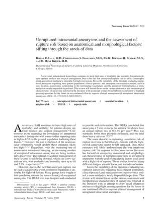
as a parameter to associate with rupture risk (Fig. 1).
Neck width has been examined in multiple studies,
most of which place the mean width for unruptured and
ruptured lesions between 2 and 3 mm, without statistical-
ly significant difference.14,16 However, due to the variabil-
ity of aneurysm height, the aspect ratio has been found Fig. 1. Diagram detailing common morphological parameters used
in most studies to be statistically significant between un- in the assessment of aneurysm rupture risk.
Neurosurg. Focus / Volume 26 / May 2009 3
4. R. Lall et al.
Aneurysm Geometry, Orientation, lature, Dhar et al.10 recently reported on 45 patients with
and Vascular Relationships terminal or sidewall aneurysms (25 ruptured and 20 un-
ruptured) whose aneurysms were analyzed with respect
Other morphological characteristics have also re- to 8 parameters (5 old and 3 novel). The more established
ceived attention with regard to assessing aneurysm rup- parameters were aspect ratio, aneurysm size, ellipticity
ture risk. More specifically, morphological parameters index, nonsphericity index, and undulation index; while
such as lobulations, daughter sacs, and surface irregular- the novel parameters, which incorporated the parent ves-
ity have been considered. Aneurysm wall irregularity and sel geometry, were vessel angle, aneurysm (inclination)
daughter sacs have long been associated with higher rup- angle, and (aneurysm-to-vessel) size ratio. Of these pa-
ture risk.5,13,22,34 rameters examined, size ratio and aneurysm angle with
Hademenos et al.13 published their account of 74 pa- respect to the parent artery had the strongest correlation to
tients with aneurysms (40 ruptured and 34 unruptured) rupture potential, although statistically significant differ-
and evaluated the location and morphological factors, for ences between ruptured and unruptured aneurysms were
example, lobulations, and reported that 16 (84%) of 19 also found for aspect ratio, undulation, ellipticity, and
multilobulated aneurysms had ruptured, compared with nonsphericity index. The fact that these novel parameters
only 24 (44%) of 55 unilobular aneurysms, a statistically involve the aneurysm’s relationship to the parent vessel
significant difference. Posterior circulation aneurysms further supports the influence of hemodynamics from the
were also noted to be of higher risk with multilobular surrounding vasculature on the behavior of the aneurysm.
posterior circulation aneurysms at the highest risk of all. Other studies have used advanced analysis of an-
Beck et al.5 detailed 147 aneurysms in 124 patients (94 giograms and CFD to further evaluate the relationships
ruptured and 53 unruptured), examining the presence of between the vasculature and the aneurysm in an attempt
lobulations, daughter sacs, and the differences in aneu- to associate specific orientations with higher risk of aneu-
rysm size. They found that multilobular aneurysms be- rysm rupture. Hassan et al.14 examined 68 aneurysms (45
tween 5 and 9 mm in dome height were more frequently ruptured and 23 unruptured) and classified them into 3 an-
ruptured than unilobular lesions (26 vs 4%). Furthermore, eurysm groups, namely sidewall, sidewall with a branch-
they could not demonstrate a significant difference based ing vessel, and endwall. They found a 100% rupture rate
on the presence of a daughter sac, but they had < 10 such for aneurysms with an aspect ratio > 1.6 and either a side-
lesions in the trial. Sampei et al.35 examined 25 aneu- wall or sidewall with branching vessel-type, as opposed
rysms found to have grown on repeat angiography and to 28.75% rate for endwall-type aneurysms. They also
found that the irregular contour and the presence of blebs found a significantly lower rupture risk and higher flow
correlated with faster growth and increased risk of rup- rates in aneurysms with wide necks and wide efferent,
ture or rebleeding. draining arteries, thus minimizing the inflow. Another
Given that intracranial aneurysms are more com- study by Castro et al.,7 evaluated 2 ACoA aneurysms by
monly found at either bi- or trifurcations or at regions of using CFD analysis of WSS and found that an unequal
high impact from flowing blood, the relationship of aneu- amount of flow in the carotid arteries could be linked to
rysm to the surrounding vasculature has been examined. an asymmetric, increased amount of WSS in the ACoA
With respect to the surrounding vasculature, Sadatomo et aneurysm, possibly rendering the aneurysm at a higher
al.34 reported on the relationship among aneurysm neck, rupture risk potential. Finally, Hoi et al.17 used CFD to
parent artery, and daughter branches in 22 consecutive evaluate the influence of variable arterial curvature on
MCA bifurcation aneurysms, which were divided into a lateral wall aneurysms and found that a greater degree of
classic-type (aneurysm at midline relative to parent ar- curvature lead to higher degrees of hemodynamic stress,
tery) and a deviating-type (aneurysm deviates to the side thus possibly increasing rupture risk.
of 1 daughter artery). They found that in all cases, the
deviating-type aneurysms were located on the side of the
daughter artery with a narrower angle to the parent ar- Aneurysm Hemodynamics
tery. Furthermore, in > 90% of the cases, the aneurysm Unfortunately, each unruptured intracranial aneu-
was located on the side of the smaller artery, suggesting rysm is a unique lesion. Thus, it is probably the individual
the dominant artery provided the hemodynamic force for flow patterns determined by the geometric relationship
aneurysm formation and likely increased rupture risk; with the surrounding vasculature as well as the anatomi-
however, this contention could not be shown statistically. cal and morphological configuration of the aneurysm that
In another report, Sadatomo’s group32 described 18 an- are the most important predictors of rupture risk. The
eurysms of the ACoA, detailing the relationships of the most recent development in the literature of unruptured
aneurysm to A1, the midline, at the junction of the ACoA, aneurysms has been the use of advanced imaging tech-
and at the A1-A2 junction. They found that for all patients niques and image postprocessing to visualize flow pat-
with codominant A1 segments, the aneurysms were always terns and hemodynamic stress in individual aneurysms.
of the classic type, where the aneurysm fundus arose in Some early studies evaluated aneurysm simulations and
the midline, as opposed to patients with a dominant A1, models, but more recent studies look at in vivo flow dy-
where the aneurysm fundus pointed to the contralateral namics. Although these imaging techniques continue to
side of the dominant A1. carry limitations, they are beginning to expand our un-
In an effort to combine the morphological charac- derstanding (or lack thereof) of aneurysm growth and its
teristics with the relationship of the surrounding vascu- potential relationship with increased rupture risk.
4 Neurosurg. Focus / Volume 26 / May 2009
5. Unruptured intracranial aneurysms and rupture risk assessment
The most commonly used techniques to evaluate an- had a single inflow jet with multiple vortices. Two small,
eurysm hemodynamics are CFD analyses of 3D digital smooth aneurysms with the smallest aspect ratios (1.1
subtraction angiography or CT angiography images and and 1.3) had single inflow jets and single vortices. De-
phase-contrast MR imaging. Shojima et al.36 evaluated the spite the fact that the WSS was underestimated due to the
CFD analyses of 3D CT angiography reconstructions of lack of inflow (parent artery) and outflow (efferent arter-
20 MCA aneurysms (3 ruptured and 17 unruptured) and ies) boundary corrections, this study showed that in vivo
found, in opposition to most previous reports, that lower phase-contrast MR imaging could correlate flow dynam-
WSS, compared with the parent vessel, was present in the ics, aspect ratio, and fundus size in a series of aneurysms.
dome of aneurysms and in the blebs and/or daughter sacs. Unfortunately, both CFD and phase-contrast MR imaging
In contrast, higher WSS values were found in ruptured techniques currently require a significant amount of im-
aneurysms. However, Castro et al.,7 in their study of the age postprocessing and computational power that neither
effects of parent and draining arteries on CFD analyses, is practical for clinical use at this time.
found that not taking into account the inflow of the parent
artery as well as outflow through the efferent arteries can
significantly underestimate the WSS values in the dome Limitations
of the aneurysm. When corrected, they found consistently Inherent limitations exist in any attempt to study fac-
higher values of WSS in the aneurysm domes and blebs. tors of aneurysm rupture risk. Specifically, most of the
Previous trials, such as that by Shojima et al.,36 which pro- previously discussed studies have examined morphologi-
moted a low WSS theory for aneurysmal rupture, must be cal characteristics of ruptured aneurysms in comparison
reexamined given the sensitivity of CFD analyses on the with unruptured aneurysms. Unfortunately, this method-
boundary conditions, namely the requirement of inflow ology does not account for the possible changes in mor-
and outflow parameters. phology that high-risk aneurysms may experience over
Cebral et al.8 evaluated 62 aneurysm models, based time, potentially evolving into the gross morphology as-
on 3D digital subtraction angiograms from patients, using sociated with ruptured aneurysms. Thus, evaluating un-
CFD. These models were divided into 4 types: 1) single ruptured aneurysms of any shape or size at only one point
inflow jet, single vortex of flow; 2) single inflow jet, mul- in time is foolhardy, because the future evolution of the
tiple vortices; 3) multiple inflow jets, single vortex; and lesion is unpredictable and likely unknown. Examining
4) multiple inflow jets, multiple vortices. Type 1 was the a population of unruptured aneurysms over time without
most frequently encountered, followed by Type 4, Type 2, treatment, regardless of size or location, monitoring he-
and Type 3. Types 4 and 2 were the most frequently multi- modynamics, and the evolution of aneurysmal morphol-
lobulated, large in size, and had a higher aspect ratio. Type ogy over time would provide the ultimate natural history
1 aneurysms, in contrast, were more likely to be smaller, study. Unfortunately, such a study will never be complet-
unilobular, and have a smaller aspect ratio. The rates of ed due to the unacceptable ethical dilemma present with
rupture for each type of aneurysm were 27, 45, 60, and regard to patient safety and previous clinical experience.
58%, respectively. Importantly, although the majority of In an effort to study ruptured and unruptured aneu-
small aneurysms were Type 1, a significant number were rysms in the same patients, Hoh,16 Baumann,3 and Nader-
also Type 3 and Type 4. Still, the only statistically signifi- Sepahi26 and their colleagues examined only patients with
cant predictor of rupture risk in this report was the size multiple aneurysms—the vast majority of patients had 1
of the flow impingement region. Aneurysms with small ruptured aneurysm, and the remainder were unruptured.
inflow jet streams or smaller flow impingement size were However, as Weir39 commented in Stroke, the ISUIA2,42
6.3 times more likely to have ruptured; however, neither results are unclear as to whether patients with multiple
large neck nor large aneurysm size statistically correlated aneurysms represent a higher risk population. Thus, it re-
with a smaller flow impingement region. mains to be seen if the aneurysms seen in patients with
In an effort to allow hemodynamic measurements multiple aneurysms behave independently or are part of
in vivo, phase-contrast MR imaging has been champi- a systemically increased risk. Most of the other studies
oned given the availability of MR imaging throughout used in evaluating the risk of aneurysm rupture and the
the world. Ahn et al.1 examined anthropomorphic in vitro factors related to such risks use a mixture of patients with
models of 2 intracranial aneurysms to show the feasibil- single and/or multiple aneurysms indiscriminately. Fur-
ity of 3D phase-contrast MR imaging as an alternative to thermore, there is a considerable variability in the ratios
CFD models. One potential advantage of phase-contrast of patients with ruptured and unruptured lesions. How-
MR imaging is the ability to visualize both the velocity ever, Beck4 and Sadatomo34 had nearly equal numbers of
and inflow hemodynamics within and around the aneu- ruptured and unruptured aneurysms in their reports and
rysm in vivo. The results showed the highest WSS at the the studies by Weir40,41 had a majority of ruptured aneu-
inflow zone of the aneurysms, but they did show a local rysms.
area in the bleb of one aneurysm and in the dome of the The literature is rather limited with regard to the evo-
other to have had constant high WSS without temporal lution of aneurysmal morphology over time. Burns et al.6
variation. These results were consistent with most high monitored 165 patients with 191 unruptured aneurysms
WSS theories. by using serial MR angiography over a median follow-
Meckel et al.24 went a step further and examined up period of 47 months. They noted that 10% of patients
cardiac-gated 3D phase-contrast MR imaging of 5 aneu- had aneurysm growth over that time period, during which
rysms in vivo and found that the highest aspect ratio (2.2) they documented 1 incident of aneurysm rupture. The
Neurosurg. Focus / Volume 26 / May 2009 5
6. R. Lall et al.
only statistically significant predictor of growth was pre- tracranial aneurysms. However, in the absence of practi-
vious aneurysm size. For aneurysms < 8, between 8 and cal, clinical applications of advanced imaging techniques,
12, and ≥ 13 mm, the frequency of enlargement was 6.9, practitioners are left to use their clinical acumen as well
25, and 83%, respectively. A significant limitation of this as the limited amount of high-quality literature to help
study was that a large number of patients were lost to fol- determine which management strategy is best for the an-
low-up, and growth and rupture rates remained unknown eurysms in question.
for these patients. Currently, the literature suggests that higher risks of
Matsubara et al.23 monitored changes in aneurysm rupture are associated with posterior circulation or poste-
morphology in 140 patients with 166 unruptured aneu- rior communicating aneurysms, size > 7 mm, high aspect
rysms by using serial CT angiography for a mean follow- ratio or bottleneck ratio, irregular surface and daughter
up of 17.7 months. They observed growth or new de- sacs, and small parent artery and/or draining vessels.
velopment of blebs/daughter sacs in 6.4% of patients (6 As imaging technology advances, including fast ac-
aneurysms grew and 4 developed blebs). Statistically sig- quisition MR imaging for hemodynamics, high-spatial
nificant predictors were aneurysm size and basilar apex resolution for aneurysm wall motion, and low signal detec-
bifurcation or internal carotid artery location. Other pre- tion for molecular imaging, the opportunities to observe
dictors of growth were patient or family history of SAH, the in vivo behavior of unruptured intracranial aneurysms
presence of a preexisting bleb, hyperlipidemia, and diabe- will likely increase. It is not unreasonable to think that the
tes. No aneurysm rupture was reported during the dura- risk of aneurysm rupture will be determined by a multi-
tion of the study; however, 7 aneurysms were treated. tude of factors, including genetic, comorbidities present,
For aneurysm growth to occur, it may be assumed precise configuration of the intracranial vasculature, and
that changes may occur in the aneurysm wall itself. the specific anatomical and morphological factors of the
Frosen et al.12 reported on the histological analyses of lesion itself. It is clear that intracranial aneurysms are
the wall tissue in 66 clipped aneurysms (24 unruptured not static but dynamic structures and the morphological
and 42 ruptured). They described the following 4 broad characteristics assessed at one point in time may not be
categories of aneurysm wall characteristics: 1) endotheli- the same ones assessed at a later time. So, to improve our
alized wall with organized smooth muscle; 2) thickened understanding of how these anatomical and morphologi-
wall with disorganized smooth muscle; 3) hypocellular cal factors relate to rupture risk, we must also examine
wall with myointimal hyperplasia or luminal thrombus; those same properties over time. But, in the end, it will
and 4) extremely thin thrombosis-lined hypocellular be a combination of factors beyond just morphological
wall. They observed progressively higher proportions of characteristics that determine the rupture risk potential
ruptured aneurysms for each of the categories studied as of unruptured intracranial aneurysms.
follows: 42, 55, 64, and 100% respectively. The authors
discussed the possibility that these 4 categories may rep- Disclaimer
resent a continuum of aneurysm wall degeneration occur-
The authors report no conflict of interest concerning the mate-
ring over time. rials or methods used in this study or the findings specified in this
These studies clearly demonstrate that aneurysms are paper.
capable of growing and evolving over time. Nevertheless,
References
the evolution of aneurysm growth and wall characteris-
tics has yet to be shown to be the absolute causative agent 1. Ahn S, Shin D, Tateshima S, et al: Fluid-induced wall shear
of increased rupture risk or simply an incidental altera- stress in anthropomorphic brain aneurysm models: MR phase-
tion in any conclusive way. The observations that patients contrast study at 3 T. J Magn Reson Imaging 25:1120–1130,
with existing blebs/daughter sacs may be more likely to 2007
2. Anonymous: Unruptured intracranial aneurysms—risk of
develop further blebs and that aneurysm walls undergo rupture and risks of surgical intervention. International Study
progressive remodeling over time both are consistent with of Unruptured Intracranial Aneurysms Investigators. N Engl
theories that lesions with unstable hemodynamic flow pat- J Med 339:1725–1733, 1998
terns ultimately present a higher risk of rupture. Further 3. Baumann F, Khan N, Yonekawa Y: Patient and aneurysm
studies examining the relationship of these characteristics characteristics in multiple intracranial aneurysms. Acta Neu-
need to be pursued. rochir Suppl (Wien) 103:19–28, 2008
4. Beck J, Rohde S, Berkefeld J, et al: Size and location of rup-
tured and unruptured intracranial aneurysms measured by
Conclusions and Future Considerations 3-dimensional rotational angiography. Surg Neurol 65:18–
25, 2006
Ultimately, every aneurysm is a unique lesion with 5. Beck J, Rohde S, el Beltagy M, et al: Difference in configura-
an individualized mixture of geometry, size, location, and tion of ruptured and unruptured intracranial aneurysms de-
relationship to its surrounding vasculature. While many termined by biplanar digital subtraction angiography. Acta
surrogates, such as size and location, have long been used Neurochir (Wien) 145:861–865, 2003
to estimate rupture risk, our clinical experience has taught 6. Burns JD, Huston J 3rd, Layton KF, et al: Intracranial aneu-
rysm enlargement on serial magnetic resonance angiography:
us that sometimes small aneurysms can lead to devastat- frequency and risk factors. Stroke 40:406–411, 2009
ing morbidity and mortality, and large aneurysms can be 7. Castro MA, Putman CM, Cebral JR: Computational fluid dy-
quite stable. Advanced imaging studies have begun to namics modeling of intracranial aneurysms: effects of par-
help elucidate the relationship between the anatomy, mor- ent artery segmentation on intra-aneurysmal hemodynamics.
phology, and hemodynamic patterns of unruptured in- AJNR Am J Neuroradiol 27:1703–1709, 2006
6 Neurosurg. Focus / Volume 26 / May 2009
7. Unruptured intracranial aneurysms and rupture risk assessment
8. Cebral JR, Castro MA, Burgess JE, et al: Characterization rysms and related factors in patients with subarachnoid hem-
of cerebral aneurysms for assessing risk of rupture by using orrhage. Surg Neurol 61:239–245, 2004
patient-specific computational hemodynamics models. AJNR 29. Raaymakers TW, Rinkel GJ, Limburg M, et al: Mortality and
Am J Neuroradiol 26:2550–2559, 2005 morbidity of surgery for unruptured intracranial aneurysms: a
9. Clarke G, Mendelow AD, Mitchell P: Predicting the risk of meta-analysis. Stroke 29:1531–1538, 1998
rupture of intracranial aneurysms based on anatomical loca- 30. Rinkel GJ: Natural history, epidemiology and screening of
tion. Acta Neurochir (Wien) 147:259–263, 2005 unruptured intracranial aneurysms. Rev Neurol (Paris)
10. Dhar S, Tremmel M, Mocco J, et al: Morphology parameters 164:781–786, 2008
for intracranial aneurysm rupture risk assessment. Neurosur- 31. Rinkel GJ: Natural history, epidemiology and screening of un-
gery 63:185–187, 2008 ruptured intracranial aneurysms. J Neuroradiol 35:99–103,
11. Forget TR Jr, Benitez R, Veznedaroglu E, et al: A review of 2008
size and location of ruptured intracranial aneurysms. Neuro- 32. Sadatomo T, Yuki K, Migita K, et al: The characteristics of the
surgery 49:1322–1326, 2001 anterior communicating artery aneurysm complex by three-
12. Frosen J, Piippo A, Paetau A, et al: Remodeling of saccular dimensional digital subtraction angiography. Neurosurg Rev
cerebral artery aneurysm wall is associated with rupture: 29:201–207, 2006
histological analysis of 24 unruptured and 42 ruptured cases. 33. Sadatomo T, Yuki K, Migita K, et al: Evaluation of relation
Stroke 35:2287–2293, 2004 among aneurysmal neck, parent artery, and daughter arteries
13. Hademenos GJ, Massoud TF, Turjman F, et al: Anatomical in middle cerebral artery aneurysms, by three-dimensional
and morphological factors correlating with rupture of intrac- digital subtraction angiography. Neurosurg Rev 28:196–200,
ranial aneurysms in patients referred for endovascular treat- 2005
ment. Neuroradiology 40:755–760, 1998 34. Sadatomo T, Yuki K, Migita K, et al: Morphological differ-
14. Hassan T, Timofeev EV, Saito T, et al: A proposed parent ves- ences between ruptured and unruptured cases in middle cere-
sel geometry-based categorization of saccular intracranial bral artery aneurysms. Neurosurgery 62:602–609, 2008
aneurysms: computational flow dynamics analysis of the risk 35. Sampei T, Mizuno M, Nakajima S, et al: [Clinical study of
factors for lesion rupture. J Neurosurg 103:662–680, 2005 growing up aneurysms: report of 25 cases.] No Shinkei Geka
15. Heiskanen O: Risk of bleeding from unruptured aneurysm 19:825–830, 1991 (Jpn)
in cases with multiple intracranial aneurysms. J Neurosurg 36. Shojima M, Oshima M, Takagi K, et al: Magnitude and role
55:524–526, 1981 of wall shear stress on cerebral aneurysm: computational
16. Hoh BL, Sistrom CL, Firment CS, et al: Bottleneck factor fluid dynamic study of 20 middle cerebral artery aneurysms.
and height-width ratio: association with ruptured aneurysms Stroke 35:2500–2505, 2004
in patients with multiple cerebral aneurysms. Neurosurgery 37. Suga M, Yamamoto Y, Sunami N, et al: Rupture of previously
61:716–722, 2007 documented asymptomatic unruptured aneurysms—aneu-
17. Hoi Y, Meng H, Woodward SH, et al: Effects of arterial geom-
rysm size: risk factor for aneurysm rupture. No Shinkei Geka
etry on aneurysm growth: three-dimensional computational
30:609–615, 2002
fluid dynamics study. J Neurosurg 101:676–681, 2004
38. Ujiie H, Tamano Y, Sasaki K, et al: Is the aspect ratio a reli-
18. Hop JW, Rinkel GJ, Algra A, et al: Case-fatality rates and
able index for predicting the rupture of a saccular aneurysm?
functional outcome after subarachnoid hemorrhage: a system-
atic review. Stroke 28:660–664, 1997 Neurosurgery 48:495–502, 2001
19. Juvela S, Porras M, Poussa K: Natural history of unruptured 39. Weir B: Patients with small, asymptomatic, unruptured intrac-
intracranial aneurysms: probability of and risk factors for an- ranial aneurysms and no history of subarachnoid hemorrhage
eurysm rupture. J Neurosurg 108:1052–1060, 2008 should be treated conservatively: against. Stroke 36:410–411,
20. Kailasnath P, Dickey P: ISUIA-II: the need to share more data. 2005
Surg Neurol 62:95, 2004 40. Weir B, Amidei C, Kongable G, et al: The aspect ratio (dome/
21. King JT Jr, Berlin JA, Flamm ES: Morbidity and mortality neck) of ruptured and unruptured aneurysms. J Neurosurg
from elective surgery for asymptomatic, unruptured, intrac- 99:447–451, 2003
ranial aneurysms: a meta-analysis. J Neurosurg 81:837–842, 41. Weir B, Disney L, Karrison T: Sizes of ruptured and unrup-
1994 tured aneurysms in relation to their sites and the ages of pa-
22. Ma B, Harbaugh RE, Raghavan ML: Three-dimensional geo- tients. J Neurosurg 96:64–70, 2002
metrical characterization of cerebral aneurysms. Ann Biomed 42. Wiebers DO, Whisnant JP, Huston J 3rd, et al: Unruptured
Eng 32:264–273, 2004 intracranial aneurysms: natural history, clinical outcome,
23. Matsubara S, Hadeishi H, Suzuki A, et al: Incidence and risk and risks of surgical and endovascular treatment. Lancet
factors for the growth of unruptured cerebral aneurysms: ob- 362:103–110, 2003
servation using serial computerized tomography angiography. 43. Winn HR, Almaani WS, Berga SL, et al: The long-term out-
J Neurosurg 101:908–914, 2004 come in patients with multiple aneurysms. Incidence of late
24. Meckel S, Stalder AF, Santini F, et al: In vivo visualization hemorrhage and implications for treatment of incidental an-
and analysis of 3-D hemodynamics in cerebral aneurysms eurysms. J Neurosurg 59:642–651, 1983
with flow-sensitized 4-D MR imaging at 3 T. Neuroradiol- 44. Yasui N, Suzuki A, Nishimura H, et al: Long term follow up of
ogy 50:473–484, 2008 unruptured intracranial aneuryms. Neurosurgery 40:1155–
25. Mira JM, Costa FA, Horta BL, et al: Risk of rupture in un- 1159, 1997
ruptured anterior communicating artery aneurysms: meta-
analysis of natural history studies. Surg Neurol 66 Suppl
3:S12–S19, 2006
26. Nader-Sepahi A, Casimiro M, Sen J, et al: Is aspect ratio a
reliable predictor of intracranial aneurysm rupture? Neuro- Manuscript submitted January 15, 2009.
surgery 54:1343–1348, 2004 Accepted February 26, 2009.
27. Nakagawa T, Hashi K: The incidence and treatment of as- Address correspondence to: Christopher S. Eddleman, M.D.,
ymptomatic, unruptured cerebral aneurysms. J Neurosurg Ph.D., Department of Neurological Surgery, Feinberg School of
80:217–223, 1994 Medicine, Northwestern University, 676 North St. Clair, Suite 2210,
28. Ohashi Y, Horikoshi T, Sugita M, et al: Size of cerebral aneu- Chicago, Illinois 60611. email: Eddleman@md.northwestern.edu.
Neurosurg. Focus / Volume 26 / May 2009 7