Stereotactic Radiosurgery of Arteriovenous Malformations Clinical White Paper
•
1 like•437 views

Learn more: https://www.brainlab.com/radiosurgery-products/ Despite the low prevalence—between 0.04% and 0.52%—in the general population, intracranial arteriovenous malformations (AVMs) are the leading cause of nontraumatic intracerebral hemorrhage in people younger than 35 years old. Intracranial AVMs are congenital anomalies developing between the fourth and eighth week of intrauterine life. They consist of the persistence of connections between an artery and a vein without the interposition of a capillary bed, typically under a high-flow regimen.
![In general, patients are immobilized with a frame and a single
mean dose of 15 to 19 Gy is prescribed to the 80% isodose
line. The dose is typically delivered by four to eight arcs—for
spherical lesions, conical collimators can be preferred over
high-resolution multi-leaf collimators.
The overall incidence of radiation-induced complications
ranges from 3 to 6% and the reported neurological deficits
10-17
.
are often of transient nature
It is therefore concluded that SRS is a safe and effective
intracranial AVM treatment option, as long as careful
neuroimaging follow-up is guaranteed to monitor the nidus
response.
Intracranial AVMs are often classified according to a grading
20
score developed by Spetzler and Martin and the prescribed
dose is related to the initial AVM grade, since complete nidus
obliteration rates were found to depend mainly on the AVM
11,13,15
.
volume and the SRS dose
Overview of the recent clinical literature on SRS for arteriovenous malformations
Institution
Pedroso
Buis
12
Scarbrough
14
Zabel-du Bois
Huang
13
16
MorenoJiménez
Raza
10
17
% Prior
Treatment
Mean Vol
(cm³)
Mean
Dose (Gy)
#
Fractions
% IDL
covering
PTV
% Complete
Obliteration
1999
50
36
23
16
1
80
45 at 20 months
2004
44
30
18
15
1
80
52 at 37 months
VU University Medical
Center, Amsterdam
15
#
Lesions
David Geffen School of
Medicine, Los Angeles
11
Mobin
Year
University of California,
Davis
Author
2005
31
32
3
19
1
80
77 at 33 months
The Melbourne Cancer
Center, Melbourne
2005
39
8
7
17
1
80
87 at 24 months
University of Heidelberg
2006
22
36
4
18
1
80
65 at 48 months
Ghang Gung Memorial
Hospital, Taiwan
2006
34
14
2
16
1
80
NA
Nat´l Institute of Neurol &
Neurosurg, Mexico
2007
40
40
8
15.4
1
80
63 at 29 months
The Johns Hopkins
Hospital, Baltimore
2007
14
47
25
36
3
NA
36 at 31 months
Complete obliteration implies that the nidus is no longer visible angiographically and that the circulation time and the afferent and efferent vessels that had supplied
the malformation have returned to normal. For angiographically occult lesions like low-flow cavernous malformations, studied by Huang et al., there is currently no
gold standard for demonstrating the obliteration.
References
[1]
[2]
[3]
[4]
[5]
[6]
[7]
[8]
[9]
[10]
Pollock B.E. et al., Stroke 27, 1, 1996
Karhunen P.J. et al., Forensic Sci Int 48, 9, 1990
Fleetwood I.G. et al., Lancet 359, 863, 2002
Thompson R.C. et al., Neurosurgery 43, 202, 1998
Nussbaum E.S. et al., Neurosurgery 43, 347, 1998
Sasaki T. et al., J Neurosurg 88, 285, 1998
Ellis T.L. et al., J Neurosurg 89, 104, 1998
Lawton M.T., Neurosurg 52, 740, 2003
Yu S.C. et al., AJNR Am J Neuroradiol 25, 1139, 2004
Moreno-Jiménez S. et al., Surg Neurol 67, 487, 2007
Europe | +49 89 99 1568 0 | de_sales@brainlab.com
North America | +1 800 784 7700 | us_sales@brainlab.com
Latin America | +55 11 3355 3370 | br_sales@brainlab.com
RT_WP_E_AVM_APR11
[11]
[12]
[13]
[14]
[15]
[16]
[17]
[18]
[19]
[20]
Mobin F. et al., Stereotact Funct Neurosurg 73, 50, 1999
Buis D.R. et al., Int J Radiat Oncol Biol Phys 62(1), 246, 2005
Zabel-du Bois A. et al., Int J Radiat Oncol Biol Phys 65(4), 1206, 2006
Scarbrough T.J. et al., Stereotact Funct Neurosurg 83, 91, 2005
Pedroso A.G. et al., J Neurosurg 101, 425, 2004
Huang Y.C. et al., Clin Neurol Neurosur 108, 750, 2006
Raza S.M. et al., Surg Neurol 68, 24, 2007
Pollock B.E. et al., Neurosurg 38, 652, 1996
Maruyama K. et al., N Engl J Med 352, 146, 2005
Spetzler et al., J Neurosurg 65, 476, 1986
Asia Pacific | +852 2417 1881 | hk_sales@brainlab.com
Japan | +81 3 3769 6900 | jp_sales@brainlab.com](data:image/gif;base64,R0lGODlhAQABAIAAAAAAAP///yH5BAEAAAAALAAAAAABAAEAAAIBRAA7)
Recommended
More Related Content
What's hot
What's hot (20)
Similar to Stereotactic Radiosurgery of Arteriovenous Malformations Clinical White Paper
Similar to Stereotactic Radiosurgery of Arteriovenous Malformations Clinical White Paper (20)
More from Brainlab
More from Brainlab (20)
Recently uploaded
Recently uploaded (20)
Stereotactic Radiosurgery of Arteriovenous Malformations Clinical White Paper
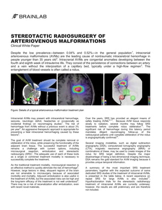
- 1. STEREOTACTIC RADIOSURGERY OF ARTERIOVENOUS MALFORMATIONS Clinical White Paper Despite the low prevalence—between 0.04% and 0.52%—in the general population 1, intracranial arteriovenous malformations (AVMs) are the leading cause of nontraumatic intracerebral hemorrhage in people younger than 35 years old 2. Intracranial AVMs are congenital anomalies developing between the fourth and eighth week of intrauterine life. They consist of the persistence of connections between an artery and a vein without the interposition of a capillary bed, typically under a high-flow regimen3. This entanglement of blood vessels is often called a nidus. Figure: Details of a typical arteriovenous malformation treatment plan Intracranial AVMs may present with intracerebral hemorrhage, seizures, neurologic deficit, headaches or occasionally as incidental findings on neuroimaging studies4. The risk of hemorrhage from AVMs without a previous event is about 3% 5 per year . An aggressive therapeutic approach is appropriate for preventing a fatal intracranial hemorrhaging caused by these 6 lesions . The goal of AVM treatment should be complete removal or obliteration of the nidus, while preserving the functionality of the adjacent brain tissue. The successful treatment of AVMs remains a challenge with current options, including microsurgical resection, embolization and stereotactic 7 radiosurgery (SRS) . The adequate use of each of these tools as a single or combined treatment modality is necessary to successfully complete the treatment. As the traditional treatment modality, microsurgical resection is an effective method to quickly eliminate the risk of hemorrhage8. However, large lesions in deep, eloquent regions of the brain are not amenable to microsurgery because of associated morbidity and mortality. Adjuvant embolization is also useful in the treatment of AVMs, but the associated risks can be high and 9 the long-term efficacy of the treatment is not well understood . There may be a risk of recanalization after embolization, even with recent novel materials. Over the years, SRS has provided an elegant means of 10-17 . Because AVM tissue responds safely treating AVMs slowly to radiation, several months may follow SRS treatments before complete nidus obliteration18. The significant risk of hemorrhage during this latency period mandates diligent neuroimaging follow-up of the radiosurgical patients until complete obliteration of the nidus 19 is angiographically confirmed . Several imaging modalities, such as digital subtraction angiography (DSA), computerized tomography angiography (CTA), magnetic resonance angiography (MRA) or combinations of these, may be used to create images of the nidus for SRS target definition. Despite the major disadvantage of being a two-dimensional imaging technique, DSA remains the gold standard for AVM imaging because it provides unique temporal information. A summary of the most important SRS treatment parameters, together with the expected outcome of some dedicated SRS studies of the treatment of intracranial AVMs, is presented in the table below. A recent experience on 17 repeat SRS for large AVMs is also included . Hypofractionation trials for the stereotactic radiotherapy treatment of intracranial AVMs are currently underway; however, the results are still preliminary and are therefore not included.
- 2. In general, patients are immobilized with a frame and a single mean dose of 15 to 19 Gy is prescribed to the 80% isodose line. The dose is typically delivered by four to eight arcs—for spherical lesions, conical collimators can be preferred over high-resolution multi-leaf collimators. The overall incidence of radiation-induced complications ranges from 3 to 6% and the reported neurological deficits 10-17 . are often of transient nature It is therefore concluded that SRS is a safe and effective intracranial AVM treatment option, as long as careful neuroimaging follow-up is guaranteed to monitor the nidus response. Intracranial AVMs are often classified according to a grading 20 score developed by Spetzler and Martin and the prescribed dose is related to the initial AVM grade, since complete nidus obliteration rates were found to depend mainly on the AVM 11,13,15 . volume and the SRS dose Overview of the recent clinical literature on SRS for arteriovenous malformations Institution Pedroso Buis 12 Scarbrough 14 Zabel-du Bois Huang 13 16 MorenoJiménez Raza 10 17 % Prior Treatment Mean Vol (cm³) Mean Dose (Gy) # Fractions % IDL covering PTV % Complete Obliteration 1999 50 36 23 16 1 80 45 at 20 months 2004 44 30 18 15 1 80 52 at 37 months VU University Medical Center, Amsterdam 15 # Lesions David Geffen School of Medicine, Los Angeles 11 Mobin Year University of California, Davis Author 2005 31 32 3 19 1 80 77 at 33 months The Melbourne Cancer Center, Melbourne 2005 39 8 7 17 1 80 87 at 24 months University of Heidelberg 2006 22 36 4 18 1 80 65 at 48 months Ghang Gung Memorial Hospital, Taiwan 2006 34 14 2 16 1 80 NA Nat´l Institute of Neurol & Neurosurg, Mexico 2007 40 40 8 15.4 1 80 63 at 29 months The Johns Hopkins Hospital, Baltimore 2007 14 47 25 36 3 NA 36 at 31 months Complete obliteration implies that the nidus is no longer visible angiographically and that the circulation time and the afferent and efferent vessels that had supplied the malformation have returned to normal. For angiographically occult lesions like low-flow cavernous malformations, studied by Huang et al., there is currently no gold standard for demonstrating the obliteration. References [1] [2] [3] [4] [5] [6] [7] [8] [9] [10] Pollock B.E. et al., Stroke 27, 1, 1996 Karhunen P.J. et al., Forensic Sci Int 48, 9, 1990 Fleetwood I.G. et al., Lancet 359, 863, 2002 Thompson R.C. et al., Neurosurgery 43, 202, 1998 Nussbaum E.S. et al., Neurosurgery 43, 347, 1998 Sasaki T. et al., J Neurosurg 88, 285, 1998 Ellis T.L. et al., J Neurosurg 89, 104, 1998 Lawton M.T., Neurosurg 52, 740, 2003 Yu S.C. et al., AJNR Am J Neuroradiol 25, 1139, 2004 Moreno-Jiménez S. et al., Surg Neurol 67, 487, 2007 Europe | +49 89 99 1568 0 | de_sales@brainlab.com North America | +1 800 784 7700 | us_sales@brainlab.com Latin America | +55 11 3355 3370 | br_sales@brainlab.com RT_WP_E_AVM_APR11 [11] [12] [13] [14] [15] [16] [17] [18] [19] [20] Mobin F. et al., Stereotact Funct Neurosurg 73, 50, 1999 Buis D.R. et al., Int J Radiat Oncol Biol Phys 62(1), 246, 2005 Zabel-du Bois A. et al., Int J Radiat Oncol Biol Phys 65(4), 1206, 2006 Scarbrough T.J. et al., Stereotact Funct Neurosurg 83, 91, 2005 Pedroso A.G. et al., J Neurosurg 101, 425, 2004 Huang Y.C. et al., Clin Neurol Neurosur 108, 750, 2006 Raza S.M. et al., Surg Neurol 68, 24, 2007 Pollock B.E. et al., Neurosurg 38, 652, 1996 Maruyama K. et al., N Engl J Med 352, 146, 2005 Spetzler et al., J Neurosurg 65, 476, 1986 Asia Pacific | +852 2417 1881 | hk_sales@brainlab.com Japan | +81 3 3769 6900 | jp_sales@brainlab.com