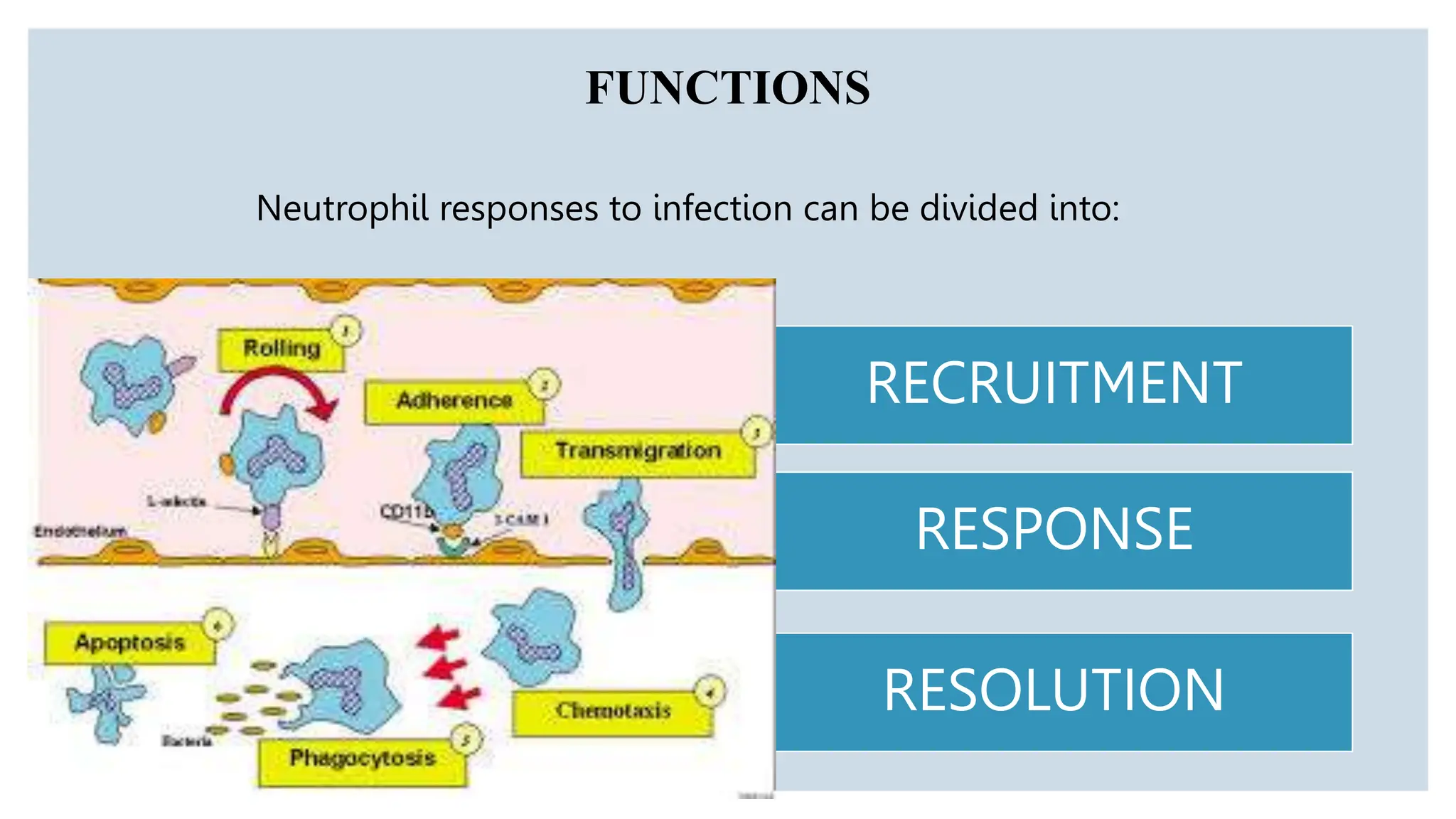
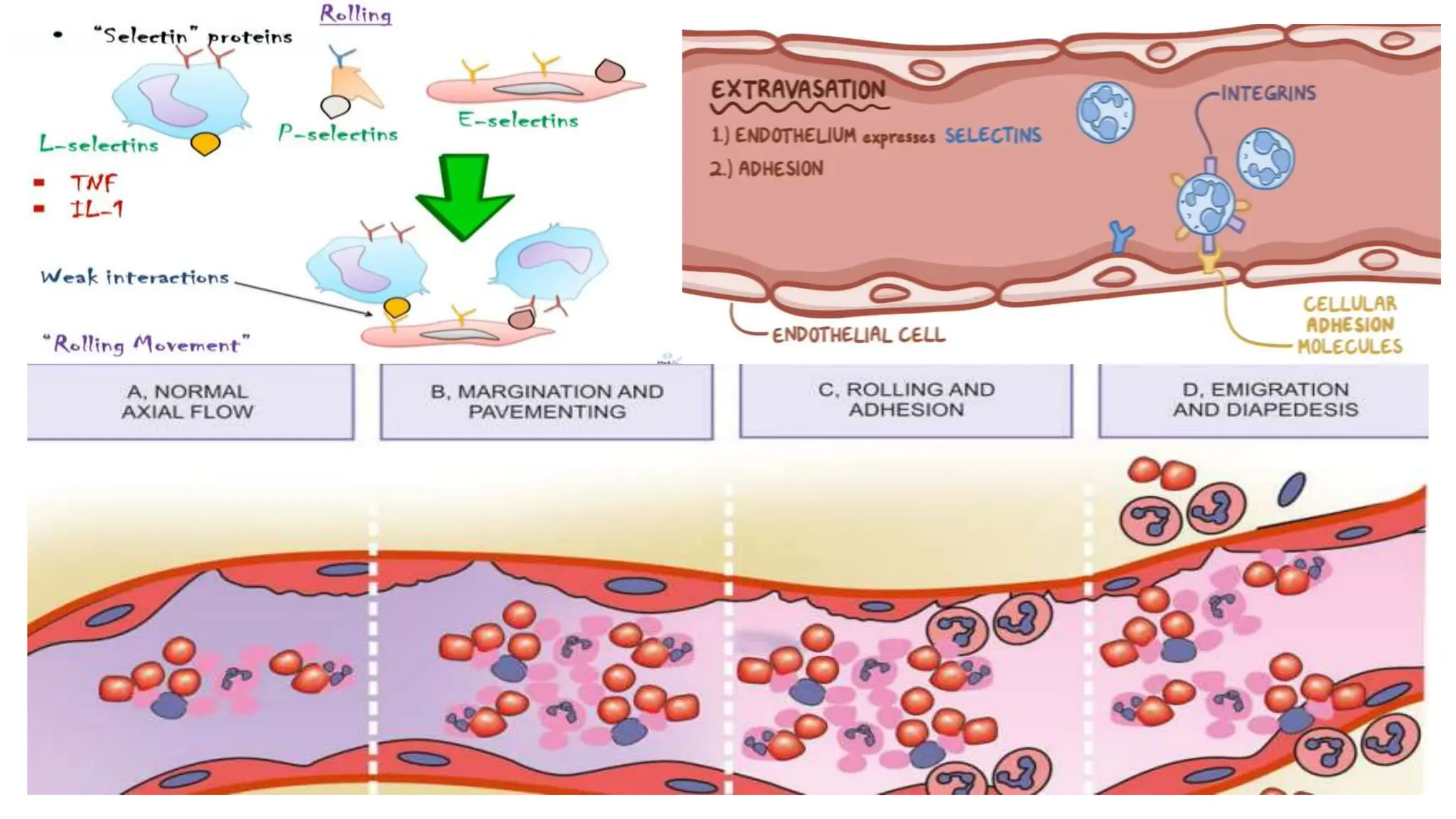
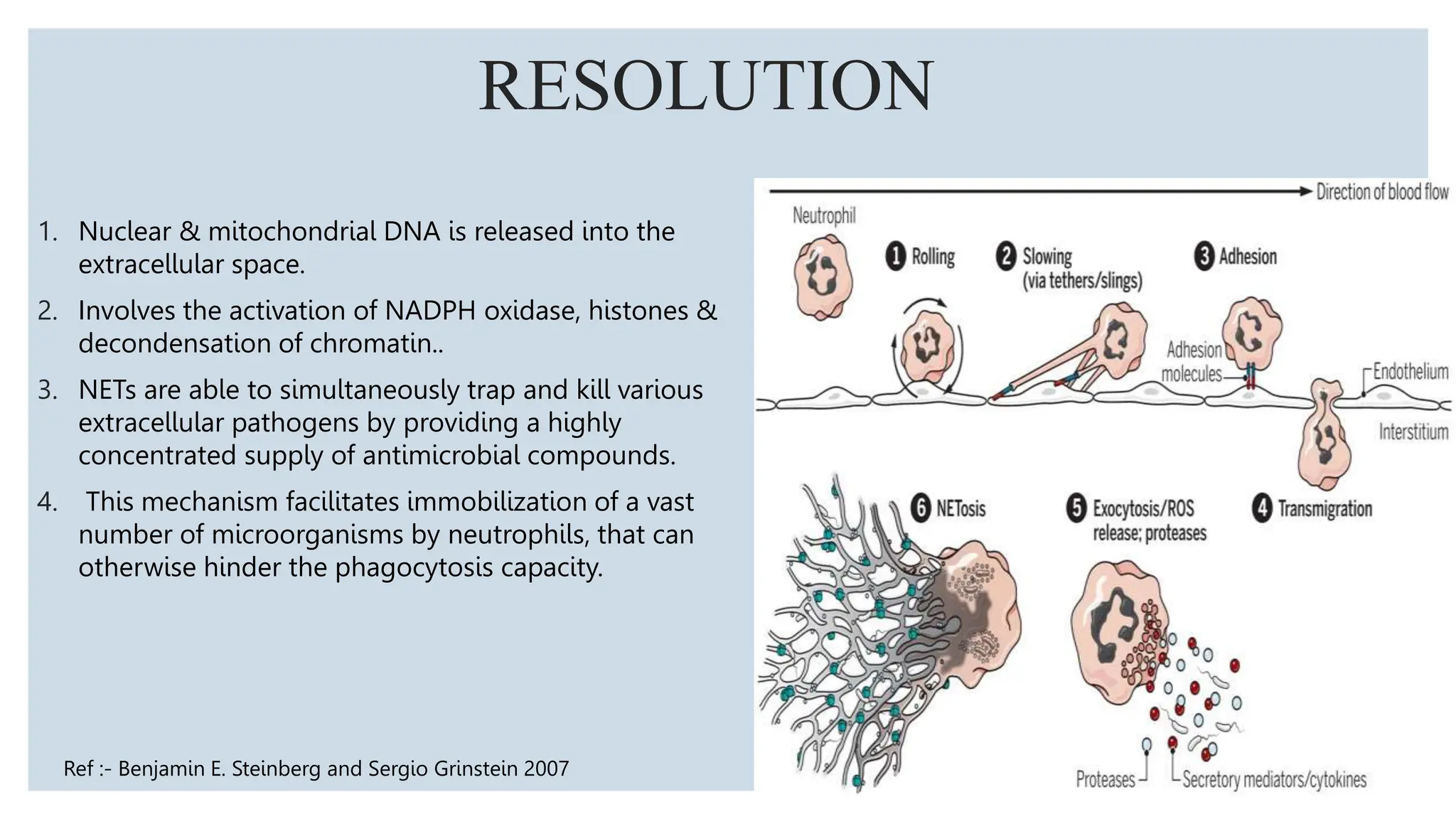
Neutrophils are the most abundant white blood cells and form the first line of defense against pathogens. They are formed in the bone marrow through a process called granulopoiesis regulated by G-CSF and migrate to sites of infection. Neutrophils phagocytose and kill microbes via oxidative and non-oxidative mechanisms. Disorders can cause quantitative or qualitative neutrophil defects, resulting in increased susceptibility to infection. Periodontal disease is more severe and widespread in patients with neutrophil disorders due to impaired host response and bacterial modulation of neutrophil functions.
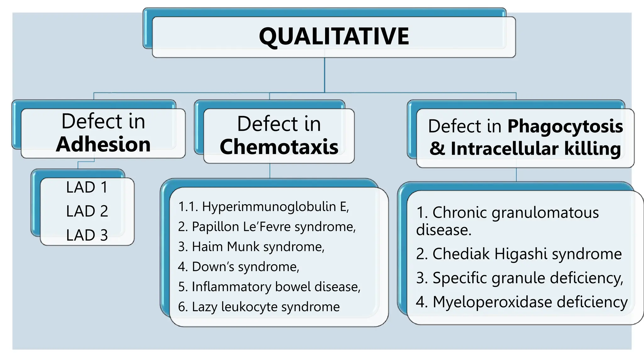
THE ROLE OF MACROPHAGE IN PERIODONTICS.pptxPrasanthThalur
The document discusses the role of macrophages and innate immunity in periodontitis. It describes how macrophages recognize pathogens via pattern recognition receptors like toll-like receptors and NLR inflammasomes. This activation of these receptors triggers signaling cascades that promote inflammation and the secretion of cytokines. Overactivation of these pathways can lead to excessive inflammation and tissue damage. The document also explains that macrophages can polarize into pro-inflammatory M1 or anti-inflammatory M2 states, and their plasticity and different states influence periodontitis development.
This document discusses neutrophil disorders and their relationship to periodontal diseases. It begins with an introduction on the role of neutrophils in the innate immune system and periodontal diseases. It then describes various quantitative and qualitative neutrophil disorders. Quantitative disorders discussed include chronic benign neutropenia, cyclic neutropenia, congenital neutropenia, agranulocytosis, and Felty's syndrome. Qualitative disorders result from defects in neutrophil functions like rolling, adhesion, chemotaxis, phagocytosis, and intracellular killing. The document examines the oral complications that can result from various neutrophil disorders like gingivitis, periodontitis, and bone loss.
Apoptosis is the programmed cell death. Aim of cancer therapy is to destroy the invading cells. Cancerous cells can be destroyed by increasing apoptosis.
it can occur in both physiological and pathological conditions. It is different from necrosis. In necrosis, the cell contents leak out and lead to inflammation. But in apoptosis there is no cellular leakage, only apoptotic bodies are formed. They are then engulfed by macrophages.
This document provides an overview of oral submucous fibrosis (OSF), including its definition, epidemiology, classification, etiology, pathogenesis, clinical features, and histopathology. OSF is a chronic disease characterized by inflammation and fibrosis of the submucosal tissues caused by chewing areca nut. It predominantly affects people from South Asia and is associated with significantly increased risk of oral cancer. The areca nut alkaloid arecoline is the main causative agent, inducing fibrosis through oxidative damage, upregulation of growth factors and cytokines, and inhibition of collagen degradation. Clinically, OSF presents with burning sensation and scarring that results in restricted mouth opening and tongue movement.
1. Generalized aggressive periodontitis is a form of periodontitis characterized by generalized attachment and bone loss affecting at least three permanent teeth other than first molars and incisors.
2. It typically affects individuals under 30 years of age and is associated with episodic periods of rapid destruction followed by periods of quiescence. Only small amounts of bacterial plaque are present relative to the degree of destruction.
3. Risk factors include certain pathogenic bacteria like P. gingivalis and A. actinomycetemcomitans, immune defects in neutrophils and monocytes, familial aggregation, and smoking can influence the extent of destruction.
This document provides information on eosinophils:
- Eosinophils are white blood cells that are characterized by distinctive granules and a bilobed nucleus. They play a role in fighting parasites and allergic responses.
- Eosinophils develop from bone marrow stem cells and are regulated by cytokines like IL-5, IL-3 and GM-CSF. Transcription factors like C/EBP, GATA-1 and PU.1 are required for eosinophil development.
- Eosinophils traffic to tissues where they can persist for over a week. Their granules contain toxic proteins that are released during degranulation which is regulated by cytokines and chemokines.
- Eosin
This document summarizes research on oral lichen planus (OLP) and its potential as a preneoplastic inflammatory model. Key points include:
- OLP is a chronic inflammatory oral condition that may carry a 1-2% risk of malignant transformation to oral squamous cell carcinoma.
- Research has explored the role of T-cells, apoptosis, cellular proliferation, p53 expression, chromosomal instability, matrix metalloproteinases, cytokines, and hepatitis C virus infection in the pathogenesis and potential malignant transformation of OLP.
- Further research is needed but OLP shows similarities to other inflammatory conditions like inflammatory bowel disease that are associated with increased cancer risk, supporting its characterization as a potentially pre
This document reviews oral manifestations and dental management considerations for patients with leukocyte alterations. Key points include:
1. Oral complications of neutropenia can include necrotic ulcers, severe gingivitis, and periodontitis. Agranulocytosis presents as necrotic ulcers without signs of inflammation. Congenital neutropenia risks periodontal disease despite treatment.
2. Dental treatment for patients with neutrophil disorders emphasizes preventive care and controlling infections to minimize surgery. Acceptable blood counts are recommended for oral surgery.
3. Leukemia can cause gingival hyperplasia from infiltration of leukemia cells. This presents as swollen, pale gingiva and
This document discusses a new taxonomy for classifying podocytopathies, diseases involving injury to podocytes. It proposes organizing podocytopathies based on their histopathology (morphological pattern of glomerular injury and podocyte number) and etiology (idiopathic, genetic, reactive). Four main morphological patterns are described: minimal change nephropathy (MCN), focal segmental glomerulosclerosis (FSGS), diffuse mesangial sclerosis (DMS), and collapsing glomerulopathy (CG). Each pattern is defined and their etiologies and clinical associations are discussed. The taxonomy aims to integrate morphological diagnoses with etiology based on current understanding of podocyte biology and
This document provides information on desquamative gingivitis, including its classification, diagnosis, and associated diseases. It classifies desquamative gingivitis into 7 categories including dermatosis, endocrine imbalance, aging, metabolic disturbances, abnormal response to irritation, chronic infection, and drug reactions. Key aspects of diagnosis include clinical history, examination, biopsy, and microscopic/immunofluorescence examination. Associated diseases discussed in detail include lichen planus, pemphigoid, pemphigus vulgaris, chronic ulcerative stomatitis, and linear IgA disease. Treatment varies depending on the underlying cause and severity of symptoms.
Chronic granulomatous disease (CGD) is an inherited immune disorder where phagocytes cannot kill ingested bacteria and fungi, leading to recurrent infections. It is caused by mutations affecting the NADPH oxidase enzyme complex needed for reactive oxygen species production. Patients experience severe lung, skin, and organ infections from catalase-positive microbes early in life. Diagnosis involves tests assessing reactive oxygen species levels. Treatment requires lifelong antibiotics, antifungals, immunoglobulin therapy, and stem cell transplantation.
This document discusses gingival inflammation and gingivitis. It begins by defining inflammation and describing the cardinal signs. It then outlines the stages of gingivitis from initial to established to advanced/periodontitis. Microorganisms attached to teeth secrete enzymes that damage tissues and widen junctional epithelium, allowing bacterial products to access connective tissue and activate immune cells. Studies showed that not practicing oral hygiene led to plaque buildup and gingivitis within 10-21 days. Gingivitis is characterized by redness, swelling, bleeding and is prevalent worldwide. The document discusses features, course, distribution and systemic influences of gingival inflammation.
1. Cells have the ability to adapt and respond to injury through processes like hypertrophy, hyperplasia, atrophy, and metaplasia. However, if the injury is too severe, it can lead to cell death through either necrosis or apoptosis.
2. Necrosis is unprogrammed cell death that results in cell contents leaking out. Apoptosis is programmed cell death where the cell activates enzymes to degrade its own DNA and proteins in a controlled manner.
3. Oxidative stress from free radicals and mitochondrial damage are key mechanisms that can lead to cell injury. If the injury is not reversible, it triggers signaling pathways to initiate necrosis or apoptosis.
1. Aggressive periodontitis is a rare, severe form of periodontitis typically affecting young individuals under 30 years of age. It is characterized by rapid attachment and bone loss.
2. It is caused by specific pathogens like Aggregatibacter actinomycetemcomitans and has a strong genetic component. Patients often exhibit impaired neutrophil function and hyper-inflammatory responses to bacterial toxins.
3. Aggressive periodontitis is classified into localized and generalized forms depending on the extent of involvement and treated through non-surgical therapies like scaling and root planing along with systemic antibiotics.
This document discusses genetic polymorphism and its relationship to periodontal disease. It begins by defining key genetic terms and describes how periodontal disease can be caused by single gene mutations (monogenic disorders) or variations in multiple genes (polygenic disorders). Several specific gene polymorphisms are examined, including genes related to cytokines, receptors, metabolism, and innate immunity. The roles of the IL-1, TNF-α, IL-10 genes and others are summarized. The document also explores several genetic syndromes associated with early-onset periodontitis, such as Papillon-Lefevre syndrome. In conclusion, genetic testing is available to assess patient susceptibility to severe periodontal disease.
This document classifies and describes the types of white blood cells (WBCs), also known as leukocytes. It discusses the normal counts of each type of WBC, their structures, functions, and variations seen in conditions like infection, malnutrition, and leukemia. The main types of WBCs are granulocytes (neutrophils, eosinophils, basophils) and agranulocytes (lymphocytes, monocytes). Neutrophils are the most abundant WBC and function in phagocytosis and inflammation. Leukemia is a cancer of the blood characterized by an abnormal increase in immature WBCs in the bloodstream.
Hematologic disorders and immune deficiencies can profoundly impact the periodontium. Disorders that affect the production or function of white blood cells like neutrophils may result in severe periodontal destruction due to lack of bacterial defense. These include neutropenia, agranulocytosis, lazy leukocyte syndrome, and chronic granulomatous disease. Leukemia involves the malignant transformation of blood cell precursors in the bone marrow, displacing normal cells and reducing blood cell production. Symptoms vary depending on the type and severity of the underlying condition. Abnormal bleeding or infections in the oral cavity can indicate an underlying hematologic or immunodeficiency disorder.
This document discusses hereditary spherocytosis (HS), an inherited disorder caused by defects in the red blood cell membrane skeleton. HS is characterized by spherical red blood cells that are less deformable and vulnerable to splenic sequestration and destruction. The defects are primarily in proteins like ankyrin, band 3, spectrin or band 4.2 that make up the membrane skeleton. This leads to a reduced red blood cell life span of 10-20 days instead of the normal 120 days. Clinical features include anemia, splenomegaly, jaundice, and hemolytic crises from infections. Diagnosis involves tests like peripheral smear, bone marrow examination, and osmotic fragility testing. Treatment
'Oral Potentially Malignant Disorders' includes a variety of lesions with risk of progression to malignancy. It is widely prevalent in the Indian population, and early diagnosis and management is the need of the hour.
Here's a discussion of the same with methods of early diagnosis of such lesions.
The gingiva provides three lines of defense against pathogens:
1. The epithelial surface acts as a mechanical and chemical barrier. Tight junctions between keratinocytes and antimicrobial peptides in epithelial layers prevent pathogen entry.
2. Components in saliva and gingival crevicular fluid (GCF) maintain tissue health and help remove debris from the sulcus. GCF is an inflammatory exudate containing enzymes, electrolytes, and host/bacterial compounds.
3. The gingival tissue mounts an innate and adaptive immune response. Langerhans cells and neutrophils present in the junctional epithelium phagocytose pathogens, while T cells and antibodies provide acquired immunity. Together,
Retinoblastoma is the most common intraocular malignancy of childhood arising from retinal cells. It can present as unilateral or bilateral tumors that are either sporadic or hereditary. The Knudson "two hit" hypothesis explains that two mutations in the RB1 gene are required for retinoblastoma to develop. Clinically, it may appear as a white pupil, strabismus, glaucoma, or proptosis. Diagnosis involves ophthalmoscopy, ultrasound, CT, MRI and biopsy if needed. Differential diagnoses include toxocariasis, persistent hyperplastic primary vitreous, and Coats' disease.
This document provides an overview of lichen planus, including:
- It is a chronic inflammatory autoimmune disease that affects the oral mucosa and skin.
- The pathogenesis involves a cell-mediated immune response targeting basal keratinocytes.
- It has various clinical presentations depending on morphology and site of involvement, such as reticular, papular, plaque-like, erosive, atrophic and bullous forms.
Dr William Barnes - The I Factor - Inflammation, Immunity, IllnessDr William Barnes
Immunity and Inflammation
Inflammation and Macrophages
Macrophage Pathology
Foam Cell, Viruses, TAMs
The Brune Theory & Nitric Oxide
Macrophages in Disease States
Treatment Strategies
Colds and Influenza
Treatment Strategies
Ageing is caused by the progressive loss of structural and functional capacity in cells over time, leading to death. It is influenced 60% by genetic factors and 40% by environmental factors such as trauma, disease, diet, and stress. The main structural and biochemical changes that occur during ageing include the accumulation of oxidative damage in cells from reactive oxygen species, advanced glycation end products, and abnormally folded proteins. Telomerase plays a role in preventing shortening of telomeres during cell division. Restricting calorie intake and maintaining good health habits can help delay the cellular ageing process.
Similar to ROLE OF NEUTROPHILS IN HEALTH & DISEASE.pptx (20)
Local Advanced Lung Cancer: Artificial Intelligence, Synergetics, Complex Sys...Oleg Kshivets
Overall life span (LS) was 1671.7±1721.6 days and cumulative 5YS reached 62.4%, 10 years – 50.4%, 20 years – 44.6%. 94 LCP lived more than 5 years without cancer (LS=2958.6±1723.6 days), 22 – more than 10 years (LS=5571±1841.8 days). 67 LCP died because of LC (LS=471.9±344 days). AT significantly improved 5YS (68% vs. 53.7%) (P=0.028 by log-rank test). Cox modeling displayed that 5YS of LCP significantly depended on: N0-N12, T3-4, blood cell circuit, cell ratio factors (ratio between cancer cells-CC and blood cells subpopulations), LC cell dynamics, recalcification time, heparin tolerance, prothrombin index, protein, AT, procedure type (P=0.000-0.031). Neural networks, genetic algorithm selection and bootstrap simulation revealed relationships between 5YS and N0-12 (rank=1), thrombocytes/CC (rank=2), segmented neutrophils/CC (3), eosinophils/CC (4), erythrocytes/CC (5), healthy cells/CC (6), lymphocytes/CC (7), stick neutrophils/CC (8), leucocytes/CC (9), monocytes/CC (10). Correct prediction of 5YS was 100% by neural networks computing (error=0.000; area under ROC curve=1.0).
- Video recording of this lecture in English language: https://youtu.be/kqbnxVAZs-0
- Video recording of this lecture in Arabic language: https://youtu.be/SINlygW1Mpc
- Link to download the book free: https://nephrotube.blogspot.com/p/nephrotube-nephrology-books.html
- Link to NephroTube website: www.NephroTube.com
- Link to NephroTube social media accounts: https://nephrotube.blogspot.com/p/join-nephrotube-on-social-media.html
Adhd Medication Shortage Uk - trinexpharmacy.comreignlana06
The UK is currently facing a Adhd Medication Shortage Uk, which has left many patients and their families grappling with uncertainty and frustration. ADHD, or Attention Deficit Hyperactivity Disorder, is a chronic condition that requires consistent medication to manage effectively. This shortage has highlighted the critical role these medications play in the daily lives of those affected by ADHD. Contact : +1 (747) 209 – 3649 E-mail : sales@trinexpharmacy.com
Histololgy of Female Reproductive System.pptxAyeshaZaid1
Dive into an in-depth exploration of the histological structure of female reproductive system with this comprehensive lecture. Presented by Dr. Ayesha Irfan, Assistant Professor of Anatomy, this presentation covers the Gross anatomy and functional histology of the female reproductive organs. Ideal for students, educators, and anyone interested in medical science, this lecture provides clear explanations, detailed diagrams, and valuable insights into female reproductive system. Enhance your knowledge and understanding of this essential aspect of human biology.
These lecture slides, by Dr Sidra Arshad, offer a quick overview of the physiological basis of a normal electrocardiogram.
Learning objectives:
1. Define an electrocardiogram (ECG) and electrocardiography
2. Describe how dipoles generated by the heart produce the waveforms of the ECG
3. Describe the components of a normal electrocardiogram of a typical bipolar lead (limb II)
4. Differentiate between intervals and segments
5. Enlist some common indications for obtaining an ECG
6. Describe the flow of current around the heart during the cardiac cycle
7. Discuss the placement and polarity of the leads of electrocardiograph
8. Describe the normal electrocardiograms recorded from the limb leads and explain the physiological basis of the different records that are obtained
9. Define mean electrical vector (axis) of the heart and give the normal range
10. Define the mean QRS vector
11. Describe the axes of leads (hexagonal reference system)
12. Comprehend the vectorial analysis of the normal ECG
13. Determine the mean electrical axis of the ventricular QRS and appreciate the mean axis deviation
14. Explain the concepts of current of injury, J point, and their significance
Study Resources:
1. Chapter 11, Guyton and Hall Textbook of Medical Physiology, 14th edition
2. Chapter 9, Human Physiology - From Cells to Systems, Lauralee Sherwood, 9th edition
3. Chapter 29, Ganong’s Review of Medical Physiology, 26th edition
4. Electrocardiogram, StatPearls - https://www.ncbi.nlm.nih.gov/books/NBK549803/
5. ECG in Medical Practice by ABM Abdullah, 4th edition
6. Chapter 3, Cardiology Explained, https://www.ncbi.nlm.nih.gov/books/NBK2214/
7. ECG Basics, http://www.nataliescasebook.com/tag/e-c-g-basics
Muktapishti is a traditional Ayurvedic preparation made from Shoditha Mukta (Purified Pearl), is believed to help regulate thyroid function and reduce symptoms of hyperthyroidism due to its cooling and balancing properties. Clinical evidence on its efficacy remains limited, necessitating further research to validate its therapeutic benefits.
3. ◦ The cellular components of blood contains :- RBC / Erythrocytes
WBC/ leucocytes
Platelet/ Thrombocytes.
◦ Neutrophils are the most abundant leucocytes, constituting 70% of WBCs .
◦ They are multilobed containing 2-5 lobes of nucleus hence the name polymorphonuclear (PMNs)
◦ They stain neutral with Wright stain hence the name neutrophils.
◦ The are the first line of defense in immunity.
INTRODUCTION
5. • Neutrophils formation in the bone marrow takes about 14days.
• Neutrophils are continuously generated in the bone marrow from myeloid precursors. Their daily production can reach up to
2 x 10¹¹ cells.
• The process is controlled by granulocyte colony stimulating factor (G-CSF).
LEUKOPOIESIS
6.
7. Mature neutrophils retained in the
bone marrow:- interaction between
the CXCCR4 on the PMN &
CXCL12 produced by stromal cells
of bone marrow
IL-17 upregulates G-
CSF which promotes
release of neutrophils
from bone marrow.
At the inflammatory site the PMNs
release chemokines CCL2 & CCL20
which are ligands for chemokine
receptors on Th17.
This attracts the Th17 cell to the site
of inflammation.
Helper T cells release IL-17
which in turn recruits more
PMNs to the site of
inflammation .
Apoptotic PMNs are
phagocytosed by the
macrophages
IL-23 released
from the
macrophages
activate Th17
IL 11 production –
upregulation of G-CSF
in fibroblasts.
Initiation of anti-
inflammatory signal-
reduction in IL-23 from
macrophages.
Reduction in IL 17
production.
Reduced G-CSF
production
Reduced production of
neutrophils.
This mechanism of
feedback control
over neutrophil
production is called
NEUTROSTAT
TRAFFICKING
10. Neutrophil responses to infection can be divided into:
RECRUITMENT
RESPONSE
RESOLUTION
FUNCTIONS
11. 1. Changes in the formed elements of blood/ Margination
2. Rolling and adhesion
3. Transmigration
4. Chemotaxis
RECRUITMENT
12.
13.
14. Phagocytosis :- 1. Recognition and attachment
2. Engulfment
3. Killing and degradation
RESPONSE
15. RECOGNITION AND ATTACHMENT
• Receptors on macrophages which recognize microorganisms: mannose receptor and scavenger receptor.
• Microorganisms are coated with specific proteins, opsonins, from the serum and the process is called opsonization.
• Opsonins establish a bond between bacteria and the cell membrane of phagocytic cell.
• The main opsonins present in the serum and their corresponding receptors on the surface of phagocytic cells (PMNs or
macrophages) are as under:
i. IgG & C3b found in serum protein - opsonin.
ii. Receptors present on the surface of the phagocyte - Fc , CR1 &3 and C1q
iii. Lectins are carbohydrate-binding proteins in the plasma which bind to bacterial cell wall.
16. KILLING AND DEGRADATION
◦ In phagosomes, PMNs kill microorganisms
through two mechanisms:
1. Oxygen dependent mechanism (ROS,
generated by NADPH oxidase)
2. Oxygen independent mechanism (granular
proteases)
17. RESOLUTION
1. Nuclear & mitochondrial DNA is released into the
extracellular space.
2. Involves the activation of NADPH oxidase, histones &
decondensation of chromatin..
3. NETs are able to simultaneously trap and kill various
extracellular pathogens by providing a highly
concentrated supply of antimicrobial compounds.
4. This mechanism facilitates immobilization of a vast
number of microorganisms by neutrophils, that can
otherwise hinder the phagocytosis capacity.
Ref :- Benjamin E. Steinberg and Sergio Grinstein 2007
18. NEUTROPHIL CLEARANCE
• Apoptotic PMNs are phagocytosed
by the macrophages.
• The senescent PMNs return to the
bone marrow for clearance after they
have increased expression of
CXCCR4.
19. NEUTROPHIL DEFECTS
QUANTITATIVE
Decreased in
number of
neutrophils –
NEUTROPENIA
Increase in
number of
neutrophils-
NEUTROPHILI
A
QUALITATIVE
Defect in
ADHESION
Defect in
CHEMOTAXIS
Defect in
PHAGOCYTOSIS
&
INTRACELLULAR
KILLING
21. NEUTROPENIA
DISEASE PMN ABNORMAILTY CLINICAL FEATURES
CYCLIC NEUTROPENIA 1. Cause – mutation for gene neutrophil
elastase (ELA2).
2. Recurring episodes of neutropenia
occurring every 21days.
3. These episodes last for 3-6 days.
1. Malaise
2. Anorexia
3. Lymphadenopathy
4. Oral ulcers resembling aphthous
ulcers.
FELTY’S SYNDROME
(Augustus Felty in 1924)
1. Triad of – rheumatoid arthritis,
splenomegaly, neutropenia
1. Anemia, mucosal and skin ulcers,
2. respiratory tract infections,
3. thrombocytopenia, and
4. lymphadenopathy
KOSTMANN’S SYNDROME
(Rolf Kostmann in 1956 )
1. Impairment of myeloid differentiation
in the bone marrow with severe
absolute neutropenia.
2. less than 500 cells/L.
1. Frequent pyogenic infections since
birth, which included oral ulceration,
stomatitis and pharyngitis.
2. Cutaneous cellulitis, furunculosis,
pneumonia and septicaemia occurred
frequently, as did chronic
periodontitis.
22. 1. The attached gingiva surrounding all of the primary teeth exhibited
severe gingivitis with ulceration (Figure 1).
2. This gingivitis extended to the mesial of the first permanent molars. The
ulcerations were more evident in the interdental papilla.
3. An obvious loss of attachment surrounding the primary teeth
4. Probing of these teeth elicited hemorrhage and revealed pocket depths
ranging from 4-10 mm.
CYCLIC NEUTROPENIA : A CASE REPORT
Linwood M. Long, Jr., DDS, MS John R. Jacoway, DDS, PhD James W. Bawden, DDS, MS, PhD
23. Oral manifestations of
congenital neutropenia or
Kostmann syndrome
Periodontal disease was so rapidly destructive in this patient that the
permanent lower anterior teeth were almost exfoliated by the age of 14
years .
26. LEUCOCYTE ADHESION DEFICIENCY (Anderson &Spring,1986)
LAD 1
• Cell surface integrin (CD18) is affected.
• ADHSION affected
LAD 2
• Defect in Saily-lewis X glycoprotein CD15 which allows neutrophils to attach to selectins CD621 on the endothelial surface.
• ROLLING is affected
CLINICAL FEATURES
1. Both the primary and the permanent teeth are affected, often resulting in early tooth loss.
2. Profound defects in peripheral blood neutrophils and monocytes and an absence of neutrophils in the gingival tissues have
been noted in patients with LAD.
LAD 3
• Defective signaling of Beta 1, 2, 3 Integrins.
• INTEGRIN SIGNALLING, therefore ADHESION affected
28. DISEASE PMN ABNORMALITY CLINICAL FEATURES
HYPERIMMUNOGLOBULIN E /
JOB’S SYNDROME
1. Increased levels of serum IgE
2. Reduced chemotaxis
1. Skin ‘‘cold’’ abscesses,
2. pneumonia,
3. deep-set eyes,
4. oral ulcerations/gingivitis
HAIM-MUNK SYNDROME 1. Mutations of a gene (known as
cathepsin C [CTSC]) located on
the long arm of chromosome 11
1. Red, scaly thickened patches of skin on the
palms of the hands and soles of the feet .
2. Frequent pyogenic skin infections.
3. Overgrowth of the fingernails and toenails.
4. Degeneration of the periodontium
LAZY LEUKOCYTE SYNDROME 1. Affect both quality and quantity
of neutrophils.
2. Defective chemotaxis along with
neutropenia is seen.
1. Destruction of bone and early tooth loss.
2. Fever skin abscess, gingivitis, periodontitis
DOWNS SYNDROME 1. Endo - lack of lip seal, tongue
thrust, malocclusion, oral
hygiene
2. Exo - neutrophil, phagocytosis,
chemo intracellular killing
(Newman & Carranza 10th edition )
1. The high prevalence and increased severity of
periodontal destruction
29. Cathepsin is present in phagocytes and epithelium.
1.In phagocytes it activates lysosomal granules resulting in phagocytosis.
Mutations in the cathepsin C gene
Non – functional cathepsin C in epithelium - palmoplantar
hyperkeratosis
Defective barrier function of JE, phagocyte defect, reduced host
response – periodontal destruction.
Hallmark feature – palmoplantar keratosis + rapid periodontal
destruction of both dentitions.
PAPILLON–LEFÈVRE SYNDROME
Vol. 6, 1994, 88-1 00
THOMAS C. HART &
LIOR SHAPIRA
31. CHEDIAK HIGASHI SYNDROME
CHÉDIAK–HIGASHI SYNDROME
Mutation in LYST gene that aids in intracellular transport of
materials into lysosomes
Azurophilic granules and specific granules unite to form,
large granules called Megabodies.
Phagosome cannot fuse with the lysosome to form
phagolysosome
Phagocytosis and intercellular killing affected
Severe periodontitis, affecting both temporary and
permanent dentition
32. CHRONIC GRANULOMATOUS DISEASE (CGD)
◦ Seen in staphylococcus, proteus ,
pseudomonas species infections.
Congenitally defective NADPH oxidase
Reduced respiratory burst
Difficulty in killing pathogens
Over a period of time, cells gather around the
pathogen to stop it from spreading ( diag)
This leads to the formation
of granulomas in many organs
33. ◦ Nicotine alters the chemotaxis, phagocytosis and respiratory burst mechanism of
neutrophils.
◦ Elevated levels of TNF a and IL- 8, PGE2, neutrophil elastase, and matrix
metalloproteinase-8 are demonstrated in GCF.
◦ IgG2 has been seen to be reduced in smokers thus making them susceptible to
periodontal bacteria.
◦ Inhibition of fibroblast growth, attachment and collagen production.
◦ Altered cytokine production.
Smoking and periodontal disease
34. Diabetes Mellitus
◦ Diabetic patients demonstrate defects in PMN activity, including impaired chemotaxis,
phagocytosis and microbicidal functions .
◦ Diabetes prolongs the inflammatory response to Porphyromonas gingivalis, with increased
production of TNF-α
35. Treponema denticola
◦ Anaerobic, gram-negative motile spirochete.
◦ T . Denticola degrades IL-8 , which disables the neutrophil chemotactic gradient.
◦ Modulates neutrophil signaling pathways involved in cytoskeletal dynamics that are relevant in
chemotaxis and phagocytosis.
36. Porphyromonas gingivalis
◦ gingival epithelial cells (GECs) secrete the chemoattractant IL-8 to recruit neutrophils.
◦ P. gingivalis inhibits the secretion of IL-8 from GECs, resulting to reduced neutrophils in the area.
◦ trypsin-like protease (TLPase), plays a role in inhibition of phagocytosis.
◦ Endogenous lipoxin A4 (LXA4) appears to inhibit the oxidative burst response and overall
activation of human neutrophils when challenged by P. gingivalis.
◦ Gingipains are also involved in the breakdown of the neutrophils granule contents.
◦ Gingipains cause cleavage of complement protein C5a, also degrades C3
37. CONCLUSION
◦ Neutrophils make up the primary line of defense and their function is important to prevent and
eliminate infection from the body.
◦ Neutrophils constantly surveil the oral tissues, in order to guarantee oral health.
◦ It has been well understood that patients with defective neutrophils, demonstrate rapid periodontal
disease.
◦ Hence a through understanding of neutrophils is important to understand the etiopathogenesis of
periodontal disease.
38. REFERENCES
◦ Harsh Mohan 7th edition , text book of pathology.
◦ Newman and Carranza’s Clinical Periodontology 13thEDITION
◦ Joerg meyl, Leukocyte adhesion deficiency and prepubertal periodontitis , Periodontology 2000, Vol. 6, 1994.
◦ Pinkerton PH, Robinson JB, Senn JS. Lazy leucocyte syndrome--disorder of the granulocyte membrane? J Clin Pathol.
1978 Apr;31(4):300-8. doi: 10.1136/jcp.31.4.300. PMID: 641207; PMCID: PMC1145266.
◦ Dale, DAVID C., and K. Welte. "Neutropenia and neutrophilia." Williams hematology 8 (2001): 939-
50.
◦ C. Scully, E. MacFadyen, A. Campbell, Oral manifestation in cyclic neutropenia, British Journal of Oral Surgery,Volume 20,
Issue 2.
39. ◦ Condliffe AM, Kitchen E, Chilvers ER. Neutrophil priming: pathophysiological consequences and
underlying mechanisms. Clin Sci (Lond). 1998 May;94(5) 461-471. doi:10.1042/cs0940461. PMID:
9682667.
◦ Pejčić, Ana, et al. "Smoking and periodontal disease: A review." Med Biol 14.2 (2007): 53-9.
◦ Brian l. Mealey & gloria l. OCAMPO, Diabetes mellitus and periodontal disease, periodontology 2000, vol. 44, 2007, 127–15
◦ Yin C, Heit B. Armed for destruction: formation, function and trafficking of neutrophil granules. Cell
Tissue Res. 2018 Mar;371(3):455-471. doi: 10.1007/s00441-017-2731-8. Epub 2017 Nov 29. PMID:
29185068.
◦ Jack Goldberg, Robert S. Pinals, Felty syndrome, Seminars in Arthritis and Rheumatism ,Volume 10, Issue 1,1980,Pages 52-
65.
◦ Z.Y. Joazlina, M.L. Wastie, A. Kamarulzaman, Kostmann's syndrome, Clinical Imaging, Volume 29, Issue 5, 2005,Pages 364-
366.
Editor's Notes
Normal blood contains 4000-10,000 WBCs / ml
circulating lifespan of 6–8 h
azurophilic granules formed first, during the pro-myelocyte phase, followed by specific, gelatinase and secretory granules during the myelocyte/metamyelocyte, band cell and segmented cell stages
receptors recognize host proteins, called opsonins, that coat microbes and target them for phagocytosis (the process called opsonization).
IgG opsonin is the Fc fragment of immunoglobulin G; C3b opsonin is the fragment generated by activation of complement pathway, Lectins are carbohydrate-binding proteins in the plasma
The senescent pmns return to the bone marrow for clearane after they have increased expression of CXCCR4
Perio 2000- vol 6 JOERG MEYLE
The lazy leucocyte syndrome may be a consequence of altered membrane microfilamentous protein structure or function, and undue rigidity of the affected neutrophils may explain the clinicopathological features of the disease.
lysosomal trafficking regulator
also known as Bridges–Good syndrome, chronic granulomatous disorder, and Quie syndrome
caused by either x linked recessive disease or autosomal recessive disease.
Perio 2000- vol 32, fransico
The presence of calcium is fundamental during the actin filament modifications that lead to cell motility; thus, by reducing cytosolic calcium, the bacteria can disrupt neutrophil movement.
Gingipains have been called the principal virulence factor created by P. gingivalis and are involved in modulation of leukocyte responses.comprises a grup of cystine endopeptidase thataccountsfor 85% of proteolytic activity & 100% expression of trypsin like activity.
C5a- chemotactic factor for pmn infiltration , c3m derived opsonins this making the p, G resistant to phagocytosis