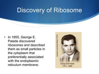
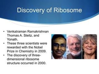
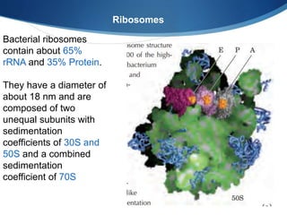
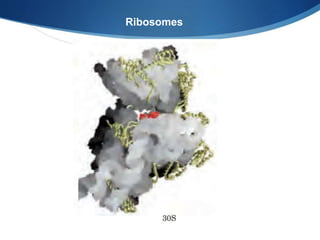
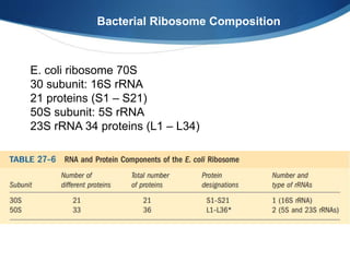
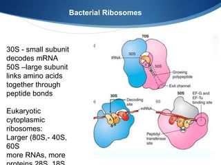
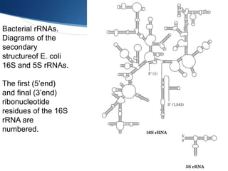
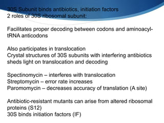
1. S
Ribosomes:
Structures- hammer, head, hairpin, biogenesis at nucleolus
Transfer RNA:
Cloverleaf structure, fidelity of proteins, genes for tRNA
processing, synthesis of
aminoacyl tRNA.
2. Discovery of Ribosome
• In 1955, George E.
Palade discovered
ribosomes and described
them as small particles in
the cytoplasm that
preferentially associated
with the endoplasmic
reticulum membrane.
3. Discovery of Ribosome
• Venkatraman Ramakrishnan
Thomas A. Steitz, and
Yonath.
• These three scientists were
rewarded with the Nobel
Prize in Chemistry in 2009.
• The discovery of three-
dimensional ribosome
structure occurred in 2000.
4. 70S ribosome with mRNA analog, 3 tRNAs
Positions, tertiary structures of all 3 rRNAs, most proteins
Shapes and locations of tRNAs in A, P, and E sites
Binding sites for tRNAs in ribosome are rRNA, not protein.
Contacts between subunits are mostly rRNA
Anticodons of tRNAs in A and P sites approach each other
closely enough to base-pair with adjacent codons bound to 30S
subunit as mRNA kinks 45°
Bacterial Thermus thermophilus crystal structure
7. Bacterial ribosomes
contain about 65%
rRNA and 35% Protein.
They have a diameter of
about 18 nm and are
composed of two
unequal subunits with
sedimentation
coefficients of 30S and
50S and a combined
sedimentation
coefficient of 70S
Ribosomes
8. Ribosomes consist of two subunits, small and large (30S
and 50S in bacteria).
The small subunit interprets the genetic information by
selecting aminoacyl-tRNAs cognate to the mRNA codons
in the decoding center.
The large subunit carries the catalytic peptidyl transferase
center (PTC) where amino acids are polymerized into a
protein.
Ribosomes
11. Bacterial Ribosomes
30S - small subunit
decodes mRNA
50S –large subunit
links amino acids
together through
peptide bonds
Eukaryotic
cytoplasmic
ribosomes:
Larger (80S,- 40S,
60S
more RNAs, more
12. Ribosome Biogenesis
It is the process of making Ribosomes in the nucleus
1. The protein parts are made in the cytoplasm
(Ribosome)
2. Then transferred to the nucleus (Nuclear Pores)
3. rRNAs are transcribed in the nucleolus
4. The ribosomal proteins and rRNAs bind together
5. Small and large subunits are made
6. They are transported out of nucleus (Pores)
14. • tRNA biogenesis occurs post-transcriptionally at numerous
distinct subcellular locations.
• In S. cerevisiae (budding yeast), pre-tRNA transcription by
Pol III and 5’ maturation, catalyzed by RNase P, are located
in the nucleolus.
• Particular tRNA modifications are added in the nucleoplasm,
and other modifications are added at the inner nuclear
membrane or in the cytoplasm after tRNA nuclear export;
• Pre-tRNA splicing occurs on the surface of mitochondria
tRNA synthesis and modification
15. Bacterial rRNAs.
Diagrams of the
secondary
structureof E. coli
16S and 5S rRNAs.
The first (5’end)
and final (3’end)
ribonucleotide
residues of the 16S
rRNA are
numbered.
16. Tranfer RNA
tRNA serves as adapter in translating the language of nucleic
acid into language of proteins.
Structure
Transfer RNAs are relatively small and consist of a single strand
of RNA folded into a precise three-dimensional structure.
Cells have at least one kind of tRNA for each amino acid; at
least 32 tRNAs are required to recognize
all the amino acid codons
17. Tranfer RNA
Yeast t-RNA the first
one to be sequenced
• 76 nucleotides
bases
• 10 of them are
modified bases.
guanylate (pG) residue
(CCA )3’ end
19. • The hydrogen-bonding pattern of all tRNAs forms a
cloverleaf structure with four arms
• The longer tRNAs have a short fifth arm.
• Two of the arms of a tRNA are critical for its adaptor
function.
Clover leaf structure of tRNA
20. 1. The amino acid arm can carry a specific amino acid
esterified by its carboxyl group to the 2’or 3’ hydroxyl
group of the A residue at the 3 end of the tRNA.
2. The anticodon arm contains the anticodon.
3. D arm- It has an unusual nucleotide dihydrouridine (D).
4. TΨC arm- which contains ribothymidine (T) and
pseudouridine.
Clover leaf structure of tRNA has four arms
21. 3-D structure of tRNA
Three-dimensional structure of yeast tRNA Phe deduced from x-ray
diffraction analysis. The shape resembles a twisted L.
24. 1. Synthesis of tRNA
Yeast tRNAs are synthesized as primary tRNAs (pre-tRNA)
in the nucleolus and undergo subsequent 5’and 3’ end
processing, modifications (such as pseudouridine
modification) and CCA addition, generating pre-tRNAs.
2. tRNA primary nuclear export.
These pre-tRNAs are then exported from the nucleus to the
cytoplasm via Los1, Mex67-Mtr2, or potentially Crm1.
tRNA biogenesis and subcellular trafficking
25. tRNA biogenesis and subcellular trafficking
3. Splicing of tRNA
tRNAs containing an intron are then spliced at the surface of
the mitochondria by the SEN complex and the two tRNA
halves are ligated by Rlg1/Trl1.
4. Nucleoside modification in cytoplasm.
The “tRNA” pseudouridine synthetases also modify mRNA,
snRNAs, and snoRNAs. Pseudouridine modification of various
RNAs is catalyzed by stand-alone proteins called
pseudouridine synthetases (Pus) or RNPs containing guide
RNAs called H/ACA snoRNPs.
28. Spliced tRNAs undergo a second trafficking step termed
retrograde nuclear import, mediated by Ssa2 and potentially
Mtr10.
Once back in the nucleus, spliced tRNAs can be modified
by enzymes that only recognize spliced tRNAs, and not
intron-containing tRNAs, such as Trm5-catalyzed
methylation of G at position 37 (m1G37; orange circle).
tRNAs are then re-exported from the nucleus to the
cytoplasm by any of the primary exporters or Msn5, which
functions solely in the re-export step, to be utilized in
translation.