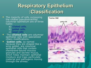
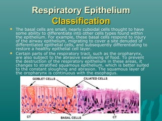
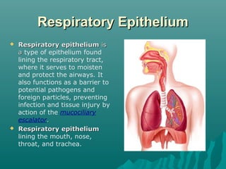
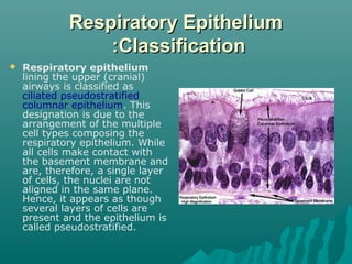
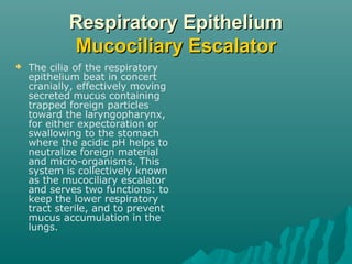
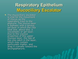
The respiratory epithelium lines the respiratory tract from the nose to the trachea. It is classified as ciliated pseudostratified columnar epithelium in the upper airways, composed mainly of ciliated cells, goblet cells, and basal cells. The cilia beat in a coordinated way to move mucus up the airways via the mucociliary escalator, trapping pathogens and particles to be expelled or swallowed, protecting the lungs.
Gas exchange between the alveoli and the pulmonary capillary blood occurs by diffusion, as will be discussed in the next chapter. Diffusion of oxygen and carbon dioxide occurs passively, according to their concentration differences across the alveolar-capillary barrier. These concentration differences must be maintained by ventilation of the alveoli and perfusion of the pulmonary capillaries.
Alveolar ventilation brings oxygen into the lung and removes carbon dioxide from it. Similarly, the mixed venous blood brings carbon dioxide into the lung and takes up alveolar oxygen. The alveolar Image not available. and Image not available. are thus determined by the relationship between alveolar ventilation and pulmonary capillary perfusion. Alterations in the ratio of ventilation to perfusion, called the Image not available., will result in changes in the alveolar Image not available. and Image not available., as well as in gas delivery to or removal from the lung.
Alveolar ventilation is normally about 4 to 6 L/min and pulmonary blood flow (which is equal to cardiac output) has a similar range, and so the Image not available. for the whole lung is in the range of 0.8 to 1.2. Image not available. However, ventilation and perfusion must be matched on the alveolar-capillary level, and the Image not available. for the whole lung is really of interest only as an approximation of the situation in all the alveolar-capillary units of the lung. For instance, suppose that all 5 L/min of the cardiac output went to the left lung and all 5 L/min of alveolar ventilation went to the right lung. The whole lung Image not available. would be 1.0, but there would be no gas exchange because there could be no gas diffusion between the ventilated alveoli and the perfused pulmonary capillaries.
Oxygen is delivered to the alveolus by alveolar ventilation, is removed from the alveolus as it diffuses into the pulmonary capillary blood, and is carried away by blood flow. Similarly, carbon dioxide is delivered to the alveolus in the mixed venous blood and diffuses into the alveolus in the pulmonary capillary. The carbon dioxide is removed from the alveolus by alveolar ventilation. As will be discussed in Chapter 6, at resting cardiac outputs the diffusion of both oxygen and carbon dioxide is normally limited by pulmonary perfusion. Thus, the alveolar partial pressures of both oxygen and carbon dioxide are determined by the Image not available. If the Image not available. in an alveolar-capillary unit increases, the delivery of oxygen relative to its removal will increase, as will the removal ...
Anatomy of Tracheobronchial Tree and Bronchopulmonary Segments with summary o...Jega Subramaniam
Edited version of my Presentation in College.
Hope its useful for you rather than sleeping in my desktop.
Sorry if there is any mistakes.
Thanks and god bless.
The mucose membrane lining of gastrointestinal tract is stratified squamous epithelium at the esophagus which slowly convert into simple columnar epithelium at the stomach until the anus it converts back into the stratified squamous epithelium at the lower half of the anal canal. The stratified epithelium is a wear and tear epithelium.
As it passes down from the small to large intestine, goblet cells increase because as it passes down water was absorb, goblet cells function to produce mucous.
This is just a rough idea, for better slides with more reference please PM the author at davidgqf@gmail.com.
Like heartbeat, breathing must occur in a continuous, cyclic pattern to sustain life processes.
Inspiratory muscles must rhythmically contract and relax to alternately fill the lungs with air and empty them.
The rhythmic pattern of breathing is established by cyclic neural activity to the respiratory muscles
Surfactant & compliance, LAW OF LAPLACE, Work of Breathing (the guyton and ha...Maryam Fida
It is a lipoprotein mixture present in thin layer of fluid lining the alveoli at the air fluid interface.
COMPOSITION
It is composed of
Apoprotein
Calcium ions
Phospholipids i.e. dipalmitoyl lecithin
Surfactant is secreted by
1. Mainly type II alveolar cells in the lungs.
2. Clara cells, which are situated in the bronchioles.
It lowers the surface tension of fluid lining the alveoli.
Surface tension is inversely proportional to surfactant concentration.
During inspiration surfactant molecules move apart as lungs are expanded and during expiration surfactant molecules become concentrated as lungs shorten.
When there is no surfactant, Surface Tension is 50 dynes/cm. when surfactant is present it is 5-30 dynes/cm depending upon the concentration
Prevents collapse of lungs
Stabilize size of alveoli
Surfactant helps to keep lungs expanded. If there is deficiency of surfactant then the pressure of -20 to -30 mm of Hg will be required to keep the lungs expanded
Surfactant also helps to keep the alveoli dry and prevent development of pulmonary edema.
Surfactant is also helpful in lung expansion at birth. If there is deficiency then there is Respiratory Distress Syndrome.
LAW OF LAPLACE:
pressure required to keep a hollow viscous distended = 2 T/R
Where T is tension and R is radius.
During expiration, size of alveoli decreases so R is decreased and if T does not decrease, much higher pressure will be required to keep the alveoli distended.
When adequate amount of surfactant is there T also decreases so increased pressure is not required. This prevents the collapse of lungs and also stabilizes the equal size of alveoli
Definition:
“Compliance is the measure of expansibility or distensibility of the lungs. It indicates with how much ease lungs can be expanded”.
Work of Breathing
In certain diseases there is increased work of breathing and depending upon the nature of breath there will be specific increase in work of breathing.
In asthma there is increase in work of breathing to overcome airway resistance
In restrictive lung diseases there is increase work of breathing in both tissue resistance and elastic recoil.
lecture 5: it's good for as to take a breif about how does atmospheric air will pass to our lungs then to blood, for transportation and utilization of oxygen and excretion of carbon dioxide. Many issue are related when gas exchange is performed.
6. What types of epithelia would be suited for the following functio.pdfalvisguyjhiy
6. What types of epithelia would be suited for the following functions? Explain why.
a. Resistance to abrasive forces in areas such as the epidermis, oral cavity, pharynx, esophagus,
anal canal, and vaginal canal.
b. Transport of substances across the epithelial sheet in areas where rapid exchange occurs, such
as with capillaries and alveoli, and where secretion and absorption occurs, such as with the
gastrointestinal tract.
c. Trapping and removal of particulates that have been inhaled into the respiratory passages (e.g.
trachea and bronchi).
Solution
Epithelial cells are composed of one or more layers of closely packed cells and will have little or
no extracellular matrix between them. These cells forms covering on body surface or lining of
body cavities and majority of glands.
a
nonkeratinized stratified squamous epithelium
These cells always remain alive way to the tissue\'s apical surface and will be maintained moist
with secretions such as saliva or mucus.
Keratin is absent here and no this layer will not be tough and non-keratinized. Here the flattened
nuclei you can see throughout the tissues as all cells are alive.
These cells are found forming lining of the oral cavity (mouth), the esophagus, part of the
pharynx (throat), the vagina, and the anus.
b
Simple Squamous Epithelium
It consist of a single layer of flattened cells and are irregular in shape and has a spherical to oval
nucleus. They are tightly bound together in a mosaic-like pattern. These cells will look like a
fried egg, with the nucleus representing the yolk. Like this, this epithelium will form an
extremely delicate and highly specialized lining so that rapid movement of molecules across its
surface by filtration, osmosis, or diffusion can take place. This also prevents friction and
abrasion and this epithelium found to be forming the lining of the lung air sacs (alveoli) allows
the gas exchange and also found in lumen of blood vessels allowing exchange of nutrients and
waste between the blood and the interstitial fluid.
c
pseudostratified ciliated columnar epithelium
On the apical surface of these cells, we can find cilia. Like this they form houses goblet cells and
these cells are specialized to secrete mucin, this will help in hydrating the mucus, and thus they
can trap foreign particles and is moved by the beating cilia. These type of cells are found air
passageways of the respiratory system (part of the pharynx, the nasal cavity, larynx, trachea, and
bronchi)
a
nonkeratinized stratified squamous epithelium
These cells always remain alive way to the tissue\'s apical surface and will be maintained moist
with secretions such as saliva or mucus.
Keratin is absent here and no this layer will not be tough and non-keratinized. Here the
flattened nuclei you can see throughout the tissues as all cells are alive.
These cells are found forming lining of the oral cavity (mouth), the esophagus, part of the
pharynx (throat), the vagina, and the anus.
b
Simple Squamous Epithelium.
The above Presentation is related to the Lungs Histology for 1st year MBBS student. it covers the trachea, lungs, bronchi upto the level of Alveoli. Also, it will help students to learn that what different type of epithelium are present at which region.
2. Respiratory Epithelium
Respiratory epithelium is
a type of epithelium found
lining the respiratory tract,
where it serves to moisten
and protect the airways. It
also functions as a barrier to
potential pathogens and
foreign particles, preventing
infection and tissue injury by
action of the mucociliary
escalator.
Respiratory epithelium
lining the mouth, nose,
throat, and trachea.
3. Respiratory Epithelium
:Classification
Respiratory epithelium
lining the upper (cranial)
airways is classified as
ciliated pseudostratified
columnar epithelium. This
designation is due to the
arrangement of the multiple
cell types composing the
respiratory epithelium. While
all cells make contact with
the basement membrane and
are, therefore, a single layer
of cells, the nuclei are not
aligned in the same plane.
Hence, it appears as though
several layers of cells are
present and the epithelium is
called pseudostratified.
4. Respiratory Epithelium
:Classification
The majority of cells composing
the ciliated pseudostratified
columnar epithelium are of three
types:
a) ciliated cells.
b) goblet cells.
c) basal cells.
The ciliated cells are columnar
epithelial cells with specialized
ciliary modifications.
Goblet cells, so named
because they are shaped like a
wine goblet, are columnar
epithelial cells that contain
membrane-bound mucous
granules and secrete mucus,
which helps maintain epithelial
moisture and traps particulate
material and pathogens moving
through the airway.
5. Respiratory Epithelium
Classification
The basal cells are small, nearly cuboidal cells thought to have
some ability to differentiate into other cells types found within
the epithelium. For example, these basal cells respond to injury
of the airway epithelium, migrating to cover a site denuded of
differentiated epithelial cells, and subsequently differentiating to
restore a healthy epithelial cell layer.
Certain parts of the respiratory tract, such as the oropharynx,
are also subject to the abrasive swallowing of food. To prevent
the destruction of the respiratory epithelium in these areas, it
changes to stratified squamous epithelium, which is better suited
to the constant sloughing and abrasion. The squamous layer of
the oropharynx is continuous with the esophagus.
6. Respiratory Epithelium
Mucociliary Escalator
The cilia of the respiratory
epithelium beat in concert
cranially, effectively moving
secreted mucus containing
trapped foreign particles
toward the laryngopharynx,
for either expectoration or
swallowing to the stomach
where the acidic pH helps to
neutralize foreign material
and micro-organisms. This
system is collectively known
as the mucociliary escalator
and serves two functions: to
keep the lower respiratory
tract sterile, and to prevent
mucus accumulation in the
lungs.
7. Respiratory Epithelium
Mucociliary Escalator
The mucocilliary escalator
is vital for the movement
of mucus up the
respiratory tract to the
pharynx. The mucus layer
is biphasic with a serous,
sol layer in which the cilia
beat and, above this, a
viscoelastic or gel layer.
Due to the viscous
properties of this upper
mucous layer, the tips of
the cilia catch in the layer,
which may contain
particulate matter, and
drag it cranially toward the
laryngopharynx.