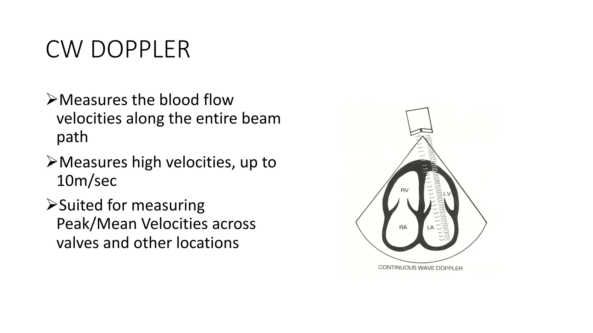
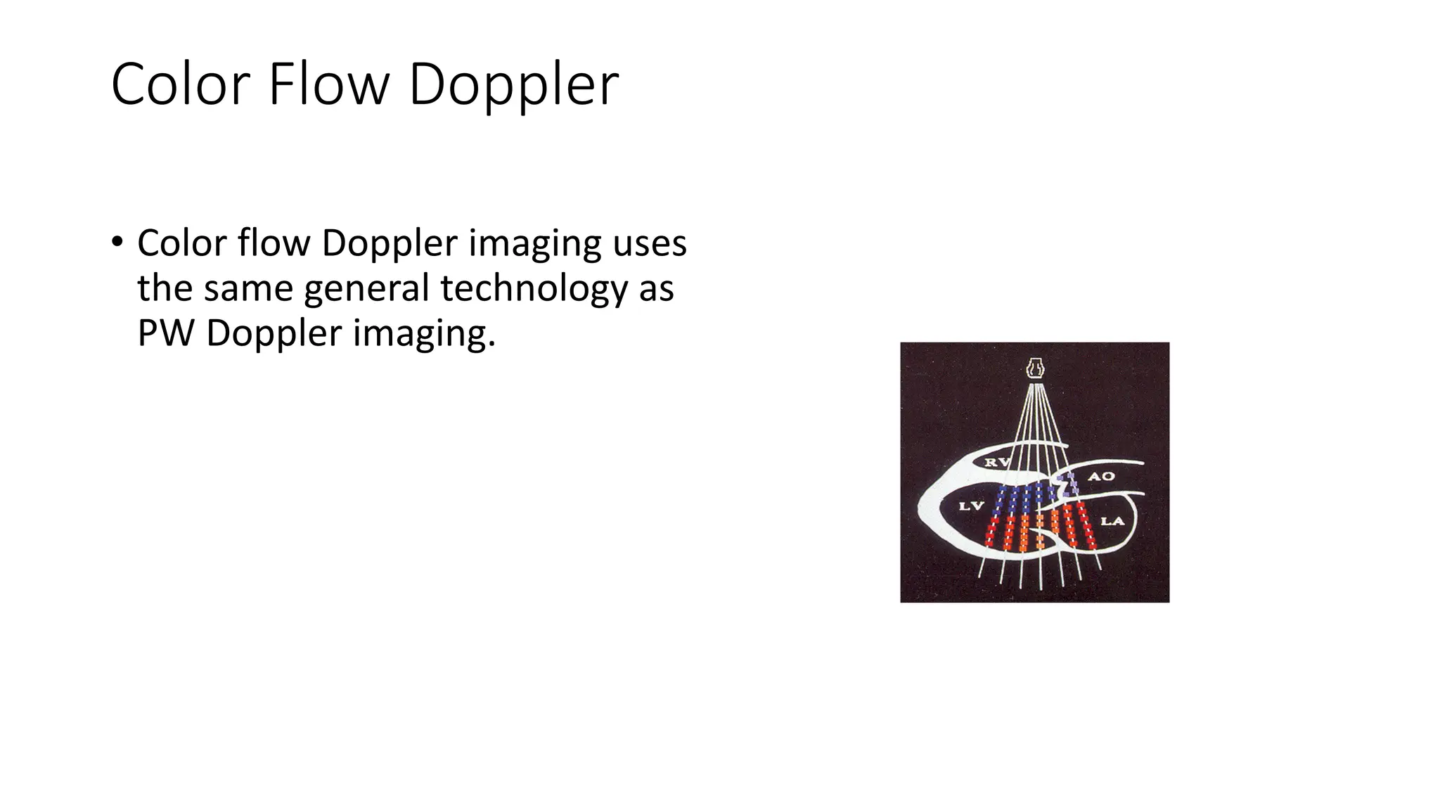
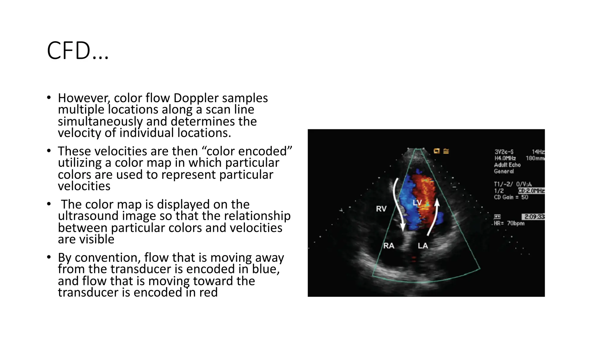
PRINCIPLES OF DOPPLER
ECHOCARDIOGRAPHY
Reuben Mutagaywa
MD, MMED, MSc (Card), Cert (Pacing), PhD cand.
Outline
• Definition DU
• Application of DU
ØDoppler effect
ØBlood velocity
ØVolumetric flow
ØPressure gradients
ØValve areas
ADULT LEARNING – what do they remember?
PRINCIPLES OF DOPPLER USS
Application:-
1. Doppler effect
2. Blood velocity
3. Volumetric flow
4. Pressure gradients
5. Valve areas
Defn
• Doppler ultrasound relies on the Doppler
principle to determine the velocity of
moving fluids or tissues.
ØBy Chritian Doppler (1803 - 1853)
ØAustrian mathematician and physicist
• The Doppler effect/shift
Øobserved frequency of a wave depends
on the relative speed of the source and
the observer
Øeg the change of pitch heard when
a vehicle sounding a horn
approaches and recedes from an
observer
Explanation of DE…
• When the source of the waves is moving towards the observer
Øeach wave crest is emitted from a position closer to the observer
than the crest of the previous wave
Øeach wave takes less time to reach the observer than the previous
wave.
Øthe time between the arrivals of successive wave crests at the
observer is reduced, causing an increase in the frequency
Øwhile they are traveling, the distance between successive wave fronts
is reduced, so the waves "bunch together”
ØConversely,…..
1.PDU: Doppler effect
2. PDU: Blood velocity/blood flows in the heart,
doppler shift
• In USS, waves are emitted from the transducer at a particular freq
and reflected off moving RBCs within the heart
• If the b/flow is moving toward the transducer, the sound waves will
be compressed (and the frequency of the returning ultrasound will be
slightly higher than the emitted ultrasound)
• The opposite is true for b/flow moving away from the transducer
• The difference between the emitted frequency and the returning
frequency is called the Doppler shift
• This relationship is described by the following equation:
Doppler Shift Frequency
D f = fr – ft
f D = shift frequency
fr = reflected frequency
ft = transmitted frequency
Df = Doppler shift frequency
ft = transmitted frequency
v = velocity of blood flow
ø = angle between sound beam and blood flow
c = speed of sound in blood ( 1,560 m/sec)
Df = 2ft v cos ø
c
Doppler Shift Frequency
FLOW VELOCITY MEASUREMENT
@ cos 00 = 1.0000
100= 0.9848
200= 0.9397
300= 0.8660
450=0.7071
600= 0.5000
900=0
Blood flow pattern
• B/flow through the heart can be laminar
or turbulent.
ØLaminar: majority of flow is moving in the
same direction at similar velocities
ØTurbulent: flow is disturbed e.g stenosis
or regurgitation
ØType of flow can be discerned from the
PW Doppler waveform.
ØLaminar appears “hollow” because b/cells
will be moving at similar velocities
ØTurbulent - filled: the velocities cover a
wider spectrum, with some b/cells
moving very rapidly and some moving
very quickly
PULSED- AND CONTINUOUS-WAVE DOPPLER
• Two modes of Doppler ultrasound are typically employed in standard
diagnostic ultrasonography
ØPW Doppler
ØCW Doppler
CW DOPPLER
ØMeasures the blood flow
velocities along the entire beam
path
ØMeasures high velocities, up to
10m/sec
ØSuited for measuring
Peak/Mean Velocities across
valves and other locations
CW…
CLINICAL APPLICATIONS
ØPeak/Mean Velocity and TVI
ØGradients across Native and Prosthetic valves
ØNative and Prosthetic valve Area
ØPressure Half-time
ØPulmonary Artery Pressure
ØLeft Ventricular dp/dt
CW…
PULSED WAVE DOPPLER
ØMeasures the blood flow velocity
at a selected location within the
heart and blood vessels
ØPerformed by duplex transducer by
placing the “Sampling Volume”
ØMaximum measurable velocity is
usually less than 2m/sec
ØSuited for measuring low velocity
at a particular intracardiac location
PW…
CLINICAL APPLICATIONS
ØLocation of flow abnormalities
ØLVOT velocity and TVI
ØLV Systolic function (SV, CO,CI measurements)
ØNative and Prosthetic valve Area
ØMitral Inflow Velocity
ØPulmonary and Hepatic Vein flow velocities
ØMitral Annulus Velocity
ØDiastolic Function
ØMR,TR,AR Assessment
ØRestrictive Cardiomyopathy Vs Constrictive Pericarditis
PW…
3. PDU: Volumetric flow
SV= AREA x VELOCITY
SV= LVOT Area x LVOT VTI= πr2 X VTI
CO= SV X HR; CI = CO/BSA
4. PDU: measuring Pressure gradient in the heart/from vel to
pressure - BERNOULLI EQUATION
Gradient= 4 X (V2- V1)2
V1 V2
P1—Pressure at location 1
P2---Pressure at location 2
p---Mass density of the
blood=1.06 x 10 3 kg/M3
V1---Velocity at location 1
V2---Velocity at location 2
Gradient Across Valve is Determined By
# Degree of orifice narrowing
# Volume of flow across valve
Eg. AS gradients by Bernoulli’s principle
• Use CW Doppler,
• For Maximum/peak gradients:
"caliper" to determine the
maximum velocity of the jet.
• To Mean gradients: "trace" the
envelope of the LVOT. The
machine will calculate the area
under the curve and will give
you the mean gradient across
the aortic valve.
} Check gradients in all views: Apical, subcostal, parasternal
5. PDU: Valve areas - CONTINUITY EQUATION
A1 x V1 = A2 x V2
A2 = A1 x V1
V2
Eg. AS Valve area by continuity equation
• Principle of Continuity
equation (Flow before the
Valve = Flow across the valve)
PRESSURE HALFTIME
Catheter derived PHT Doppler derived PHT
A---Maximum velocity (meters/sec)
B---Maximum velocity/1.4 or square root of 2
t ½- Pressure Half Time in milli sec
Time taken for the velocity to decrease from A to B
Examples of Doppler derived PHT & PGs
Color Flow Doppler
• Color flow Doppler imaging uses
the same general technology as
PW Doppler imaging.
CFD…
• However, color flow Doppler samples
multiple locations along a scan line
simultaneously and determines the
velocity of individual locations.
• These velocities are then “color encoded”
utilizing a color map in which particular
colors are used to represent particular
velocities
• The color map is displayed on the
ultrasound image so that the relationship
between particular colors and velocities
are visible
• By convention, flow that is moving away
from the transducer is encoded in blue,
and flow that is moving toward the
transducer is encoded in red
CFD…
NYQUIST LIMIT & ALIASING IMAGE
• Shades of red and blue indicate flow
toward and away from the transducer,
respectively, becoming progressively
brighter as flow velocity approaches
the Nyquist limit of 64 cm/s.
• A black band represents zero velocity
or no flow.
• Shades of yellow and green are
added to each color Doppler sample
volume to represent turbulence.
• When the Nyquist limit is exceeded
aliasing occurs
• Reds become blues or blues become
reds.
CFI…
CLINICAL APPLICATIONS
ØDiagnosis of Native and Prosthetic valve Stenosis and Regurgitation
ØDiagnosis of Shunts
ØSemiquantitative assessment of Regurgitation
ØPISA Method
Doppler Cautions and Caveats
• Doppler cannot:-
Ømeasure pressure directly
Ø measure “flow.”
• Doppler measures the velocity of b/flow.
• For this reason, much of the information that is derived from Doppler
measurements has to be inferred.
• Eg, for estimation of RVSP by TR jet we have to define the RAP first
THANK YOU FOR LISTENING

Principle of Doppler Echocardiography.pdf

  • 1.
    PRINCIPLES OF DOPPLER ECHOCARDIOGRAPHY ReubenMutagaywa MD, MMED, MSc (Card), Cert (Pacing), PhD cand.
  • 2.
    Outline • Definition DU •Application of DU ØDoppler effect ØBlood velocity ØVolumetric flow ØPressure gradients ØValve areas
  • 3.
    ADULT LEARNING –what do they remember?
  • 4.
    PRINCIPLES OF DOPPLERUSS Application:- 1. Doppler effect 2. Blood velocity 3. Volumetric flow 4. Pressure gradients 5. Valve areas
  • 5.
    Defn • Doppler ultrasoundrelies on the Doppler principle to determine the velocity of moving fluids or tissues. ØBy Chritian Doppler (1803 - 1853) ØAustrian mathematician and physicist • The Doppler effect/shift Øobserved frequency of a wave depends on the relative speed of the source and the observer Øeg the change of pitch heard when a vehicle sounding a horn approaches and recedes from an observer
  • 6.
    Explanation of DE… •When the source of the waves is moving towards the observer Øeach wave crest is emitted from a position closer to the observer than the crest of the previous wave Øeach wave takes less time to reach the observer than the previous wave. Øthe time between the arrivals of successive wave crests at the observer is reduced, causing an increase in the frequency Øwhile they are traveling, the distance between successive wave fronts is reduced, so the waves "bunch together” ØConversely,…..
  • 7.
  • 8.
    2. PDU: Bloodvelocity/blood flows in the heart, doppler shift • In USS, waves are emitted from the transducer at a particular freq and reflected off moving RBCs within the heart • If the b/flow is moving toward the transducer, the sound waves will be compressed (and the frequency of the returning ultrasound will be slightly higher than the emitted ultrasound) • The opposite is true for b/flow moving away from the transducer • The difference between the emitted frequency and the returning frequency is called the Doppler shift • This relationship is described by the following equation:
  • 9.
    Doppler Shift Frequency Df = fr – ft f D = shift frequency fr = reflected frequency ft = transmitted frequency
  • 10.
    Df = Dopplershift frequency ft = transmitted frequency v = velocity of blood flow ø = angle between sound beam and blood flow c = speed of sound in blood ( 1,560 m/sec) Df = 2ft v cos ø c Doppler Shift Frequency
  • 11.
    FLOW VELOCITY MEASUREMENT @cos 00 = 1.0000 100= 0.9848 200= 0.9397 300= 0.8660 450=0.7071 600= 0.5000 900=0
  • 12.
    Blood flow pattern •B/flow through the heart can be laminar or turbulent. ØLaminar: majority of flow is moving in the same direction at similar velocities ØTurbulent: flow is disturbed e.g stenosis or regurgitation ØType of flow can be discerned from the PW Doppler waveform. ØLaminar appears “hollow” because b/cells will be moving at similar velocities ØTurbulent - filled: the velocities cover a wider spectrum, with some b/cells moving very rapidly and some moving very quickly
  • 13.
    PULSED- AND CONTINUOUS-WAVEDOPPLER • Two modes of Doppler ultrasound are typically employed in standard diagnostic ultrasonography ØPW Doppler ØCW Doppler
  • 14.
    CW DOPPLER ØMeasures theblood flow velocities along the entire beam path ØMeasures high velocities, up to 10m/sec ØSuited for measuring Peak/Mean Velocities across valves and other locations
  • 15.
    CW… CLINICAL APPLICATIONS ØPeak/Mean Velocityand TVI ØGradients across Native and Prosthetic valves ØNative and Prosthetic valve Area ØPressure Half-time ØPulmonary Artery Pressure ØLeft Ventricular dp/dt
  • 16.
  • 17.
    PULSED WAVE DOPPLER ØMeasuresthe blood flow velocity at a selected location within the heart and blood vessels ØPerformed by duplex transducer by placing the “Sampling Volume” ØMaximum measurable velocity is usually less than 2m/sec ØSuited for measuring low velocity at a particular intracardiac location
  • 18.
    PW… CLINICAL APPLICATIONS ØLocation offlow abnormalities ØLVOT velocity and TVI ØLV Systolic function (SV, CO,CI measurements) ØNative and Prosthetic valve Area ØMitral Inflow Velocity ØPulmonary and Hepatic Vein flow velocities ØMitral Annulus Velocity ØDiastolic Function ØMR,TR,AR Assessment ØRestrictive Cardiomyopathy Vs Constrictive Pericarditis
  • 19.
  • 20.
    3. PDU: Volumetricflow SV= AREA x VELOCITY SV= LVOT Area x LVOT VTI= πr2 X VTI CO= SV X HR; CI = CO/BSA
  • 21.
    4. PDU: measuringPressure gradient in the heart/from vel to pressure - BERNOULLI EQUATION Gradient= 4 X (V2- V1)2 V1 V2 P1—Pressure at location 1 P2---Pressure at location 2 p---Mass density of the blood=1.06 x 10 3 kg/M3 V1---Velocity at location 1 V2---Velocity at location 2
  • 22.
    Gradient Across Valveis Determined By # Degree of orifice narrowing # Volume of flow across valve
  • 23.
    Eg. AS gradientsby Bernoulli’s principle • Use CW Doppler, • For Maximum/peak gradients: "caliper" to determine the maximum velocity of the jet. • To Mean gradients: "trace" the envelope of the LVOT. The machine will calculate the area under the curve and will give you the mean gradient across the aortic valve. } Check gradients in all views: Apical, subcostal, parasternal
  • 24.
    5. PDU: Valveareas - CONTINUITY EQUATION A1 x V1 = A2 x V2 A2 = A1 x V1 V2
  • 25.
    Eg. AS Valvearea by continuity equation • Principle of Continuity equation (Flow before the Valve = Flow across the valve)
  • 26.
    PRESSURE HALFTIME Catheter derivedPHT Doppler derived PHT A---Maximum velocity (meters/sec) B---Maximum velocity/1.4 or square root of 2 t ½- Pressure Half Time in milli sec Time taken for the velocity to decrease from A to B
  • 27.
    Examples of Dopplerderived PHT & PGs
  • 28.
    Color Flow Doppler •Color flow Doppler imaging uses the same general technology as PW Doppler imaging.
  • 29.
    CFD… • However, colorflow Doppler samples multiple locations along a scan line simultaneously and determines the velocity of individual locations. • These velocities are then “color encoded” utilizing a color map in which particular colors are used to represent particular velocities • The color map is displayed on the ultrasound image so that the relationship between particular colors and velocities are visible • By convention, flow that is moving away from the transducer is encoded in blue, and flow that is moving toward the transducer is encoded in red
  • 30.
  • 31.
    NYQUIST LIMIT &ALIASING IMAGE • Shades of red and blue indicate flow toward and away from the transducer, respectively, becoming progressively brighter as flow velocity approaches the Nyquist limit of 64 cm/s. • A black band represents zero velocity or no flow. • Shades of yellow and green are added to each color Doppler sample volume to represent turbulence. • When the Nyquist limit is exceeded aliasing occurs • Reds become blues or blues become reds.
  • 32.
    CFI… CLINICAL APPLICATIONS ØDiagnosis ofNative and Prosthetic valve Stenosis and Regurgitation ØDiagnosis of Shunts ØSemiquantitative assessment of Regurgitation ØPISA Method
  • 33.
    Doppler Cautions andCaveats • Doppler cannot:- Ømeasure pressure directly Ø measure “flow.” • Doppler measures the velocity of b/flow. • For this reason, much of the information that is derived from Doppler measurements has to be inferred. • Eg, for estimation of RVSP by TR jet we have to define the RAP first
  • 34.
    THANK YOU FORLISTENING