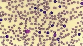
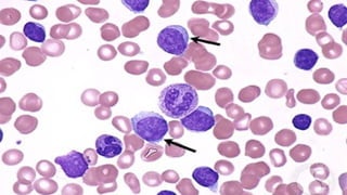
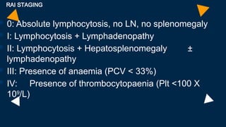
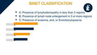
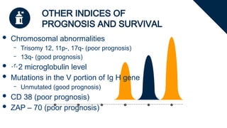
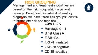
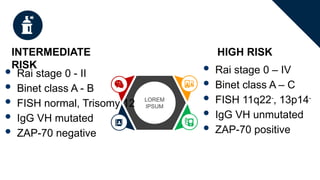
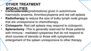
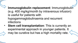
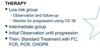
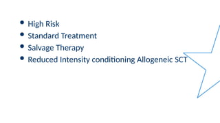
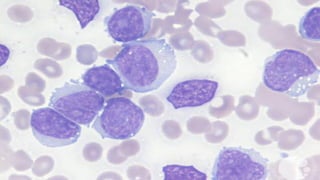
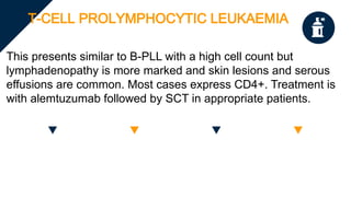
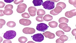
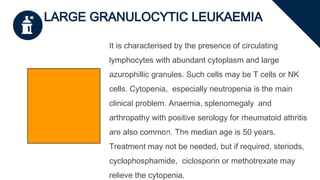
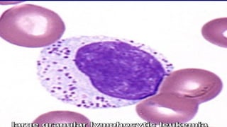
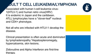
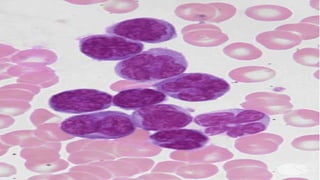
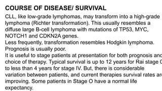
CLINICAL FEATURES
• Early stage is asymptomatic and 80% of cases are diagnosed from routine
blood tests.
• Symmetrical enlargement of cervical, axillary, or inguinal lymph nodes is the
most frequent clinical sign. The nodes are usually discrete and non-tender.
• Clinical features of anaemia may be present and patients with
thrombocytopaenia may show bruising or purpura.
CLINICAL FEATURES
• Splenomegaly and less commonly, hepatomegaly are
often seen in later stages.
• Immunosuppression is often a significant problem
resulting from hypogammaglobulinaemia and cellular
immune dysfunction. Early in the disease course, bacterial
infection predominate but with advanced disease, viral
infections and fungal are also seen.
DIAGNOSIS
Lymphocytosis
The absolute clonal
B-cell lymphocyte
count is >5 x10^9/L
and may be
Lymphadenopathy
Symmetrical
enlargement
Morphology on
Peripheral blood fim
and bone marrow
Between 70-90% of WBC appear as
small lympocytes. Smudge/smear cells
are also present. Bone marrow
aspiration shows lymphocytic
replacement of normal marrow
Anaemia/Thrombocytopaenia
Normocytic normochromic
anaemia is present in later
stages as a result of marrow
infiltration or hypersplenism.
Slenomegaly
Seen in 50% of
patients
MOLECULAR DIAGNOSIS
 Flow cytometry
 Chromosomal abnormality using the FISH technique
 Identification of mutations in the variable region of the
heavy chain on the immunoglobulin gene
 ZAP-70 (zeta associated protein 70kDa TK)
 CD38 cell marker
IMMUNOLOGY AND CELL MARKERS
• Low density of surface Ig (IgM or IgD) with κ or λ light chains
• B-cell surface antigens (CD19, CD20, CD23) CD20 dim
• CD5 surface antigen
STAGING AND
CLASSIFICATION
 Involves the Rai Staging and Binet
Classification
 Rai Staging – 0 to IV
 Binet Classification – A to C
 They form part of the criteria for risk
grouping in CLL
RAI STAGING
 0: Absolute lymphocytosis, no LN, no splenomegaly
 I: Lymphocytosis + Lymphadenopathy
 II: Lymphocytosis + Hepatosplenomegaly ±
lymphadenopathy
 III: Presence of anaemia (PCV < 33%)
 IV: Presence of thrombocytopaenia (Plt <100 X
109
/L)
BINET CLASSIFICATION
 A: Presence of lymphadenopathy in less than 3 regions
 B: Presence of lymph node enlargement in 3 or more regions
 C: Presence of anaemia, and, or thrombocytopaenia
OTHER INDICES OF
PROGNOSIS AND SURVIVAL
 Chromosomal abnormalities
– Trisomy 12, 11p-, 17q- (poor prognosis)
– 13q- (good prognosis)
 2 microglobulin level
 Mutations in the V portion of Ig H gene
– Unmutated (good prognosis)
 CD 38 (poor prognosis)
 ZAP – 70 (poor prognosis)
RISK GROUPING
Management and treatment modalities are
based on the risk group which a patient
belongs. Based on clinical and laboratory
diagnosis, we have three risk groups: low risk,
intermediate risk and high risk.
LOW RISK
 Rai stage 0 – I
 Binet Class A
 FISH 13q-,
 IgG VH mutated
 ZAP-70 negative
 CD 38 negative
LOREM
IPSUM
INTERMEDIATE
RISK
HIGH RISK
 Rai stage 0 - II
 Binet class A - B
 FISH normal, Trisomy 12
 IgG VH mutated
 ZAP-70 negative
 Rai stage 0 – IV
 Binet class A – C
 FISH 11q22-
, 13p14-
 IgG VH unmutated
 ZAP-70 positive
TREATMENT MODALITIES
 It's difficult to cure CLL except with allogeneic stem cell
transplant(rarely employed due to tha advanced age of most
patients and the availability of excellent safer therapies).
 Therefore, the approach to therapy is generally conservative,
aiming for symptom control rather than a normal blood cell
count. Treatment given too early in the disease can shorten
rather than prolong life expectancy.
 Many patients won't require treatment. Treatment is basically
given for troublesome symptoms like enlarged spleen or
lymph nodes.
CHEMOTHERAPY
However, in the recent years, several new
and highly effective therapies have emerged
in the treatment of lymphoid disorders.
Beginning in the 90's, the most commonly used first-line treatment
for younger patients was FCR/R-FC. The average time to disease
progression after treatment with FCR was 4.5 years .
• Fludarabine (adenosine analogue) 25mg/m2 x4 wks
• Cyclophosphamide & Chlorambucil
• Rituximab (monoclonal antibody) 375mg/m2 /wk x 4wks.
NEWER CHEMOTHERAPEUTIC AGENTS
Drugs which suppress signalling through the B-cell receptor
Drugs have been introduced that block two of the B-cell signalling
proteins, Bruton Kinase(BTK) and phsosphoinositide 3-
kinase(PI3K), both of which act downstream of the B-Cell Receptor.
• Ibrutinib and acalabrutinib are oral drugs which inactivate BTK
and lead to B-Cell apoptosis.
• Idelasib is an oral drug which blocks PI3K.
• A feature of both drugs is that they initially cause an increase in
lymphocyte count in peripheral blood. This reflects a
redistribution from lymph nodes and bone marrow.
Drugs which suppress the activity of BCL-2.
BCL-2 is expressed at a high level in most CLL cells
and has potent anti-apoptotic effects, leading to
abnormally prolonged cell survival. Venetoclax, a
direct inhibitor of BCL-2, is highly active
Effective monoclonal antibodies include
ofatumumab and obinutuzumab, which are CD20
specific . The combination of obinutuzumab with
chlorambucil is excellent first-line treatment for older
and less fit patients.
OTHER TREATMENT
MODALITIES
• Corticosteroids(prednisolone) given in autoimmune
haemolytic anaemia, thrombocytopenia and red cell aplasia
• Radiotherapy to reduce the size of bulky lymph node groups
that are unresponsive to chemotherapy.
• Ciclosporin- Red cell aplasia may respond to ciclosporin.
• Splenectomy -This is generally reserved for those patients
with immune - mediated cytopenias that do not respond to
short courses of steroids or those with symptomatic
enlargement of the spleen unresponsive to other therapy.
• Immunoglobulin replacement: Immunoglobulin
(e.g. 400 mg/kg/month by intravenous infusion)
is useful for patients with
hypogammaglobulinaemia and recurrent
infections
• Stem cell transplantation: This is currently an
experimental approach in younger patients. It
may be curative but has a high mortality rate.
THERAPY
Low risk group
– Observation and follow-up
– Monitor for progression using CD 38
Intermediate group
Initial Observation until progression
Then: Standard Treatment with FC,
FCR, PCR, CHOPR
 High Risk
 Standard Treatment
 Salvage Therapy
 Reduced Intensity conditioning Allogeneic SCT
OTHER TYPES OF CHRONIC
LYMPHOCYTIC LEUKEMIAS
HAIRY CELL LEUKAEMIA
It is an uncommon disease with a male to female
ratio of 4:1. Patients typically present with
infections, anaemia or splenomegaly.
Lympadenopathy is uncommon.
Pancytopenia is usual at
presentation and the lymphocyte
count rarely exceeds 20x10^9/L.
Monocytopenia is a distinctive
feature..
Peripheral blood film shows ‘Hairy’ cells with oval nuclei and finely mottled
pale grey–blue cytoplasm with an irregular edge (lymphocytes with villous
cytoplasmic projections). Treatment of choice is deoxocoformycin or 2-
chlorodeoxyadenosine.
It may appear similar to CLL, but its
diagnosis is made by the appearance
of a majority of prolymphocytes in the
blood. The prolymphocye is twice the
size of a CLL lymphocyte and has a
large central nucleus.
B-CELL PROLYMPHOCYTIC LEUKAEMIA
It typically presents with splenomegaly without
lymphadenopathy and with a high and rapidly rising
lymphocyte count. Anaemia is a poor prognostic feature. It
has a worse prognosis than CLL(median survival is 3-5
years). Treatment of choice is Alemtuzumab.
T-CELL PROLYMPHOCYTIC LEUKAEMIA
This presents similar to B-PLL with a high cell count but
lymphadenopathy is more marked and skin lesions and serous
effusions are common. Most cases express CD4+. Treatment is
with alemtuzumab followed by SCT in appropriate patients.
LARGE GRANULOCYTIC LEUKAEMIA
It is characterised by the presence of circulating
lymphocytes with abundant cytoplasm and large
azurophillic granules. Such cells may be T cells or NK
cells. Cytopenia, especially neutropenia is the main
clinical problem. Anaemia, splenomegaly and
arthropathy with positive serology for rheumatoid athritis
are also common. The median age is 50 years.
Treatment may not be needed, but if required, steriods,
cyclophosphamide, ciclosporin or methotrexate may
relieve the cytopenia.
Enter title
ADULT T CELL LEUKAEMIA/LYMPHOMA
Associated with human t-cell leukemia virus-
1(HTLV-1) and human retro virus(HRV)
It is endemic in Japan and the caribbean.
ATLL lymphocytes have a "clover-leaf" nucleus
and CD4+ phenotype.
Not all who are infected with HTLV-1 develop the
disease.
Clinical presentation is often acute and dominated
by lymphadenopathy 'Hepatosplenomegaly,
hypercalcemia, skin lesions.
Zidovudine and Alpha Interferon are first-line
therapy.
COURSE OF DISEASE/ SURVIVAL
CLL, like low-grade lymphomas, may transform into a high-grade
lymphoma (Richter transformation). This usually resembles a
diffuse large B-cell lymphoma with mutations of TP53, MYC,
NOTCH1 and CDKN2A genes.
Less frequently, transformation resembles Hodgkin lymphoma.
Prognosis is usually poor.
It is useful to stage patients at presentation for both prognosis and
choice of therapy. Typical survival is up to 12 years for Rai stage 0
to less than 4 years for stage IV. But, there is considerable
variation between patients, and current therapies survival rates are
improving. Some patients in Stage O have a normal life
expectancy.
THANKS
FOR YOUR ATTENTION

PRESENTATION _ CLL clinical features.pptx

  • 1.
    CLINICAL FEATURES • Earlystage is asymptomatic and 80% of cases are diagnosed from routine blood tests. • Symmetrical enlargement of cervical, axillary, or inguinal lymph nodes is the most frequent clinical sign. The nodes are usually discrete and non-tender. • Clinical features of anaemia may be present and patients with thrombocytopaenia may show bruising or purpura.
  • 2.
    CLINICAL FEATURES • Splenomegalyand less commonly, hepatomegaly are often seen in later stages. • Immunosuppression is often a significant problem resulting from hypogammaglobulinaemia and cellular immune dysfunction. Early in the disease course, bacterial infection predominate but with advanced disease, viral infections and fungal are also seen.
  • 3.
    DIAGNOSIS Lymphocytosis The absolute clonal B-celllymphocyte count is >5 x10^9/L and may be Lymphadenopathy Symmetrical enlargement Morphology on Peripheral blood fim and bone marrow Between 70-90% of WBC appear as small lympocytes. Smudge/smear cells are also present. Bone marrow aspiration shows lymphocytic replacement of normal marrow Anaemia/Thrombocytopaenia Normocytic normochromic anaemia is present in later stages as a result of marrow infiltration or hypersplenism. Slenomegaly Seen in 50% of patients
  • 8.
    MOLECULAR DIAGNOSIS  Flowcytometry  Chromosomal abnormality using the FISH technique  Identification of mutations in the variable region of the heavy chain on the immunoglobulin gene  ZAP-70 (zeta associated protein 70kDa TK)  CD38 cell marker IMMUNOLOGY AND CELL MARKERS • Low density of surface Ig (IgM or IgD) with κ or λ light chains • B-cell surface antigens (CD19, CD20, CD23) CD20 dim • CD5 surface antigen
  • 9.
    STAGING AND CLASSIFICATION  Involvesthe Rai Staging and Binet Classification  Rai Staging – 0 to IV  Binet Classification – A to C  They form part of the criteria for risk grouping in CLL
  • 10.
    RAI STAGING  0:Absolute lymphocytosis, no LN, no splenomegaly  I: Lymphocytosis + Lymphadenopathy  II: Lymphocytosis + Hepatosplenomegaly ± lymphadenopathy  III: Presence of anaemia (PCV < 33%)  IV: Presence of thrombocytopaenia (Plt <100 X 109 /L)
  • 11.
    BINET CLASSIFICATION  A:Presence of lymphadenopathy in less than 3 regions  B: Presence of lymph node enlargement in 3 or more regions  C: Presence of anaemia, and, or thrombocytopaenia
  • 12.
    OTHER INDICES OF PROGNOSISAND SURVIVAL  Chromosomal abnormalities – Trisomy 12, 11p-, 17q- (poor prognosis) – 13q- (good prognosis)  2 microglobulin level  Mutations in the V portion of Ig H gene – Unmutated (good prognosis)  CD 38 (poor prognosis)  ZAP – 70 (poor prognosis)
  • 13.
    RISK GROUPING Management andtreatment modalities are based on the risk group which a patient belongs. Based on clinical and laboratory diagnosis, we have three risk groups: low risk, intermediate risk and high risk. LOW RISK  Rai stage 0 – I  Binet Class A  FISH 13q-,  IgG VH mutated  ZAP-70 negative  CD 38 negative
  • 14.
    LOREM IPSUM INTERMEDIATE RISK HIGH RISK  Raistage 0 - II  Binet class A - B  FISH normal, Trisomy 12  IgG VH mutated  ZAP-70 negative  Rai stage 0 – IV  Binet class A – C  FISH 11q22- , 13p14-  IgG VH unmutated  ZAP-70 positive
  • 15.
    TREATMENT MODALITIES  It'sdifficult to cure CLL except with allogeneic stem cell transplant(rarely employed due to tha advanced age of most patients and the availability of excellent safer therapies).  Therefore, the approach to therapy is generally conservative, aiming for symptom control rather than a normal blood cell count. Treatment given too early in the disease can shorten rather than prolong life expectancy.  Many patients won't require treatment. Treatment is basically given for troublesome symptoms like enlarged spleen or lymph nodes.
  • 16.
    CHEMOTHERAPY However, in therecent years, several new and highly effective therapies have emerged in the treatment of lymphoid disorders. Beginning in the 90's, the most commonly used first-line treatment for younger patients was FCR/R-FC. The average time to disease progression after treatment with FCR was 4.5 years . • Fludarabine (adenosine analogue) 25mg/m2 x4 wks • Cyclophosphamide & Chlorambucil • Rituximab (monoclonal antibody) 375mg/m2 /wk x 4wks.
  • 17.
    NEWER CHEMOTHERAPEUTIC AGENTS Drugswhich suppress signalling through the B-cell receptor Drugs have been introduced that block two of the B-cell signalling proteins, Bruton Kinase(BTK) and phsosphoinositide 3- kinase(PI3K), both of which act downstream of the B-Cell Receptor. • Ibrutinib and acalabrutinib are oral drugs which inactivate BTK and lead to B-Cell apoptosis. • Idelasib is an oral drug which blocks PI3K. • A feature of both drugs is that they initially cause an increase in lymphocyte count in peripheral blood. This reflects a redistribution from lymph nodes and bone marrow.
  • 18.
    Drugs which suppressthe activity of BCL-2. BCL-2 is expressed at a high level in most CLL cells and has potent anti-apoptotic effects, leading to abnormally prolonged cell survival. Venetoclax, a direct inhibitor of BCL-2, is highly active Effective monoclonal antibodies include ofatumumab and obinutuzumab, which are CD20 specific . The combination of obinutuzumab with chlorambucil is excellent first-line treatment for older and less fit patients.
  • 19.
    OTHER TREATMENT MODALITIES • Corticosteroids(prednisolone)given in autoimmune haemolytic anaemia, thrombocytopenia and red cell aplasia • Radiotherapy to reduce the size of bulky lymph node groups that are unresponsive to chemotherapy. • Ciclosporin- Red cell aplasia may respond to ciclosporin. • Splenectomy -This is generally reserved for those patients with immune - mediated cytopenias that do not respond to short courses of steroids or those with symptomatic enlargement of the spleen unresponsive to other therapy.
  • 20.
    • Immunoglobulin replacement:Immunoglobulin (e.g. 400 mg/kg/month by intravenous infusion) is useful for patients with hypogammaglobulinaemia and recurrent infections • Stem cell transplantation: This is currently an experimental approach in younger patients. It may be curative but has a high mortality rate.
  • 21.
    THERAPY Low risk group –Observation and follow-up – Monitor for progression using CD 38 Intermediate group Initial Observation until progression Then: Standard Treatment with FC, FCR, PCR, CHOPR
  • 22.
     High Risk Standard Treatment  Salvage Therapy  Reduced Intensity conditioning Allogeneic SCT
  • 23.
    OTHER TYPES OFCHRONIC LYMPHOCYTIC LEUKEMIAS
  • 24.
    HAIRY CELL LEUKAEMIA Itis an uncommon disease with a male to female ratio of 4:1. Patients typically present with infections, anaemia or splenomegaly. Lympadenopathy is uncommon. Pancytopenia is usual at presentation and the lymphocyte count rarely exceeds 20x10^9/L. Monocytopenia is a distinctive feature.. Peripheral blood film shows ‘Hairy’ cells with oval nuclei and finely mottled pale grey–blue cytoplasm with an irregular edge (lymphocytes with villous cytoplasmic projections). Treatment of choice is deoxocoformycin or 2- chlorodeoxyadenosine.
  • 26.
    It may appearsimilar to CLL, but its diagnosis is made by the appearance of a majority of prolymphocytes in the blood. The prolymphocye is twice the size of a CLL lymphocyte and has a large central nucleus. B-CELL PROLYMPHOCYTIC LEUKAEMIA It typically presents with splenomegaly without lymphadenopathy and with a high and rapidly rising lymphocyte count. Anaemia is a poor prognostic feature. It has a worse prognosis than CLL(median survival is 3-5 years). Treatment of choice is Alemtuzumab.
  • 28.
    T-CELL PROLYMPHOCYTIC LEUKAEMIA Thispresents similar to B-PLL with a high cell count but lymphadenopathy is more marked and skin lesions and serous effusions are common. Most cases express CD4+. Treatment is with alemtuzumab followed by SCT in appropriate patients.
  • 30.
    LARGE GRANULOCYTIC LEUKAEMIA Itis characterised by the presence of circulating lymphocytes with abundant cytoplasm and large azurophillic granules. Such cells may be T cells or NK cells. Cytopenia, especially neutropenia is the main clinical problem. Anaemia, splenomegaly and arthropathy with positive serology for rheumatoid athritis are also common. The median age is 50 years. Treatment may not be needed, but if required, steriods, cyclophosphamide, ciclosporin or methotrexate may relieve the cytopenia. Enter title
  • 32.
    ADULT T CELLLEUKAEMIA/LYMPHOMA Associated with human t-cell leukemia virus- 1(HTLV-1) and human retro virus(HRV) It is endemic in Japan and the caribbean. ATLL lymphocytes have a "clover-leaf" nucleus and CD4+ phenotype. Not all who are infected with HTLV-1 develop the disease. Clinical presentation is often acute and dominated by lymphadenopathy 'Hepatosplenomegaly, hypercalcemia, skin lesions. Zidovudine and Alpha Interferon are first-line therapy.
  • 34.
    COURSE OF DISEASE/SURVIVAL CLL, like low-grade lymphomas, may transform into a high-grade lymphoma (Richter transformation). This usually resembles a diffuse large B-cell lymphoma with mutations of TP53, MYC, NOTCH1 and CDKN2A genes. Less frequently, transformation resembles Hodgkin lymphoma. Prognosis is usually poor. It is useful to stage patients at presentation for both prognosis and choice of therapy. Typical survival is up to 12 years for Rai stage 0 to less than 4 years for stage IV. But, there is considerable variation between patients, and current therapies survival rates are improving. Some patients in Stage O have a normal life expectancy.
  • 35.