2. tory process of the distal convoluted tubule and segments of
the collecting duct in the cortex and outer medulla (the cor-
tical collecting duct and the outer medullary collecting duct,
respectively) (Figure 2). The cortical collecting duct and outer
medullary collecting duct consist of at least 2 very different
cell types, termed principal cells and intercalated cells (Figure
2). Principal cells, which comprise approximately 70% to
75% of collecting duct cells, mediate sodium reabsorption
and potassium secretion and are targets for angiotensin II (11,
12), aldosterone, aldosterone receptor antagonists, and
potassium-sparing diuretics (Figure 2). Principal cells exploit
the electrochemical gradient established by sodium entry into
the cell through a sodium channel at the luminal membrane
(the molecular target of amiloride) and the basolateral mem-
brane Na,K-ATPase to drive potassium secretion through 2
classes of luminal membrane potassium channels (13). One of
these, the renal outer medullary potassium (known among
renal physiologists as “ROMK”) channels, secrete potassium
under normal tubular fluid flow conditions and are inserted
into or internalized from the luminal membrane, depending
on the demand for potassium secretion. The other class of
potassium channels are the “big” conductance channels
(known as “BK” channels), which are relatively inactive under
normal conditions but exhibit increased activity during high
tubular flow or high-potassium conditions (13). Factors that
regulate principal cell potassium secretion include previous
potassium intake; intracellular potassium level; sodium deliv-
ery to the cells; urine flow rate; and hormones, such as aldo-
sterone and -catecholamines (14). The other collecting duct
cell type, intercalated cells, mediate acid–base transport but
upregulate expression of luminal H,K-ATPases (see Glossary)
during potassium depletion to enhance potassium reabsorp-
tion (1) (Figure 2).
To summarize, mammalian cells require a steep con-
centration gradient of potassium between ICF and extra-
cellular fluid to function properly, which requires primary
active transport by Na,K-ATPase. The kidney excretes suf-
ficient amounts of potassium to maintain total body ho-
meostasis. Although the proximal nephron reabsorbs the
bulk of the potassium filtered at the glomerulus, the col-
lecting duct fine-tunes potassium excretion and is subject
to several regulatory influences.
FEEDBACK CONTROL OF POTASSIUM BALANCE
The use of thermostats to adjust heating or cooling is
a common example of feedback control, a control mecha-
nism in a homeostatic system that uses the consequences or
outputs of a process to “feed back” and regulate the process
itself. The thermostat detects the “error” (for example, the
room is too hot) and signals for the air conditioner to provide
cool air. Once the room reaches the temperature set at the
thermostat (the room becomes cool enough), the air condi-
tioner turns off. This example of feedback control also applies
to potassium homeostasis. Although feedback control of po-
tassium balance has been recognized for decades, only recently
have some of its secrets been discovered.
In response to a high-potassium meal that includes glu-
cose, pancreatic insulin secretion activates skeletal muscle and
liver Na,K-ATPase, which pumps potassium from the plasma
to the ICF of these cells. This mechanism minimizes the post-
prandial increase in plasma potassium concentration (15).
With muscle activity, potassium is released into the plasma
and filtered at the glomerulus. To maintain balance, the
amount of potassium consumed in the meal (minus the small
amount lost in the feces) is secreted into the urine. When
potassium consumption increases plasma potassium concen-
tration enough, it triggers aldosterone synthesis and release
from the adrenals, which stimulates the activity and synthesis
of Na,K-ATPase and luminal potassium channels in collecting
duct principal cells to secrete the excess potassium (16) (Fig-
ures 1 and 2). Aldosterone also enhances potassium secretion
Figure 1. Integrated model of the regulation of body
potassium balance.
CNS ϭ central nervous system. Left. Classic mechanisms. Right. Addi-
tional putative mechanisms.
Glossary
Na,K-ATPase: Plasma membrane protein that pumps 3 sodium ions into the
cell and 2 potassium ions out of the cell.
H,K-ATPase: Plasma membrane protein that pumps 1 hydrogen ion into the
cell and 1 potassium ion out of the cell.
NKCC1: Isoform 1 of a plasma membrane protein that cotransports 1
sodium ion, 1 potassium ion, and 2 chloride ions into the cell.
NKCC2: Isoform 2 of a plasma membrane protein that cotransports 1
sodium ion, 1 potassium ion, and 2 chloride ions into the cell.
Review Evolving Concepts in Potassium Homeostasis and Hypokalemia
620 5 May 2009 Annals of Internal Medicine Volume 150 • Number 9 www.annals.org
3. in the distal colon (17), which can be especially important
when renal function is compromised.
Conversely, if potassium intake is very low or its out-
put is very high, plasma potassium concentration decreases
and feedback regulation redistributes potassium from ICF
to plasma and minimizes renal potassium excretion. Skel-
etal muscle becomes insulin-resistant to potassium (but not
glucose) uptake even before plasma potassium concentration
decreases, which blunts the shift of potassium from plasma
into the cell (18). After hypokalemia ensues, the expression of
skeletal muscle Na,K-ATPase ␣2 isoform decreases, which al-
lows a net potassium “leak” from ICF to the plasma (19). The
low plasma potassium concentration suppresses adrenal aldo-
sterone release so that the kidney can reclaim all but about 1%
of the filtered potassium (Figure 2).
This renal potassium conservation involves downregu-
lation of potassium secretion by means of the ROMK
channels in cortical collecting duct principal cells. Chronic
potassium depletion activates a renal NADPH oxidase (a
relative of the enzyme that produces the “respiratory burst”
in neutrophils) that produces reactive oxygen species to
signal for the ROMK channels to be internalized and thus
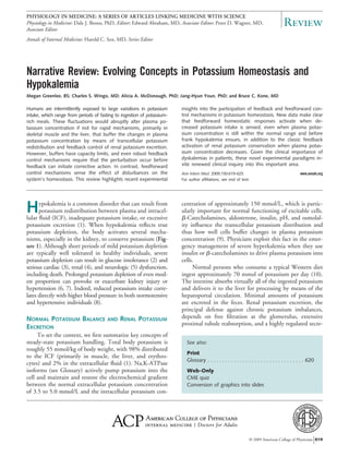
Figure 2. Segmental handling of potassium excretion along the nephron and collecting duct cell types under normal conditions or
conditions of potassium excess or deficiency.
Blood
Lumen
Lumen
Blood
We present a simplified model of potassium handling by collecting duct cell types. Principal cells in the collecting duct are responsible for
secretion of excess potassium in the circulation into the tubule lumen and thus into the urine. This secretion is accomplished by luminal
membrane potassium channels responding primarily to the electrochemical gradient for potassium generated by the combined actions of the
basolateral membrane Na,K-ATPase and a luminal membrane sodium channel (the target of the potassium-sparing diuretic amiloride). In states
of potassium depletion, potassium secretion by the principal cells is inhibited and the luminal membrane H,K-ATPase is activated in the
intercalated cells to reclaim the potassium that remains in the tubular fluid, thereby limiting urinary potassium wasting. ADP ϭ adenosine
diphosphate; ATP ϭ adenosine triphosphate; CCD ϭ cortical collecting duct; DT ϭ distal tubule; Glom ϭ glomerulus; IMCD ϭ inner
medullary collecting duct; MTAL ϭ medullary thick ascending limb of Henle loop; OMCD ϭ outer medullary collecting duct; Pi ϭ inorganic
phosphate.
ReviewEvolving Concepts in Potassium Homeostasis and Hypokalemia
www.annals.org 5 May 2009 Annals of Internal Medicine Volume 150 • Number 9 621
4. incompetent to conduct membrane transport of potassium
(20, 21). In addition, an H,K-ATPase (a relative of the
proton pump active in the gastric mucosa) is activated in
outer medullary collecting duct intercalated cells to reclaim
any remaining filtered potassium into the plasma. Figure 2
summarizes the responses of the different nephron seg-
ments to accommodate changes in potassium intake.
FEEDFORWARD CONTROL OF POTASSIUM BALANCE
Feedforward control refers to a pathway in a homeo-
static system that responds to a signal in the environment
in a predetermined manner, without responding to how
the system subsequently reacts (that is, without responding
to feedback). The most famous example of feedforward
control is the conditioned salivation of Pavlov’s dogs in
anticipation of food (22). Pavlov implanted small stomach
pouches in dogs to measure salivation. He and his assis-
tants would ring bells to call the dogs to their food and
measure the dogs’ increase in salivation. After several rep-
etitions of this experiment, the dogs increased their saliva-
tion when they heard the bell, without any food being
presented. The dogs had been trained to “sense” the im-
pending arrival of a meal and prepare for it, in a physio-
logic sense, by increasing the production of saliva.
Physiologists studying potassium homeostasis were
surprised to discover that even minor changes in dietary
potassium intake, insufficient to change plasma concentra-
tions of either potassium (23) or aldosterone (24) and
therefore too minor to activate feedback control, evoked
rapid changes in renal potassium excretion through feed-
forward mechanisms. Twenty-five years ago, Rabinowitz
and colleagues (25) challenged feedback regulation as the
sole mechanism for compensatory renal potassium excre-
tion and proposed a feedforward kaliuretic reflex, in which
potassium sensors in the splanchnic vascular bed (the gut,
portal vein, or liver) detect local changes in potassium con-
centration resulting from potassium ingestion and signal
the kidney to alter potassium excretion to restore balance
(Figure 1). These investigators showed that sheep that in-
gested a potassium-rich meal over 1 hour rapidly and
appropriately increased renal potassium excretion, even
though plasma concentrations of potassium and aldoste-
rone did not increase sufficiently to account for the ensu-
ing kaliuresis. Subsequent studies in adrenalectomized rats
proved that this rapid urinary potassium excretion was not
mediated by aldosterone, because doses of aldosterone
much higher than the physiologic range were required (26,
27). Studies in normal humans undergoing water diuresis
corroborated these findings by showing that ingestion
of potassium salts promoted urinary potassium excretion
within 20 minutes, before any increase in plasma con-
centrations of potassium or aldosterone (23), which
means that it was not mediated through classical feedback
regulation.
What is the potassium sensor in feedforward regula-
tion, and where is it located in the body? Studies in rats
done in 2 of the reviewers’ laboratories (28) demonstrated
that a gut sensor detects potassium intake during a meal
and triggers a signal to the kidneys to increase potassium
excretion. An intragastric potassium infusion given with a
meal to unfed animals led to greater renal clearance of
plasma potassium than did the same intragastric infusion
given without the meal or given by systemic infusion of
potassium (28). The fact that plasma potassium clearance
was enhanced in response to a potassium load only when
food was present in the stomach provides strong evidence
that the sensor in this feedforward pathway is in the gut.
Whether mechanical factors associated with the bulk of
food (such as stretching of the stomach) play a role in the
gut potassium sensing, and how such mechanical sensors
might initiate the kaliuretic reflex signal, remain to be es-
tablished. A hepatoportal potassium sensor may also par-
ticipate in the kaliuretic reflex. Morita and colleagues (29)
found that intraportal infusion of potassium chloride in
animals yielded greater urinary potassium excretion than
did an intravenous infusion. Severing the periarterial he-
patic nervous plexus attenuated the kaliuresis. Further-
more, an acute potassium chloride infusion directly into
the hepatoportal circulation stimulated hepatic afferent
nerve activity and increased urinary potassium excretion in
the absence of changes in plasma potassium concentration
(29). Bumetanide, a clinically used loop diuretic that in-
hibits cation/chloride cotransporters (including the Naϩ
,
Kϩ
, 2ClϪ
cotransporter NKCC1 [see Glossary]), ablated
the activity of this potassium sensor. This suggests that the
hepatoportal Naϩ
, Kϩ
, 2ClϪ
cotransporter, or a related
bumetanide-sensitive cotransporter, sensed an increase in
portal venous potassium and activated the periarterial he-
patic nerve complex, which sent a neurally mediated signal
transmission to the kidneys with consequent kaliuresis
(30). Consistent with these studies, the NKCC1 knockout
mouse (which lacks the NKCC1 cotransporter and there-
fore lacks this feedforward mechanism) exhibits hyper-
kalemia after ingesting a potassium load and inappropri-
ately low urinary potassium excretion (31, 32).
We have not fully determined the molecular events
involved in the kaliuretic reflex. However, several prece-
dents for feedforward regulation exist: the related Naϩ
,
Kϩ
, 2ClϪ
cotransporter isoform NKCC2 (see Glossary)
senses NaCl delivery to the macula densa segment of the
glomerulus and signals for afferent arteriolar constriction at
the glomerulus (which decreases glomerular filtration)
(33); several tissues express Ca2ϩ
-sensing receptors (34);
glucose sensors operate in the portal vein (35); and diverse
subpopulations of afferent nerves coordinate reflex control
of the local mechanical and chemical environment in the
liver (36), gut mucosa, muscle, and mesentery (37). These
examples of sensors in other feedforward mechanisms make
it plausible that potassium sensors govern feedforward reg-
ulation of potassium in the skeletal muscle and kidney.
Review Evolving Concepts in Potassium Homeostasis and Hypokalemia
622 5 May 2009 Annals of Internal Medicine Volume 150 • Number 9 www.annals.org
5. Feedforward regulation in these tissues also occurs
with chronic restriction of potassium intake, again even
before hypokalemia occurs. As noted, when dietary potas-
sium restriction is prolonged and severe enough to cause
hypokalemia, Na,K-ATPase pumps become less numerous
and less active in the skeletal muscle to protect against
further decrements in muscle potassium. In contrast, ani-
mals chronically fed a moderately potassium-restricted diet
that did not produce hypokalemia maintained normal
muscle potassium levels and normal amounts and activity
of Na,K-ATPase pumps in their membranes. However,
potassium-conserving mechanisms were activated: Insulin-
stimulated clearance of potassium from the plasma was
blunted (evidence of insulin resistance to cellular potas-
sium uptake) (38), and renal potassium excretion was re-
duced to 20% of that of control participants (indicating
potassium conservation). The decrease in renal potassium
excretion was apparently the result of enhanced phosphor-
ylation and consequent internalization of ROMK channels
from the plasma membrane (rendering them inaccessible to
conduct potassium secretion) in the collecting duct principal
cells (38). These results indicate that even before the plasma
potassium concentration begins to decrease, feedforward acti-
vation of potassium-conserving mechanisms acts to maintain
plasma potassium concentration. The error signals that incite
and maintain these responses are unknown but may be related
to gastric or hepatoportal sensing of potassium intake.
METABOLIC CONTROL OF POTASSIUM BALANCE
Intense exercise or ischemia significantly alters potas-
sium homeostasis. The 2 situations share similarities,
which include elevated extracellular fluid potassium
concentration caused by an increase in potassium efflux
relative to influx and an increased ratio of intracellular
adenosine monophosphate to adenosine triphosphate. Ho-
meostasis is restored in both cases by net potassium uptake
into the cells through mediators that have not been clearly
identified.
A good candidate mediator for this net potassium up-
take is cellular adenosine monophosphate–activated pro-
tein kinase. This enzyme, which activates when it detects
an increase in the cellular ratio of adenosine monophos-
phate to adenosine triphosphate, mediates increased glu-
cose transport in exercising muscles to replete cellular
adenosine triphosphate (39). Indeed, 2 reviewers estab-
lished that chemical activation of adenosine monophos-
phate–activated protein kinase in conscious rats acutely de-
creased plasma potassium concentration without increasing
urinary potassium excretion, which indicates that it shifted
potassium to the ICF (40). In support of this concept,
mutant mice with inactive muscle adenosine monophos-
phate–activated protein kinase exhibited impaired potas-
sium uptake into the cells in response to an activator of
adenosine monophosphate–activated protein kinase.
Whether active potassium uptake by cells or depressed po-
tassium efflux from cells mediates this potassium redistri-
bution remains to be determined, but this novel mecha-
nism—activated by metabolic stress—operates in parallel
to the potassium uptake pathway stimulated by insulin
(40).
CLINICAL IMPLICATIONS
The data we reviewed indicate that plasma potas-
sium concentration is a relatively insensitive marker for
the integrated homeostatic responses that are activated
by changes in potassium intake or renal excretion. These
responses occur while plasma potassium concentration is
still within the normal range and before frank hypokalemia
and potassium depletion ensue. Studies in rats that were
fed a modestly potassium-restricted diet (38) indicate that
feedforward adjustments to low-potassium diets occur in
both muscle and kidney, operate independently of changes
in plasma potassium concentration, and can persist in the
long term without hypokalemia. The classic paradigm for
diagnosis and treatment of hypokalemia (1) distinguishes
only between transcellular potassium shifts and alterations
in total body potassium due to altered intake or feedback
regulation of potassium excretion. Feedforward regulation
of potassium buffering and renal potassium excretion
changes this paradigm, which may change the treatment of
some clinical conditions.
The metabolic syndrome and type 2 diabetes mellitus are
both associated with insulin resistance of muscle, fat, and liver
cells. Recent animal studies (18) demonstrating that skeletal
muscle becomes insulin-resistant to cellular potassium (but
not glucose) uptake even before plasma potassium concentra-
tion decreases may lead to new strategies to optimize potas-
sium homeostasis—and thus, to a degree, modulate cardiovas-
cular risk—in these patients. Moreover, the finding that
adenosine monophosphate–activated protein kinase activation
promotes not only cellular glucose uptake (a key mechanism
of action of the antidiabetic drugs metformin and thiazo-
lidinediones) but also potassium uptake in experimental ani-
mals may drive clinical investigation to consider expanded
uses of these or closely related drugs. For example, serious
hyperkalemia can occur in patients with heart failure who
receive spironolactone (41). In hyperkalemic spironolactone-
treated rats, pharmacologic stimulation of adenosine mono-
phosphate–activated protein kinase with 5-aminoimidazole-
4-carboxamide ribonucleoside quickly restored plasma potas-
sium concentration to baseline (40). This observation suggests
that co-administering such agents with medications that can
increase plasma potassium concentration—such as spironolac-
tone—might be clinically useful. On a cautionary note, infus-
ing 5-aminoimidazole-4-carboxamide ribonucleoside into
potassium-depleted hypokalemic rats induced profound hy-
pokalemia (40). This observation suggests that metformin, a
commonly used adenosine monophosphate–activated protein
kinase activator, could provoke dangerous hypokalemia in pa-
tients susceptible to potassium depletion from taking
ReviewEvolving Concepts in Potassium Homeostasis and Hypokalemia
www.annals.org 5 May 2009 Annals of Internal Medicine Volume 150 • Number 9 623
6. potassium-wasting diuretics or fasting (2). We need to explore
these clinical scenarios.
Enteral feeding is an increasingly common and gener-
ally safe practice in acutely ill patients and those who
chronically cannot eat. Feedforward regulation of renal po-
tassium excretion by the putative gut or hepatoportal sen-
sors may help to ensure the safety of potassium replace-
ment by the enteral route. The growing use of enteral
feeding tubes in patients who chronically cannot eat offers an
excellent investigative opportunity to identify the optimal
route of enteral potassium administration in these patients
and to confirm the existence of the putative gut potassium
sensor in humans. When we understand the afferent and ef-
ferent mechanisms that respond to potassium intake by means
of the hepatoportal and gut potassium sensors, we may find
new targets for treating hypokalemia.
CONCLUSION
Given the importance of keeping the plasma potassium
concentration within a narrow range, it is not surprising that
distinct but integrated mechanisms of feedback and feedfor-
ward modes have evolved that act on skeletal muscle and the
liver and kidneys to regulate potassium balance. These dual
mechanisms provide the exquisite and efficient control needed
to maintain and restore potassium homeostasis in response to
acute or chronic changes in body potassium levels. Although
humans clearly have a feedforward kaliuretic reflex, the exis-
tence and location of the potassium sensors remain to be es-
tablished. Moreover, we do not know the specific renal effec-
tor mechanisms downstream of these potassium sensors in
animals or in humans. Despite the remaining gap between the
experimental physiology of these pathways in animals and
clinical practice in humans, these new paradigms of body po-
tassium metabolism provide exciting new insights that may
eventually guide diagnosis and treatment.
From the University of Florida College of Medicine and the Department
of Veterans Affairs Medical Center, Gainesville, Florida, and the Keck
School of Medicine of the University of Southern California, Los Ange-
les, California.
Grant Support: By National Institutes of Health grants R01 DK47981
and R01 DK075065 (Dr. Kone), R01 DK49750 and the Department of
Veterans Affairs (Dr. Wingo), R01 DK34316 (Dr. McDonough), and
R21 DK080233 (Dr. Youn).
Potential Financial Conflicts of Interest: None disclosed.
Requests for Single Reprints: Bruce C. Kone, MD, University of Flor-
ida College of Medicine, 1600 Southwest Archer Road, Gainesville, FL
32610; e-mail, bkone@ufl.edu.
Current author addresses are available at www.annals.org.
References
1. Kone BC. Hypokalemia. In: Hamm LL, Dubose, TD, eds. Acid–Base and
Electrolyte Disorders: A Companion to Brenner and Rector’s The Kidney. Phil-
adelphia: Saunders; 2002:381-94.
2. Zillich AJ, Garg J, Basu S, Bakris GL, Carter BL. Thiazide diuretics, potas-
sium, and the development of diabetes: a quantitative review. Hypertension.
2006;48:219-24. [PMID: 16801488]
3. Gheeraert PJ, De Buyzere ML, Taeymans YM, Gillebert TC, Henriques JP,
De Backer G, et al. Risk factors for primary ventricular fibrillation during acute
myocardial infarction: a systematic review and meta-analysis. Eur Heart J. 2006;
27:2499-510. [PMID: 16952926]
4. Reungjui S, Roncal CA, Sato W, Glushakova OY, Croker BP, Suga S, et al.
Hypokalemic nephropathy is associated with impaired angiogenesis. J Am Soc
Nephrol. 2008;19:125-34. [PMID: 18178802]
5. Riggs JE. Neurologic manifestations of electrolyte disturbances. Neurol Clin.
2002;20:227-39, vii. [PMID: 11754308]
6. Khosla N, Hogan D. Mineralocorticoid hypertension and hypokalemia. Se-
min Nephrol. 2006;26:434-40. [PMID: 17275580]
7. Krishna GG, Kapoor SC. Potassium depletion exacerbates essential hyperten-
sion. Ann Intern Med. 1991;115:77-83. [PMID: 2058867]
8. Whelton PK, He J, Cutler JA, Brancati FL, Appel LJ, Follmann D, et al.
Effects of oral potassium on blood pressure. Meta-analysis of randomized con-
trolled clinical trials. JAMA. 1997;277:1624-32. [PMID: 9168293]
9. Gennari FJ. Hypokalemia. N Engl J Med. 1998;339:451-8. [PMID:
9700180]
10. Elliott P, Dyer A, Stamler R. The INTERSALT study: results for 24 hour
sodium and potassium, by age and sex. INTERSALT Co-operative Research
Group. J Hum Hypertens. 1989;3:323-30. [PMID: 2810328]
11. Peti-Peterdi J, Warnock DG, Bell PD. Angiotensin II directly stimulates
ENaC activity in the cortical collecting duct via AT(1) receptors. J Am Soc
Nephrol. 2002;13:1131-5. [PMID: 11960999]
12. Wei Y, Zavilowitz B, Satlin LM, Wang WH. Angiotensin II inhibits the
ROMK-like small conductance K channel in renal cortical collecting duct during
dietary potassium restriction. J Biol Chem. 2007;282:6455-62. [PMID:
17194699]
13. Sansom SC, Welling PA. Two channels for one job. Kidney Int. 2007;72:
529-30. [PMID: 17713560]
14. Field MJ, Stanton BA, Giebisch GH. Differential acute effects of aldoste-
rone, dexamethasone, and hyperkalemia on distal tubular potassium secretion in
the rat kidney. J Clin Invest. 1984;74:1792-802. [PMID: 6501571]
15. DeFronzo RA, Felig P, Ferrannini E, Wahren J. Effect of graded doses of
insulin on splanchnic and peripheral potassium metabolism in man. Am J
Physiol. 1980;238:E421-7. [PMID: 6990783]
16. Wang W. Regulation of renal K transport by dietary K intake. Annu Rev
Physiol. 2004;66:547-69. [PMID: 14977413]
17. Giebisch G, Krapf R, Wagner C. Renal and extrarenal regulation of potas-
sium. Kidney Int. 2007;72:397-410. [PMID: 17568786]
18. McDonough AA, Youn JH. Role of muscle in regulating extracellular potas-
sium. Semin Nephrol. 2005;25:335-42. [PMID: 16139689]
19. McDonough AA, Thompson CB, Youn JH. Skeletal muscle regulates extra-
cellular potassium. Am J Physiol Renal Physiol. 2002;282:F967-74. [PMID:
11997312]
20. Babilonia E, Lin D, Zhang Y, Wei Y, Yue P, Wang WH. Role of
gp91phox-containing NADPH oxidase in mediating the effect of K restriction on
ROMK channels and renal K excretion. J Am Soc Nephrol. 2007;18:2037-45.
[PMID: 17538186]
21. Zhang Y, Lin DH, Wang ZJ, Jin Y, Yang B, Wang WH. K restriction
inhibits protein phosphatase 2B (PP2B) and suppression of PP2B decreases
ROMK channel activity in the CCD. Am J Physiol Cell Physiol. 2008;294:
C765-73. [PMID: 18184875]
22. Pavlov IP. Conditioned Reflexes: An Investigation of the Physiological Ac-
tivity of the Cerebral Cortex. Anrep GV, trans. London: Oxford Univ Pr;
1927.
23. Calo` L, Borsatti A, Favaro S, Rabinowitz L. Kaliuresis in normal
subjects following oral potassium citrate intake without increased plasma
potassium concentration. Nephron. 1995;69:253-8. [PMID: 7753258]
24. Rabinowitz L, Denham SC, Gunther RA. Aldosterone and postprandial
renal excretion of sodium and potassium in sheep. Am J Physiol. 1977;233:
F213-6. [PMID: 910916]
25. Rabinowitz L, Green DM, Sarason RL, Yamauchi H. Homeostatic potas-
sium excretion in fed and fasted sheep. Am J Physiol. 1988;254:R357-80.
[PMID: 3344840]
26. Campen TJ, Vaughn DA, Fanestil DD. Mineralo- and glucocorticoid effects
on renal excretion of electrolytes. Pflugers Arch. 1983;399:93-101. [PMID:
Review Evolving Concepts in Potassium Homeostasis and Hypokalemia
624 5 May 2009 Annals of Internal Medicine Volume 150 • Number 9 www.annals.org
7. 6647008]
27. Stanton B, Pan L, Deetjen H, Guckian V, Giebisch G. Independent effects
of aldosterone and potassium on induction of potassium adaptation in rat kidney.
J Clin Invest. 1987;79:198-206. [PMID: 3793923]
28. Lee FN, Oh G, McDonough AA, Youn JH. Evidence for gut factor in Kϩ
homeostasis. Am J Physiol Renal Physiol. 2007;293:F541-7. [PMID: 17522262]
29. Morita H, Fujiki N, Miyahara T, Lee K, Tanaka K. Hepatoportal
bumetanide-sensitive K(ϩ)-sensor mechanism controls urinary K(ϩ) excretion.
Am J Physiol Regul Integr Comp Physiol. 2000;278:R1134-9. [PMID:
10801279]
30. Tsuchiya Y, Nakashima S, Banno Y, Suzuki Y, Morita H. Effect of high-
NaCl or high-KCl diet on hepatic Naϩ- and Kϩ-receptor sensitivity and
NKCC1 expression in rats. Am J Physiol Regul Integr Comp Physiol. 2004;286:
R591-6. [PMID: 14656769]
31. Meyer JW, Flagella M, Sutliff RL, Lorenz JN, Nieman ML, Weber CS,
et al. Decreased blood pressure and vascular smooth muscle tone in mice lacking
basolateral Na(ϩ)-K(ϩ)-2Cl(-) cotransporter. Am J Physiol Heart Circ Physiol.
2002;283:H1846-55. [PMID: 12384462]
32. Wall SM, Knepper MA, Hassell KA, Fischer MP, Shodeinde A, Shin W,
et al. Hypotension in NKCC1 null mice: role of the kidneys. Am J Physiol Renal
Physiol. 2006;290:F409-16. [PMID: 16159893]
33. Schnermann J, Briggs JP. Tubuloglomerular feedback: mechanistic
insights from gene-manipulated mice. Kidney Int. 2008;74:418-26.
[PMID: 18418352]
34. Brown EM, Gamba G, Riccardi D, Lombardi M, Butters R, Kifor O, et al.
Cloning and characterization of an extracellular Ca(2ϩ)-sensing receptor from
bovine parathyroid. Nature. 1993;366:575-80. [PMID: 8255296]
35. Hevener AL, Bergman RN, Donovan CM. Hypoglycemic detection does
not occur in the hepatic artery or liver: findings consistent with a portal vein
glucosensor locus. Diabetes. 2001;50:399-403. [PMID: 11272153]
36. Uno K, Katagiri H, Yamada T, Ishigaki Y, Ogihara T, Imai J, et al.
Neuronal pathway from the liver modulates energy expenditure and systemic
insulin sensitivity. Science. 2006;312:1656-9. [PMID: 16778057]
37. Grundy D. Sensory signals from the gastrointestinal tract. J Pediatr Gastro-
enterol Nutr. 2005;41 Suppl 1:S7-9. [PMID: 16131978]
38. Chen P, Guzman JP, Leong PK, Yang LE, Perianayagam A, Babilonia E,
et al. Modest dietary Kϩ restriction provokes insulin resistance of cellular Kϩ
uptake and phosphorylation of renal outer medulla Kϩ channel without fall in
plasma Kϩ concentration. Am J Physiol Cell Physiol. 2006;290:C1355-63.
[PMID: 16354756]
39. Towler MC, Hardie DG. AMP-activated protein kinase in metabolic control
and insulin signaling. Circ Res. 2007;100:328-41. [PMID: 17307971]
40. Zheng D, Perianayagam A, Lee DH, Brannan MD, Yang LE, Tellalian D,
et al. AMPK activation with AICAR provokes an acute fall in plasma potassium.
Am J Physiol Cell Physiol. 2008;294:C126-35. [PMID: 18003746]
41. Juurlink DN, Mamdani MM, Lee DS, Kopp A, Austin PC, Laupacis A,
et al. Rates of hyperkalemia after publication of the Randomized Aldactone Eval-
uation Study. N Engl J Med. 2004;351:543-51. [PMID: 15295047]
NOW AVAILABLE EARLY ONLINE AT WWW.ANNALS.ORG
Stent Placement in Patients With Atherosclerotic Renal Artery Stenosis
and Impaired Renal Function
Bax, Woittiez, Kouwenberg, Mali, and others
Sign up to receive e-mail alerts for early-release articles at www.annals
.org/subscriptions/etoc.shtml.
ReviewEvolving Concepts in Potassium Homeostasis and Hypokalemia
www.annals.org 5 May 2009 Annals of Internal Medicine Volume 150 • Number 9 625
8. Current Author Addresses: Ms. Greenlee and Drs. Wingo and Kone:
University of Florida College of Medicine, 1600 Southwest Archer Road,
Gainesville, FL 32610.
Drs. McDonough and Youn: University of Southern California
Keck School of Medicine, 1333 San Pablo Street, Los Angeles, CA
90089.
Annals of Internal Medicine
W-110 5 May 2009 Annals of Internal Medicine Volume 150 • Number 9 www.annals.org