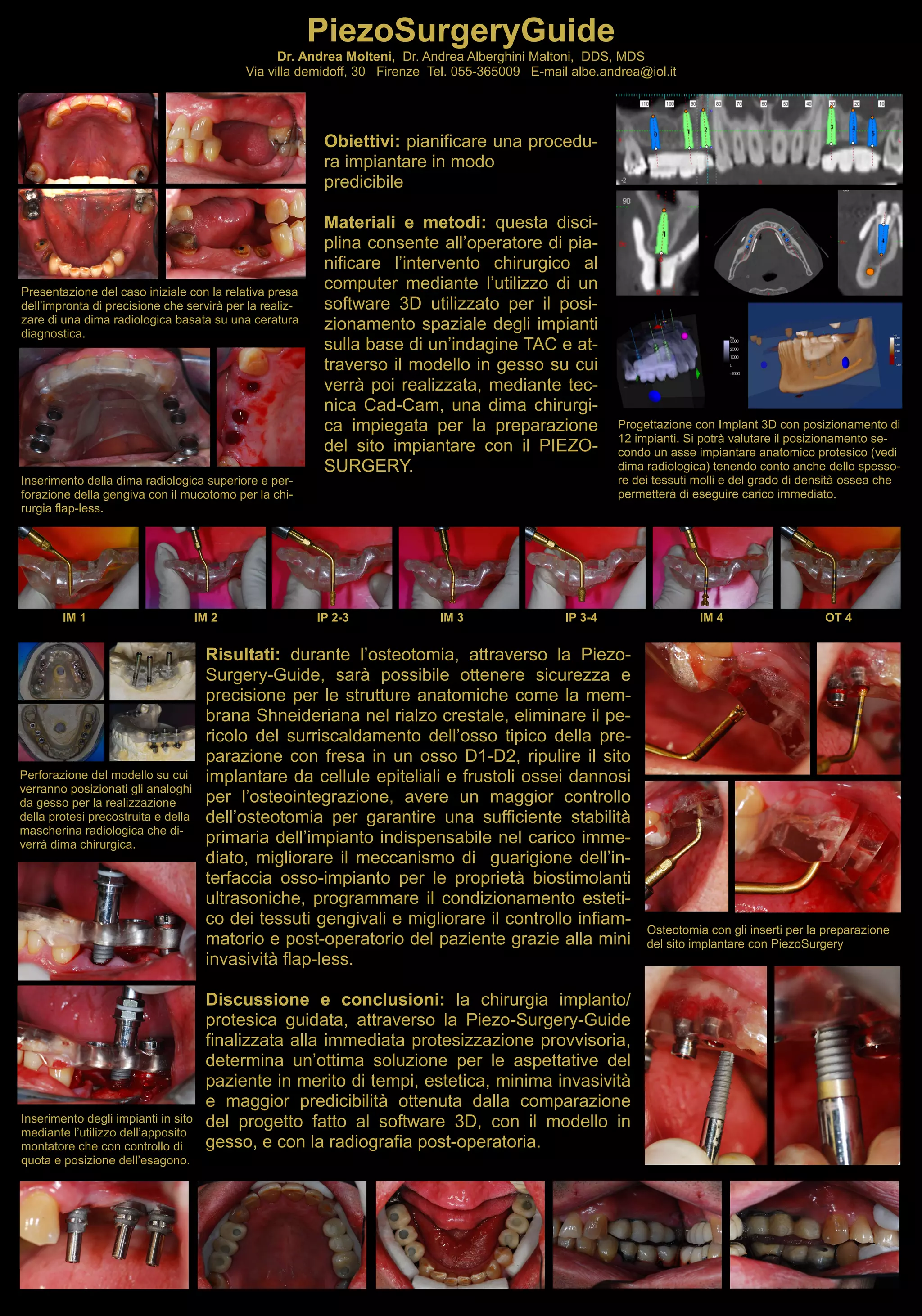
L’innovazione organizzativa attraverso il Project Cycle Management: l’esperienza dell’ASL Città di Torino - Premio Qualità 2017/2018 (*) - Prima edizione elettronica: Dicembre 2018 | ISBN: 978-88-943964-0-9 | Edizioni Asl Città di Torino | WWW.ASLCITTADITORINO.IT
Il progetto “Progettazione e realizzazione di tessuti e endoprotesi su misura mediante tecnologie sottrattive e additive” (Custom Implants) si inserisce nel campo della chirurgia personalizzata di precisione e in particolare:
Precision Grafting: produzione di tessuti umani mediante manifattura sottrattiva robotizzata
Personalized-Scaffolds: realizzazione di dispositivi costituiti da biomateriali e/o cellule umane tramite stampa 3D e cellularizzazione in-vitro
Custom-Endoprotesi: realizzazione in stampa 3D di protesi in leghe metalliche e in polietilene per la chirurgia protesica sostitutiva
Principali filiere coinvolte: Biomedicale, Farmaceutica, Chimica, Biologica, Ingegneria tissutale, Clinica, Meccanica, Automazione, Packaging, ICT, Manifatturiera
Sito web del progetto: www.custom-implants.it
Presentazioni e video: cerr.eu/what-s-going-on/357-materiali-dei-seminari-disponibili-online
Laser Banca delle cornee Emilia-Romagna - Congresso SITRAC (Società italiana ...Luca Avoni
Il trapianto di cornea (cheratoplastica) endoteliale è ormai il gold standard per la maggior parte delle malattie corneali con indicazione di trapianto nelle quali l'endotelio (strato più interno della cornea) non funziona correttamente.
La letteratura internazionale evidenzia gli indubbi vantaggi di tale tecnica e il crescente numero di interventi effettuati a livello mondiale.
Tra le indicazioni più frequenti di questo intervento sono la cheratopatia bollosa e la distrofia corneale endoteliale di Fuchs.
La ricerca è particolarmente attenta allo sviluppo di questa metodica di trapianto corneale per migliorare i risultati. Le direzioni sono varie, dall'UTDSAEK (ultra thin DSAEK che prevede la creazione di lembi endoteliali particolarmente sottili per poi impiantarli) alla DMEK che prevede l'impianto del solo endotelio-descemet.
Altra linea di ricerca è il trattamento della cornea con laser ad eccimeri prima del taglio col microcheratomo al fine di ottenere lembi endoteliali sottili e a facce parallele.
Piezosurgery in oral and maxillofacial surgeryArjun Shenoy
This document discusses piezoelectricity and its use in maxillofacial surgery procedures through a tool called piezosurgery. Piezoelectricity was discovered in 1880 and involves generating electric charges in response to mechanical stress in certain solid materials. Piezosurgery uses this principle with piezoelectric ceramic materials in surgical instruments to precisely cut bone. It allows for selective cutting of hard tissues while sparing soft tissues. Some advantages of piezosurgery include clean cuts with limited damage to bone cells and bloodless surgery. The document outlines several maxillofacial procedures where piezosurgery has been applied and its benefits compared to traditional techniques, such as reduced blood loss and nerve injury.
A diplomate of the Board of Periodontology, Perry Westbrook, DMD, is also one of the nation’s first Kois Center Recognized Periodontist Specialists. Dr. Perry Westbrook has trained in piezosurgery, an advanced type of bone surgery, and holds a membership with the International Piezosurgery Academy.
In an age where every teeny tiny bit of electricity is valued, conservation is much talked about, can piezoelectricity be the messiah to ease the burden off the conventional energy sources?
Who says it cannot?
--
Presentation as a part of seminar coursework.
Piezoelectric electric based energy harvestingSubash John
Piezoelectric materials can generate an electric charge when subjected to mechanical stress. This phenomenon known as the piezoelectric effect enables piezoelectric materials to convert mechanical vibrational energy into electrical energy through a process known as energy harvesting. Common sources of vibration that can be used for piezoelectric energy harvesting include footsteps on sidewalks, movements from gym equipment, and vibrations from vehicles. The electric energy produced can be stored in batteries or capacitors and used to power small electronic devices. Piezoelectric materials have applications in various technologies including ultrasound imaging, sensors, musical instruments, and automotive engine management systems.
The document discusses piezoelectric surgery, which uses ultrasonic microvibrations from piezoelectric inserts to cut bone precisely while sparing soft tissues. Key points:
1. Piezoelectric surgery was invented in 1988 and uses modulated ultrasonic frequencies (25-30 kHz) to cut mineralized tissue selectively.
2. It allows for precise cuts with less bleeding and better visibility due to cavitation. Post-operative pain and healing time are reduced compared to traditional techniques.
3. Indications include various dental and medical procedures like sinus lifts, grafting and extractions where selective bone cuts are needed to protect adjacent soft tissues.
Los autores presentan su experiencia utilizando un bisturí piezoeleéctrico para realizar osteotomías extraorales como condilectomías, eminectomías, toma de injertos óseos de la calota craneal y osteotomías para rinoplastia. Este instrumento permite cortes óseos precisos sin riesgo de lesionar tejidos blandos vecinos. Se discuten las ventajas e inconvenientes del bisturí piezoeleéctrico para diferentes procedimientos.
This document discusses piezosurgery, which uses ultrasonic vibrations from piezoelectric inserts to cut bone. Piezosurgery offers several advantages over traditional rotary cutting instruments, including more precise cuts with less risk of soft tissue or nerve damage due to lower temperatures. It allows for selective cutting of mineralized tissue without affecting elastic structures. Piezosurgery is used in various oral and maxillofacial procedures like dental implant placement, sinus lifts, orthognathic surgery, and bone harvesting. It provides better visibility, less bleeding, and more precise cuts compared to conventional techniques.
This document discusses surgical treatment options for sinusitis. It notes that surgery should only be considered if conservative treatment fails or if signs of sinusitis or intracranial complications emerge. Several surgical procedures are described for treating maxillary sinus disease, including antral lavage, intranasal antrostomy, sinoscopy, and Caldwell-Luc or radical antrostomy operations. The Caldwell-Luc procedure involves a sublabial incision to access and clear out the maxillary sinus. Post-operative care and potential complications are also outlined.
Osseous resective surgery involves recontouring bone through ostectomy or osteoplasty to establish a healthier periodontal pocket depth. The goal is to create a shallower pocket that is easier to maintain. Indications for osseous resection include shallow infrabony defects, one-walled angular defects, and furcation involvement. Contraindications are deep isolated defects and advanced periodontitis. The surgery involves raising a mucoperiosteal flap and reshaping the bone using burs and chisels, then suturing the flap.
B.tech sem i engineering physics u v chapter 2-ultrasonic wavesRai University
This document discusses ultrasonic waves and their properties and production methods. It describes how ultrasonic waves are produced using magnetostriction and piezoelectric generators. The magnetostriction method uses the magnetostrictive effect in ferromagnetic rods to produce ultrasonic vibrations, while the piezoelectric method uses the inverse piezoelectric effect in quartz crystals. The document also covers the detection of ultrasonic waves and some of their applications, including non-destructive testing of metals and ultrasonic drilling.
This document discusses osseous surgery and the treatment of bone defects caused by periodontitis. It begins with an overview of normal bone topography and how bone loss from periodontal disease can result in abnormal architectures like interproximal craters and angular bony defects. The rationale for surgical correction of these bone defects is explained, along with the objectives, techniques, and healing process of osseous surgery. Specific challenges like furcation invasions are also addressed. The goal of osseous surgery is to reshape damaged bone in order to reduce pockets and allow for periodontal regeneration.
This document discusses crown lengthening procedures and biological width. It notes that crown lengthening surgery should remove enough bone to allow for 2mm of tooth structure, the biological width of 2mm, and 1mm sulcus depth for non-post-core teeth. For teeth requiring posts, an additional 1.5mm ferrule effect space is needed. The document outlines factors to consider for crown lengthening like tissue biotype, tooth anatomy, and restorability. It recommends more conservative bone removal lingually and waiting at least 20 weeks after surgery before final crown preparation.
This document provides an overview of the maxillary sinus, including its discovery, anatomy, development, functions, and associated pathologies. Some key points:
- The maxillary sinus was first discovered and illustrated by Leonardo da Vinci, but was described in detail by Nathaniel Highmore in 1651.
- It is a pyramid-shaped air space within the body of the maxilla. It is bounded by the zygomatic process, nasal surface, orbital surface, and alveolar process.
- Development begins in the newborn as a tubular structure, becoming ovoid in childhood and pyramidal in adults.
- Pathologies associated with the maxillary sinus include sinusitis, cyst
This document provides an overview of periodontal considerations for restorative dentistry. It discusses the normal periodontium, biologic width, and factors that can irritate the periodontium during restorative procedures. Margin placement is an important consideration, as subgingival margins pose the greatest biologic risk. Contour, contacts, embrasures and overhangs can also impact the periodontium. Proper evaluation and correction of biologic width violations is discussed to minimize risks to periodontal health from restorative work.
Surgical procedures in fixed partial denture prostheses/ General orthodonticsIndian dental academy
Indian Dental Academy: will be one of the most relevant and exciting training center with best faculty and flexible training programs for dental professionals who wish to advance in their dental practice,Offers certified courses in Dental implants,Orthodontics,Endodontics,Cosmetic Dentistry, Prosthetic Dentistry, Periodontics and General Dentistry.
A 40-year-old female presented with an inability to eat properly due to a fractured crown. Upon examination, the remaining tooth structure was below the gumline and not accessible. The procedure involves adjusting the gum tissue and bone around the tooth to provide better access and ensure a proper restoration fit. Biologic width and ferrule effect must be considered to prevent inflammation, bone loss, and future issues. Complications can include an unaesthetic appearance, sensitivity, and loss of bone or papilla.
CHIRURGIA GUIDATA: GUIDE-D.SIGN
Guide-D.Sign è un sistema di progettazione, simulazione e realizzazione di guide per il chirurgo che deve riabilitare un paziente con una edentulia parziale o totale.
Perché è necessario progettare un intervento chirurgico?
La posizione degli impianti è un molto importante per l'affidabilità del risultato. Un impianto inserito senza una posizione prestabilita, potrebbe diventare un problema ingestibile dal punto di vista estetico e funzionale.
Per aiutare il chirurgo in questo delicato compito, abbiamo ideato il sistema Guide-D.Sign.
Guide-D.Sign ha il suo punto di partenza dalla ricostruzione delle strutture anatomiche mancanti: partendo dai tessuti ossei e mucosi per arrivare al posizionamento dei denti mancanti.
Il sistema Guide-D.Sign è composto da 4 prodotti:
1. La dima radiologica: è un dispositivo che si inserisce nella bocca del paziente e ricostruisce i volumi e le forme che avranno le protesi definitive. Gli speciali materiali con cui sono realizzate, permettono di comunicare al chirurgo la posizione dei denti della futura protesi.
2. La pianificazione chirurgica: grazie al team di chirurghi esperti, Keys DS è in grado di fornire anche una pianificazione chirurgica a tutti coloro che non sono dotati di un software di pianificazione, riducendo i tempi necessari alla pianificazione e i costi della stessa.
3. Il modello prechirurgico: è un modello in resina che trasferisce al laboratorio odontotecnico la posizione degli impianti così come sono stati previsti dal progetto dell'odontoiatra. Grazie a questo prodotto, l'odontotecnico può confezionare un provvisorio prima dell'intervento e permettere all'odontoiatra di caricare immediatamente gli impianti.
4. La dima chirurgica: in base alla progettazione dell'intervento, realizziamo un dispositivo che permette al chirurgo di posizionare gli impianti in modo semplice e completamente guidato dal progetto prestabilito. Con le nostre guide, il chirurgo riduce i tempi di intervento con evidenti vantaggi per il paziente e la ottimizzazione del risultato funzionale ed estetico.
L’invenzione consta in un dispositivo per video-endoscopia odontoiatrica intraorale ideato per migliorare la qualità dell’esame o dell’intervento odontoiatrico del paziente attraverso la riduzione dell’affaticamento posturale e oculare dell’operatore
Grazie alla moderna tecnologia è ormai possibile avere la realtà muscolo scheletrica virtuale
di ogni paziente.
Questo ci permette di progettare e costruire protesi dentali personalizzate e quindi più performanti
dal punto di vista estetico e funzionale, con l’ottimizzazione di tempi e costi di
lavorazione.
Procedimento e sistema per rappresentazione tridimensionale di una scena a re...Toscana Open Research
La presente invenzione riguarda un procedimento ed un sistema per la rappresentazione tridimensionale di una scena a realtà aumentata pensato soprattutto per essere impiegato come ausilio chirurgico
Questa affermazione può essere applicata alle più
svariate circostanze della nostra vita quotidiana.
Nel caso particolare della professione medica, ottenere
informazioni diagnostiche accurate rappresenta
un presupposto prioritario al fine di fornire ai propri
pazienti un piano di trattamento/terapia
con la massima probabilità di esito positivo.
Tale obiettivo può essere raggiunto utilizzando
tecnologie e metodiche diagnostiche di ultima
generazione in grado di garantire al paziente
il miglior compromesso tra la riduzione significativa
della radiazione ricevuta e la necessità
del professionista odontoiatrico di disporre
di informazioni non ottenibili con altra
metodologia.
Il progetto Custom Implants si inserisce nel campo della chirurgia personalizzata di precisione e in particolare:
Precision Grafting: produzione di tessuti umani mediante manifattura sottrattiva robotizzata
Personalized-Scaffolds: realizzazione di dispositivi costituiti da biomateriali e/o cellule umane tramite stampa 3D e cellularizzazione in-vitro
Custom-Endoprotesi: realizzazione in stampa 3D di protesi in leghe metalliche e in polietilene per la chirurgia protesica sostitutiva
Principali filiere coinvolte: Biomedicale, Farmaceutica, Chimica, Biologica, Ingegneria tissutale, Clinica, Meccanica, Automazione, Packaging, ICT, Manifatturiera
Sito web del progetto: www.custom-implants.it
Presentazioni e video: cerr.eu/what-s-going-on/357-materiali-dei-seminari-disponibili-online
Buongiorno! Siamo al decimo appuntamento con "Benvenuti dal dentista".
Questa settimana vi presentiamo lo Studio Dentistico del dr. Gianluca Marini a Firenze nel quartiere 4.
#dentista #firenze #gianlucamarini #firenzedentista #dentcard
Buongiorno!! Siamo al sesto appuntamento con "Benvenuti dal dentista".
Questa settimana vi presentiamo lo Studio dentistico associato dei Dottori Cito e Corigliano.
Terzo appuntamento con "Benvenuti dal dentista". Questa settimana protagonista lo Studio Dentistico del dr. Jacopo Bacherini.
Lo studio si trova a campi Bisenzio in Via Ponchielli n 56
1. PiezoSurgeryGuide
Dr. Andrea Molteni, Dr. Andrea Alberghini Maltoni, DDS, MDS
Via villa demidoff, 30 Firenze Tel. 055-365009 E-mail albe.andrea@iol.it
Obiettivi: pianificare una procedu-
ra impiantare in modo
predicibile
Materiali e metodi: questa disci-
plina consente all’operatore di pia-
nificare l’intervento chirurgico al
computer mediante l’utilizzo di un
software 3D utilizzato per il posi-
zionamento spaziale degli impianti
sulla base di un’indagine TAC e at-
traverso il modello in gesso su cui
verrà poi realizzata, mediante tec-
nica Cad-Cam, una dima chirurgi-
ca impiegata per la preparazione
del sito impiantare con il PIEZO-
SURGERY.
Presentazione del caso iniziale con la relativa presa
dell’impronta di precisione che servirà per la realiz-
zare di una dima radiologica basata su una ceratura
diagnostica.
Progettazione con Implant 3D con posizionamento di
12 impianti. Si potrà valutare il posizionamento se-
condo un asse impiantare anatomico protesico (vedi
dima radiologica) tenendo conto anche dello spesso-
re dei tessuti molli e del grado di densità ossea che
permetterà di eseguire carico immediato.
Inserimento della dima radiologica superiore e per-
forazione della gengiva con il mucotomo per la chi-
rurgia flap-less.
Risultati: durante l’osteotomia, attraverso la Piezo-
Surgery-Guide, sarà possibile ottenere sicurezza e
precisione per le strutture anatomiche come la mem-
brana Shneideriana nel rialzo crestale, eliminare il pe-
ricolo del surriscaldamento dell’osso tipico della pre-
parazione con fresa in un osso D1-D2, ripulire il sito
implantare da cellule epiteliali e frustoli ossei dannosi
per l’osteointegrazione, avere un maggior controllo
dell’osteotomia per garantire una sufficiente stabilità
primaria dell’impianto indispensabile nel carico imme-
diato, migliorare il meccanismo di guarigione dell’in-
terfaccia osso-impianto per le proprietà biostimolanti
ultrasoniche, programmare il condizionamento esteti-
co dei tessuti gengivali e migliorare il controllo infiam-
matorio e post-operatorio del paziente grazie alla mini
invasività flap-less.
Discussione e conclusioni: la chirurgia implanto/
protesica guidata, attraverso la Piezo-Surgery-Guide
finalizzata alla immediata protesizzazione provvisoria,
determina un’ottima soluzione per le aspettative del
paziente in merito di tempi, estetica, minima invasività
e maggior predicibilità ottenuta dalla comparazione
del progetto fatto al software 3D, con il modello in
gesso, e con la radiografia post-operatoria.
IM 1 IP 2-3 IM 3 IP 3-4 IM 4 OT 4IM 2
Perforazione del modello su cui
verranno posizionati gli analoghi
da gesso per la realizzazione
della protesi precostruita e della
mascherina radiologica che di-
verrà dima chirurgica.
Inserimento degli impianti in sito
mediante l’utilizzo dell’apposito
montatore che con controllo di
quota e posizione dell’esagono.
Osteotomia con gli inserti per la preparazione
del sito implantare con PiezoSurgery