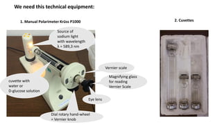
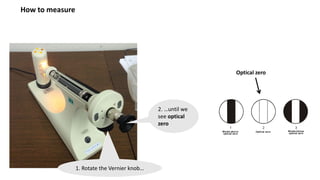
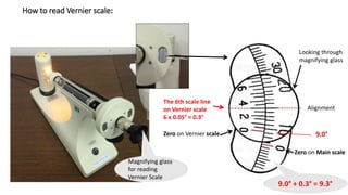
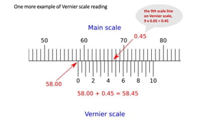
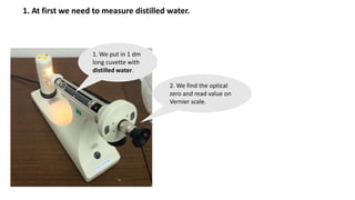
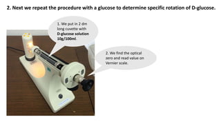
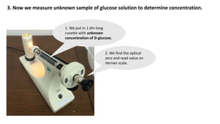
3rd Practical Exercise
Polarimetry
Second Faculty of Medicine, Charles University
General Medicine, 1st year
Biophysics
Practical Exercises
This slides show you how we get a record from a polarimeter.
We need this technical equipment:
1. Manual Polarimeter Krüss P1000
Source of
sodium light
with wavelength
λ = 589,3 nm
2. Cuvettes
cuvette with
water or
D-glucose solution
Vernier scale
Dial rotary hand-wheel
= Vernier knob
Magnifying glass
for reading
Vernier Scale
Eye lens
How to measure
2. …until we
see optical
zero
1. Rotate the Vernier knob…
Optical zero
How to read Vernier scale:
Magnifying glass
for reading
Vernier Scale
Looking through
magnifying glass
9.0° + 0.3° = 9.3°
Alignment
Zero on Main scale
Zero on Vernier scale 9.0°
The 6th scale line
on Vernier scale
6 x 0.05° = 0.3°
One more example of Vernier scale reading
the 9th scale line
on Vernier scale,
9 x 0.05 = 0.45
1. At first we need to measure distilled water.
1. We put in 1 dm
long cuvette with
distilled water.
2. We find the optical
zero and read value on
Vernier scale.
2. Next we repeat the procedure with a glucose to determine specific rotation of D-glucose.
1. We put in 2 dm
long cuvette with
D-glucose solution
10g/100ml.
2. We find the optical
zero and read value on
Vernier scale.
3. Now we measure unknown sample of glucose solution to determine concentration.
1. We put in 1 dm long
cuvette with unknown
concentration of D-glucose.
2. We find the optical
zero and read value on
Vernier scale.
3. Fill the Protocol. Task 1.
FILL IN
THE YELLOW
CELLS ONLY.
You can find it in
this presentation.
You will receive data
from measurement.
Formula:
100 * α = [α] * l * c
Compare your calculated
value with a value from a
table (cite source).
Why do they differ?
(What are next parameters?)
Units. Be careful.
4. Fill the Protocol. Task 2.
You can find it in
this presentation.
You will receive data
from measurement.
Formula:
100 * α = [α] * l * c
Units. Be careful.
Compare your calculated value of D-
glucose concentration with physiological
values of D-glucose concentration in
blood (cite source) in terms of
hypoglycemia or hyperglycemia.

Polarimetry

  • 1.
    3rd Practical Exercise Polarimetry SecondFaculty of Medicine, Charles University General Medicine, 1st year Biophysics Practical Exercises
  • 2.
    This slides showyou how we get a record from a polarimeter.
  • 3.
    We need thistechnical equipment: 1. Manual Polarimeter Krüss P1000 Source of sodium light with wavelength λ = 589,3 nm 2. Cuvettes cuvette with water or D-glucose solution Vernier scale Dial rotary hand-wheel = Vernier knob Magnifying glass for reading Vernier Scale Eye lens
  • 4.
    How to measure 2.…until we see optical zero 1. Rotate the Vernier knob… Optical zero
  • 5.
    How to readVernier scale: Magnifying glass for reading Vernier Scale Looking through magnifying glass 9.0° + 0.3° = 9.3° Alignment Zero on Main scale Zero on Vernier scale 9.0° The 6th scale line on Vernier scale 6 x 0.05° = 0.3°
  • 6.
    One more exampleof Vernier scale reading the 9th scale line on Vernier scale, 9 x 0.05 = 0.45
  • 7.
    1. At firstwe need to measure distilled water. 1. We put in 1 dm long cuvette with distilled water. 2. We find the optical zero and read value on Vernier scale.
  • 8.
    2. Next werepeat the procedure with a glucose to determine specific rotation of D-glucose. 1. We put in 2 dm long cuvette with D-glucose solution 10g/100ml. 2. We find the optical zero and read value on Vernier scale.
  • 9.
    3. Now wemeasure unknown sample of glucose solution to determine concentration. 1. We put in 1 dm long cuvette with unknown concentration of D-glucose. 2. We find the optical zero and read value on Vernier scale.
  • 10.
    3. Fill theProtocol. Task 1. FILL IN THE YELLOW CELLS ONLY. You can find it in this presentation. You will receive data from measurement. Formula: 100 * α = [α] * l * c Compare your calculated value with a value from a table (cite source). Why do they differ? (What are next parameters?) Units. Be careful.
  • 11.
    4. Fill theProtocol. Task 2. You can find it in this presentation. You will receive data from measurement. Formula: 100 * α = [α] * l * c Units. Be careful. Compare your calculated value of D- glucose concentration with physiological values of D-glucose concentration in blood (cite source) in terms of hypoglycemia or hyperglycemia.