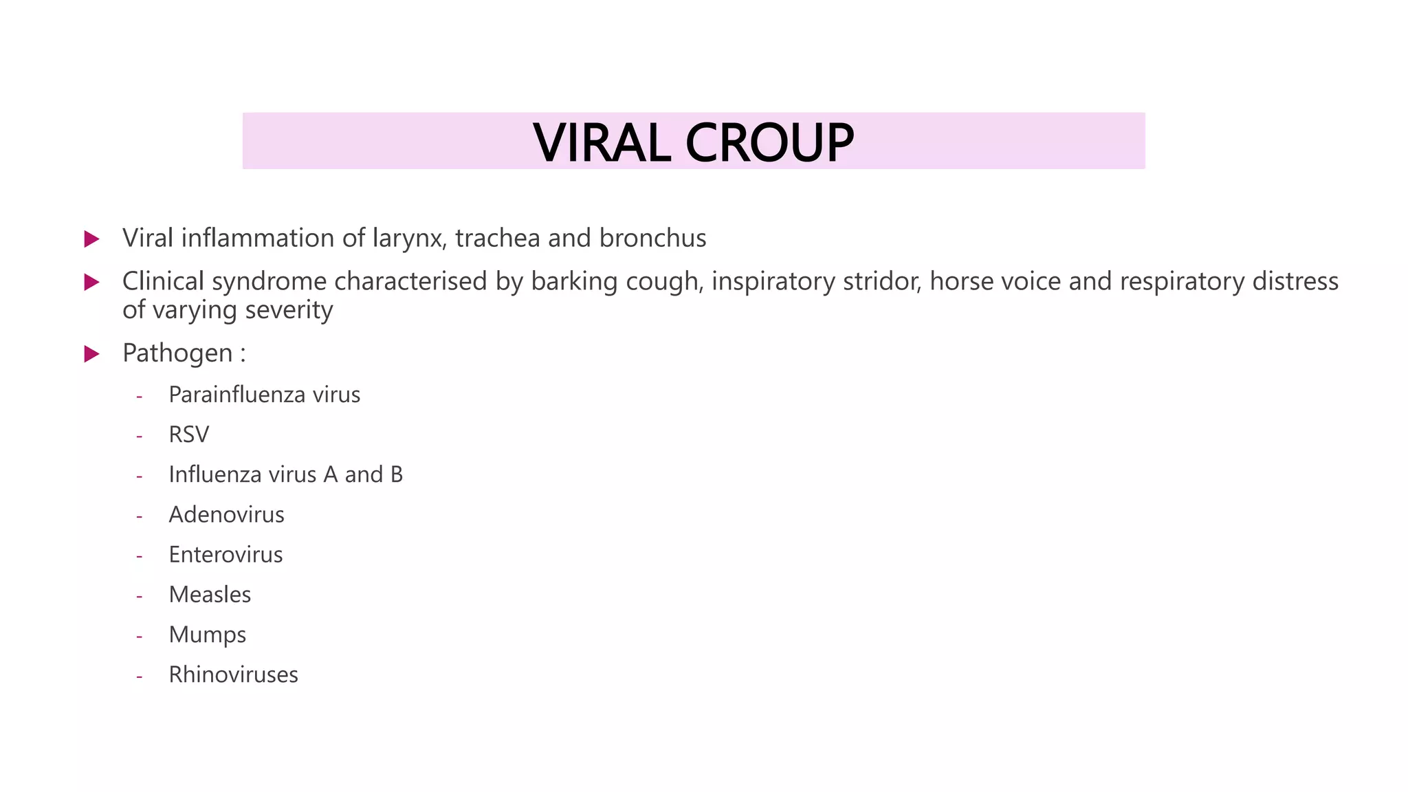
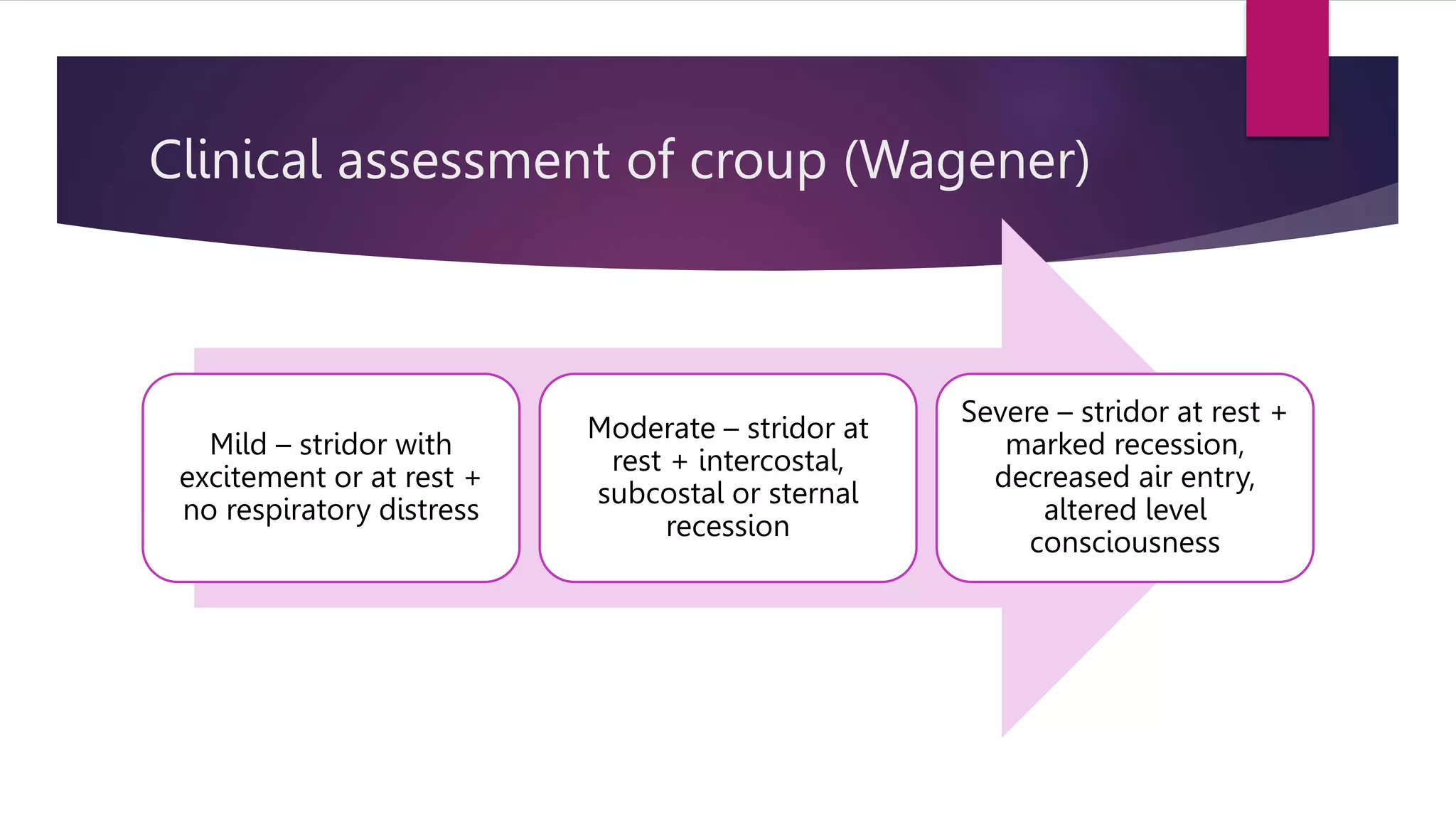
ACUTE RESPIRATORY
INFECTION
PRESENTERS :
AININ SORAYA BINTI MOHAMAD
NUR ATIKAH BINTI HARUN
SUPERVISOR :
DR TEH SC
CONTENT
 Pneumonia
 Viral bronchiolitis
 Viral croup
 Pertussis
Case scenario
 2 year 5 month old, boy was brought to emergency department
with a complaint of,
 Fever for 1 week, on and off fever, highest documented temperature 39C
 Cough for 1 week, chesty cough with whitish sputum
 a/w runny nose and post tussive vomiting
 Rapid breathing for 2 day
 No history of taken any antibiotic before
 Otherwise, good oral intake, no loose stool, no sick contact
 On examination,
 He was alert, pink, fretful,
tachypneic with subcostal recession, RR 48
 Lungs: equal air entry, no rhonchi
 CVS : DRNM
 Abdomen : soft, not distended
 CXR: Bilateral lungs patchy consolidation.
Investigations
 FBC
 Wcc 10.49, (P 46.2%, L 39.1)
 Hb 11.7
 Plt 394
 CRP 41.7
 RP
 Na 135, K 4.1, Cl 97, urea 2.5, creat 18
Impression : Bronchopneumonia
PNEUMONIA
Definitions
 Pneumonia is an inflammatory conditions of the lung- especially
affecting the alveoli and the parenchyma of the lung.
 There are two clinical definitions of pneumonia:
Bronchopneumonia: a febrile illness with cough, respiratory distress
with evidence of localised or generalised patchy infiltrates.
Lobar pneumonia: similar to bronchopneumonia except that the
physical findings and radiographs indicate lobar consolidation.
Aetiology
 Specific aetiological agents are not identified in 40% to 60% of
cases.
 It is often difficult to distinguish viral from bacterial disease.
 The majority of lower respiratory tract infections are viral in origin
e.g. Respiratory syncytial virus, Influenza A or B, Adenovirus,
Parainfluenza virus.
Clinical manifestations
SYMPTOMS SIGN
Fever Tachypnea
Fast and difficult breathing Chest recession
Cough Grunting and stridor
Chest pain Nasal flaring
Abdominal pain Cyanosis
Poor feeding Dullness on percussion
Irritability Diminished breath sound, rhonchi,
crepitation on auscultation
Criteria for hospitalization
 The following indicators can be used as a guide for admission:
 Children aged 3 months and below, whatever the severity of
pneumonia.
 Fever ( more than 38.5 ⁰C ), refusal to feed and vomiting
 Fast breathing with or without cyanosis
 Failure of previous antibiotic therapy
 Recurrent pneumonia
 Severe underlying disorder, e.g. Immunodeficiency
Investigations
 Chest radiograph
 Full blood count
 Blood culture
 Non-invasive gold standard for determining the precise aetiology.
 Sensitivity is low: Positive blood cultures only in 10%-30% of patients.
 Do cultures in severe pneumonia or if poor response to first line antibiotics.
 Pleural fluid analysis
 If there is significant pleural effusion, a diagnostic pleural tap will be helpful.
 Serological tests
 Serology is performed in patients with suspected atypical pneumonia, i.e. Mycoplasma pneumoniae,
Chlamydia, Legionella, Moraxella catarrhalis
MANAGEMENT
 Fluids
 Withhold oral intake when a child is in severe respiratory distress.
 Oxygen
 Oxygen reduces mortality associated with severe pneumonia.
 It should be given especially to children who are restless, and tachypnoeic with severe chest
indrawing, cyanosis, or is not tolerating feeds.
 Maintain the SpO₂ > 95%.
 Cough medication
 Not recommended as it causes suppression of cough and may interfere with airway clearance.
Adverse effects and overdosage have been reported.
MANAGEMENT
 Temperature control
 Reduces discomfort from symptoms, as paracetamol will not
abolish fever.
 Chest physiotherapy
 This assists in the removal of tracheobronchial secretions:
removes airway obstruction, increase gas exchange and reduce
the work of breathing.
ANTIBIOTICS
ANTIBIOTICS
OUTPATIENT MANAGEMENT
 In children with mild pneumonia, their breathing is fast but there is
no chest indrawing.
 Oral antibiotics can be prescribed.
 Educate parents/caregivers about management of fever, preventing
dehydration and identifying signs of deterioration.
 The child should return in two days for reassessment, or earlier if
the condition is getting worse.
VIRAL BRONCHIOLITIS
 Common respiratory illness in infants aged 1 to 6 months old
 Commonest cause – Respiratory syncytial virus (RSV)
 Majority of children has mild ilness
 Symptoms
 Mild coryza and nasal congestion
 Low grade fever
 Cough
 Laboured breathing
 Signs
 Tachypneoa
 Chest wall recession
 Wheezing
 Hyperinflated chest
 Fine crepitation/rhonchi
GUIDELINES FOR HOSPITALISATION Home management Hospital management
Age <3 Months No Yes
Toxic looking No Yes
Chest recession Mild Moderate/severe
Central cyanosis No Yes
Wheeze Yes Yes
Crepitation Yes Yes
Feeding Well Difficult
Apnea No Yes
SPO2 >95% <93%
Chest X ray :
- Hyperinflated lungs
- Segmental collapse/consolidation
- Lobar collapse/consolidation
Normal CXR Hyperinflated lungs Right upper lobe collapse
Management
- Supplemental oxygen
- Maintain SPO2 >95%
- Maintain nutrition and hydration
- Nebulised saline – mucus clearance
VIRAL CROUP
 Viral inflammation of larynx, trachea and bronchus
 Clinical syndrome characterised by barking cough, inspiratory stridor, horse voice and respiratory distress
of varying severity
 Pathogen :
- Parainfluenza virus
- RSV
- Influenza virus A and B
- Adenovirus
- Enterovirus
- Measles
- Mumps
- Rhinoviruses
Clinical features
- Low grade fever
- Cough
- Coryza
- Increasingly bark-like cough and hoarseness
- Inspiratory stridor
- Respiratory distress of varying degree
Clinical assessment of croup (Wagener)
Mild – stridor with
excitement or at rest +
no respiratory distress
Moderate – stridor at
rest + intercostal,
subcostal or sternal
recession
Severe – stridor at rest +
marked recession,
decreased air entry,
altered level
consciousness
Management
 Indication for hospitalisation
- Moderate to severe croup
- Age <6 months
- Poor oral intake
- Toxic, sick looking
- Lives long distance from hospital
PERTUSSIS
 Also known as whooping cough
 Caused by bacterium Bordatella pertussis (gram negative bacilli)
STAGE CLINICAL FEATURES
1
Catarrhal
•Coryza, Low-grade fever
•Mild, occasional cough (which gradually becomes more severe)
2
Paroxysmal*
•Paroxysms of numerous, rapid coughs due to difficulty expelling thick mucus from the tracheobronchial
tree
•Long inspiratory effort accompanied by a high-pitched “whoop” at the end of the paroxysms
•Cyanosis
•Post-tussive vomiting
Paroxysmal attacks:
•Occur frequently at night, with an average of 15 attacks per 24 hours
•Increase in frequency during the first 1-2 weeks, remain at the same frequency for 2-3 weeks, and then
gradually decrease
3
Convalescent
•Gradual recovery
•Less persistent, paroxysmal coughs that disappear in 2-3 weeks
Diagnosis
 Isolation of bordatella pertussis in culture
 Nasopharyngeal swab for PCR
 FBC – absolute lymphocytosis
Management
 Adequate hydration and oral intake
 Antibiotic choice (Macrolides) : Erythromycin, Clarithromycin, Azithromycin
 Prevention
- Postexposure prophylaxis – antibiotic within 21 days of exposure
- Immunization
Thank you

PNEUMONIA CME PAED.pptx

  • 1.
    ACUTE RESPIRATORY INFECTION PRESENTERS : AININSORAYA BINTI MOHAMAD NUR ATIKAH BINTI HARUN SUPERVISOR : DR TEH SC
  • 2.
    CONTENT  Pneumonia  Viralbronchiolitis  Viral croup  Pertussis
  • 3.
    Case scenario  2year 5 month old, boy was brought to emergency department with a complaint of,  Fever for 1 week, on and off fever, highest documented temperature 39C  Cough for 1 week, chesty cough with whitish sputum  a/w runny nose and post tussive vomiting  Rapid breathing for 2 day  No history of taken any antibiotic before  Otherwise, good oral intake, no loose stool, no sick contact
  • 4.
     On examination, He was alert, pink, fretful, tachypneic with subcostal recession, RR 48  Lungs: equal air entry, no rhonchi  CVS : DRNM  Abdomen : soft, not distended
  • 5.
     CXR: Bilaterallungs patchy consolidation.
  • 6.
    Investigations  FBC  Wcc10.49, (P 46.2%, L 39.1)  Hb 11.7  Plt 394  CRP 41.7  RP  Na 135, K 4.1, Cl 97, urea 2.5, creat 18 Impression : Bronchopneumonia
  • 7.
  • 8.
    Definitions  Pneumonia isan inflammatory conditions of the lung- especially affecting the alveoli and the parenchyma of the lung.  There are two clinical definitions of pneumonia: Bronchopneumonia: a febrile illness with cough, respiratory distress with evidence of localised or generalised patchy infiltrates. Lobar pneumonia: similar to bronchopneumonia except that the physical findings and radiographs indicate lobar consolidation.
  • 10.
    Aetiology  Specific aetiologicalagents are not identified in 40% to 60% of cases.  It is often difficult to distinguish viral from bacterial disease.  The majority of lower respiratory tract infections are viral in origin e.g. Respiratory syncytial virus, Influenza A or B, Adenovirus, Parainfluenza virus.
  • 11.
    Clinical manifestations SYMPTOMS SIGN FeverTachypnea Fast and difficult breathing Chest recession Cough Grunting and stridor Chest pain Nasal flaring Abdominal pain Cyanosis Poor feeding Dullness on percussion Irritability Diminished breath sound, rhonchi, crepitation on auscultation
  • 15.
    Criteria for hospitalization The following indicators can be used as a guide for admission:  Children aged 3 months and below, whatever the severity of pneumonia.  Fever ( more than 38.5 ⁰C ), refusal to feed and vomiting  Fast breathing with or without cyanosis  Failure of previous antibiotic therapy  Recurrent pneumonia  Severe underlying disorder, e.g. Immunodeficiency
  • 16.
    Investigations  Chest radiograph Full blood count  Blood culture  Non-invasive gold standard for determining the precise aetiology.  Sensitivity is low: Positive blood cultures only in 10%-30% of patients.  Do cultures in severe pneumonia or if poor response to first line antibiotics.  Pleural fluid analysis  If there is significant pleural effusion, a diagnostic pleural tap will be helpful.  Serological tests  Serology is performed in patients with suspected atypical pneumonia, i.e. Mycoplasma pneumoniae, Chlamydia, Legionella, Moraxella catarrhalis
  • 17.
    MANAGEMENT  Fluids  Withholdoral intake when a child is in severe respiratory distress.  Oxygen  Oxygen reduces mortality associated with severe pneumonia.  It should be given especially to children who are restless, and tachypnoeic with severe chest indrawing, cyanosis, or is not tolerating feeds.  Maintain the SpO₂ > 95%.  Cough medication  Not recommended as it causes suppression of cough and may interfere with airway clearance. Adverse effects and overdosage have been reported.
  • 18.
    MANAGEMENT  Temperature control Reduces discomfort from symptoms, as paracetamol will not abolish fever.  Chest physiotherapy  This assists in the removal of tracheobronchial secretions: removes airway obstruction, increase gas exchange and reduce the work of breathing.
  • 19.
  • 20.
  • 21.
    OUTPATIENT MANAGEMENT  Inchildren with mild pneumonia, their breathing is fast but there is no chest indrawing.  Oral antibiotics can be prescribed.  Educate parents/caregivers about management of fever, preventing dehydration and identifying signs of deterioration.  The child should return in two days for reassessment, or earlier if the condition is getting worse.
  • 22.
    VIRAL BRONCHIOLITIS  Commonrespiratory illness in infants aged 1 to 6 months old  Commonest cause – Respiratory syncytial virus (RSV)  Majority of children has mild ilness  Symptoms  Mild coryza and nasal congestion  Low grade fever  Cough  Laboured breathing  Signs  Tachypneoa  Chest wall recession  Wheezing  Hyperinflated chest  Fine crepitation/rhonchi
  • 23.
    GUIDELINES FOR HOSPITALISATIONHome management Hospital management Age <3 Months No Yes Toxic looking No Yes Chest recession Mild Moderate/severe Central cyanosis No Yes Wheeze Yes Yes Crepitation Yes Yes Feeding Well Difficult Apnea No Yes SPO2 >95% <93%
  • 24.
    Chest X ray: - Hyperinflated lungs - Segmental collapse/consolidation - Lobar collapse/consolidation Normal CXR Hyperinflated lungs Right upper lobe collapse
  • 25.
    Management - Supplemental oxygen -Maintain SPO2 >95% - Maintain nutrition and hydration - Nebulised saline – mucus clearance
  • 26.
    VIRAL CROUP  Viralinflammation of larynx, trachea and bronchus  Clinical syndrome characterised by barking cough, inspiratory stridor, horse voice and respiratory distress of varying severity  Pathogen : - Parainfluenza virus - RSV - Influenza virus A and B - Adenovirus - Enterovirus - Measles - Mumps - Rhinoviruses
  • 27.
    Clinical features - Lowgrade fever - Cough - Coryza - Increasingly bark-like cough and hoarseness - Inspiratory stridor - Respiratory distress of varying degree
  • 28.
    Clinical assessment ofcroup (Wagener) Mild – stridor with excitement or at rest + no respiratory distress Moderate – stridor at rest + intercostal, subcostal or sternal recession Severe – stridor at rest + marked recession, decreased air entry, altered level consciousness
  • 29.
    Management  Indication forhospitalisation - Moderate to severe croup - Age <6 months - Poor oral intake - Toxic, sick looking - Lives long distance from hospital
  • 31.
    PERTUSSIS  Also knownas whooping cough  Caused by bacterium Bordatella pertussis (gram negative bacilli) STAGE CLINICAL FEATURES 1 Catarrhal •Coryza, Low-grade fever •Mild, occasional cough (which gradually becomes more severe) 2 Paroxysmal* •Paroxysms of numerous, rapid coughs due to difficulty expelling thick mucus from the tracheobronchial tree •Long inspiratory effort accompanied by a high-pitched “whoop” at the end of the paroxysms •Cyanosis •Post-tussive vomiting Paroxysmal attacks: •Occur frequently at night, with an average of 15 attacks per 24 hours •Increase in frequency during the first 1-2 weeks, remain at the same frequency for 2-3 weeks, and then gradually decrease 3 Convalescent •Gradual recovery •Less persistent, paroxysmal coughs that disappear in 2-3 weeks
  • 32.
    Diagnosis  Isolation ofbordatella pertussis in culture  Nasopharyngeal swab for PCR  FBC – absolute lymphocytosis
  • 33.
    Management  Adequate hydrationand oral intake  Antibiotic choice (Macrolides) : Erythromycin, Clarithromycin, Azithromycin  Prevention - Postexposure prophylaxis – antibiotic within 21 days of exposure - Immunization
  • 34.

Editor's Notes

  • #14 A helpful indicator in predicting aetiological agents is the age group. The predominant bacterial pathogens are shown in the table below:
  • #15 The predictive value of respiratory rate for the diagnosis of pneumonia may be improved by making it age specific. Tachypnoea is defined as follows : < 2 months age: > 60 /min 2- 12 months age: > 50 /min 12 months – 5 years age: > 40 /min
  • #17 Community acquired pneumonia can be treated at home • Identify indicators of severity in children who need admission, as pneumonia can be fatal.
  • #21 • Second line antibiotics need to be considered when : • There are no signs of recovery • Patients remain toxic and ill with spiking temperature for 48 - 72 hours • A macrolide antibiotic is used in pneumonia from Mycoplasma or Chlamydia. • A child admitted with severe community acquired pneumonia must receive parenteral antibiotics. In severe cases of pneumonia, give combination therapy with a second or third generation cephalosporins and macrolide. • Staphylococcal infections and infections caused by Gram negative organisms such as Klebsiella have been frequently reported in malnourished children.