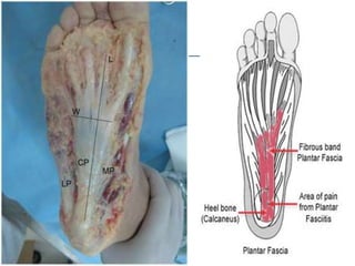
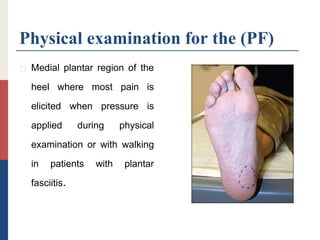
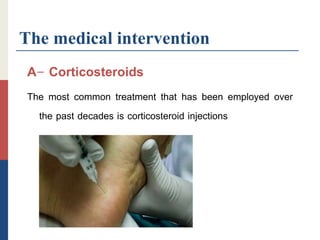
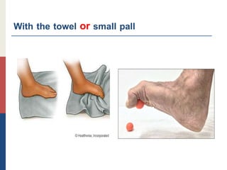
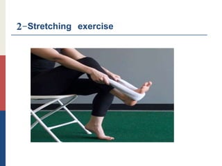
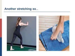
Planter fasciitis
The student  Sohaib Shehada Alashqar
3rd level of physiotherapy
Orthopedics course
Supervisor Dr Fadel Naim
2nd March 2019
Introduction
The aponeurosis of the foot or plantar fascia is comprised of
three distinct bands of dense connective tissue which support
the arch of the foot The plantar fascia originates at the medial
tubercle of the calcaneus extends distally into the five
metatarsophalangeal joints and ends at the proximal
phalanges of each digit.
Definition of the planter fasciitis
Plantar fasciitis has commonly been defined as an
inflammatory process resulting from repetitive microtrauma
leading to micro-tears of the plantar fascia However current
literature suggests plantar fasciitis is better referred to as
“plantar fasciosis” (PF) due to the chronic and degenerative
processes evident in the tissues rather than inflammation.
THE (PF)
Plantar fasciitis (PF) is the most common cause of chronic
pain beneath the heel in adults and may be treated using
different therapeutic strategies.
Conservative treatments have always been the first approach
for treating PF as recommended by the APTA (American
physical therapy association ).
THAT’S MEAN
The current literature has confirmed its degenerative rather
than inflammatory pathology and coined the term plantar
fasciosis instead of PF because of the histological evidence of
chronic inflammatory changes without fibroblastic proliferation
suggestive of degenerative changes.
CON..
During locomotion and standing plantar fascia acts as shock
absorbers and supports the arch of the foot It is an important
static stabilizer of the longitudinal arch of the foot.
However, when the pressure on the plantar ligament is
important in overweight or obese people or increases with
sudden weight gain (pregnant women, etc.) or with repetitive
activity (long-distance runner activity involving being on feet
often, etc.) the risk factor related to this overuse increases.
The causes
A- The lesion is usually an overuse phenomenon occurring in
the presence of predisposing anatomical biomechanical or
environmental factors that put too much strain on the plantar
fascia.
B-The condition seems to be more common in people with a
valgus deformity because this flattens the foot and puts more
strain on the fascia.
CON..
C- Short calf muscles can also be the cause of an overstrained
fascia.
In this is condition the Achilles tendon tends to pull the hell
upwards during standing which stresses the led longitudinal
arch an the fascia .
The risk factor
Common risk factors include
1-obesity
2-decreased ankle flexion
3- running
4- shortened or tight achilles tendon
5- high arches
6- flat feet
Women aged 40–60 years are more frequently diagnosed than
other demographics.
Statistic
The human foot has 26 bones 33 joints 107 ligaments 19
muscles and tendons.
That’s 52 bones in your feet which make up about 25 percent
of all the bones in your body.
CON..
Since a quarter of all the bones in the human body are down in
your feet when a great deal of force weight and pressure are
placed on them your chance of injury increases
CON..
In fact the American Podiatric Medical Association)APMA)
conducted a study that revealed 77 percent of Americans (18
and up) suffer from foot pain.
Approximately 2 million people in the United States will suffer
from Plantar Fasciitis one of the most common foot pains.
CON…
1- About 83 percent of active adults ages 25 to 65 will
experience plantars
2-Ten percent of runners will suffer from it
3-Fourteen percent reported improved symptoms simply from
changing their shoe .
The symptoms
The primary symptom complaint is pain in the arch of the foot
when bearing weight which is worse during the first few steps
in the morning and improves with rest .
Symptom of the (PF)
1-Tightness in the fascia
2- increased discomfort with passive dorsiflexion of great toe
3- standing on the tip of toe In majority it is a self-limiting
disease .
The Diagnosis
1-The diagnosis of PF is exclusively based on clinical history
2-physical examination it usually presents with severe sharp
early morning first step inferior heel pain that improves with
movements but aggravated by weight-bearing activities.
3- Imaging by (ultrasound  MRI  X- Rays)
CON…
Use of ultrasonography and magnetic resonance imaging is
reserved for recalcitrant cases or to rule out other heel
pathology findings of increased plantar fascia thickness and
abnormal tissue signal the diagnosis of plantar fasciitis.
Clinical Diagnosis
Diagnosis of plantar fasciitis is based on patient history risk
factors and physical examination findings.
Most patients have heel pain and tightness after standing up from
bed in the morning or after they have been seated for a
prolonged time.
CON..
Typically the heel pain will improve with ambulation but could
intensify by day's end if the patient continues to walk or stand
for a long time.
On physical examination patients may walk with their affected
foot in an equine position to avoid placing pressure on the
painful heel.
CON…
Palpation of the medial plantar calcaneal region will elicit a sharp
stabbing pain Passive ankle first toe dorsiflexion can cause
discomfort in the proximal plantar fascia it can also assess
tightness of the Achilles tendon.
Other causes of heel pain should be sought if history and
physical examination findings are atypical for plantar fasciitis.
Physical examination for the (PF)
 Medial plantar region of the
heel where most pain is
elicited when pressure is
applied during physical
examination or with walking
in patients with plantar
fasciitis.
Imaging
Imaging can aid in the diagnosis of plantar fasciitis.
Although not routinely needed initially imaging can be used to
confirm recalcitrant plantar fasciitis or to rule out other heel
pathology.
X-rays for the (PF)
 Lateral radiography of the
foot showing a large heel
spur
Ultrasonography
Ultrasonography is inexpensive and useful in ruling out soft
tissue pathology of the heel.
Findings that support the diagnosis of plantar fasciitis include
proximal plantar fascia thickness greater than 4 mm and
areas of hypoechogenicity.
MRI
Magnetic resonance imaging although expensive is a valuable
tool for assessing causes of recalcitrant heel pain.
Diagnostic findings include increased proximal plantar fascia
thickening with increased signal intensity on T2-weighted and
short tau inversion recovery images.
MRI for the (PF)
 Sagittal T2-weighted
magnetic resonance
imaging showing thickening
of the plantar fascia (short
arrows) and increased
signal intensity (long arrow).
The treatment
Include two type of management
1-medical management
2- physical therapy management
1- Medical Management
When conservative measures fail, surgical plantar fasciotomy
with or without heel spur removal may be employed.
There is a method through an open procedure
percutaneously or most common endoscopic ally that
releases the plantar fascia this is an effective treatment.
Red flag
Surgery for plantar fasciitis should be considered only after all
other forms of treatment have failed.
The medical intervention
A- Corticosteroids
The most common treatment that has been employed over
the past decades is corticosteroid injections
B- Botulinum toxin Type-A
Traditionally botulinum toxin has been used in the treatment of
spasticity and nerve blocks.
Only recently has it found its way into musculoskeletal medicine.
CON..
Three RCTs compared the effect of botulinum toxin type-A
(BTA) on heel pain with steroids
The studies reported significant improvements with BTA.
Furthermore patients with plantar fasciitis who received BTA
had significantly longer lasting relief of dysfunction and pain
than those who received placebo further comparative studies
are needed with larger sample sizes.
C- Autologous platelet-rich plasma therapy
Platelet-rich plasma (PRP) therapy showed significant
improvements in the 3month follow-up.
The use of PRP improves blood flow at the site of injection
which aids in the regeneration at the site of pain and
inflammation and the boost that occurs after the injections
help the regeneration of the site of pain and inflammation.
D- Polydeoxyribonucleotide (PDRN)
injections
Clinical efficacy with no notable
complications and were associated with symptomatic
improvement in refractory plantar fasciitis.
E-Endoscopic plantar fasciotomy (EPF)
Is a minimally invasive and minimally traumatic surgical
treatment for the common problem of chronic plantar fasciitis.
This procedure is indicated only for the release of the proximal
medial aspect of the fascia in cases that do not respond to
aggressive conservative nonsurgical treatment.
Endoscopic plantar fasciotomy
2-physical therapy management
A-Therapeutic exercise
The most common treatments include:
1 strengthening exercise for the fascia .
2stretchingexercise of the gastroconemious
– soleus- plantar fascia.
3 joint mobilization- manipulation.
1-Strength exercise
Similar to tendinopathy management high-load strength training
appears to be effective in the treatment of plantar fasciitis.
High-load strength training may aid in a quicker reduction in pain
and improvements in function.
With the towel or small pall
2-Stretching exercise
Another stretching ex..
3-Mobilizations and manipulations
have also been shown to decrease pain and relieve symptoms
in some cases.
Posterior talocrural joint mobs and subtalar joint distraction
manipulation have been performed with the hypomobile
talocrural joint.
Massage for the sole is very effected in this is case.
B-The physical modalities
 1- Therapeutic Ultrasound
 2-Contras bath
 3- shock wave
 4-Intophoresis
 5-TENS
C-Foot orthoses
Foot orthotics are commonly recommended for persons with
plantar fasciitis to aid in preventing overpronation of the foot
and to unload tensile forces on the plantar fascia.
There are many different orthotics available.
CON…
Produce small short-term benefits in function and may also
produce small reductions in pain for people with plantar
fasciitis but they do not have long-term beneficial effects.
D-Posterior-night splints
Maintain ankle dorsiflexion and toe extension allowing for a
constant stretch on the plantar fascia.
Summery
Plantar fasciitis accounts for a large percentage of cases seen
by podiatric physicians and is often seen by general
practitioners and orthopedic surgeons.
Although most cases respond to 4–6 months of conservative
nonsurgical treatment 10–15% require surgery.
Summary
If aggressive conservative treatment for plantar fasciitis fails and
surgery is indicated the endoscopic approach is superior to
conventional open procedures and significantly minimizes
surgical trauma resulting in an earlier return to regular
activities with fewer complications.
The References
1-Lee, Tamsin L., and Benjamin L. Marx. "Noninvasive, Multimodality Approach
to Treating Plantar Fasciitis: A Case Study." Journal of acupuncture and
meridian studies 11.4 (2018): 162-164
2- Gonnade, Nitesh. "Regenerative efficacy of therapeutic quality platelet-rich
plasma injections versus phonophoresis with kinesiotaping for the treatment
of chronic plantar fasciitis: A prospective randomized pilot study." Asian
Journal of Transfusion Science 12.2 (2018): 105
CON..
3- Al- Boloushi, Z. "Minimally invasive non-surgical management of plantar fasciitis:
A systematic review." Journal of bodywork and movement therapies 2018
4- Hake, Daniel H. "Endoscopic plantar fasciotomy: A minimally traumatic procedure
for chronic plantar fasciitis." The Ochsner Journal 2.3 (2000): 175-178
5- Moyne-Bressand, Sébastien. "Effectiveness of Foot Biomechanical Orthoses to
Relieve Patients Suffering from Plantar Fasciitis: Is the Reduction of Pain Related
to Change in Neural Strategy?." BioMed research international 2018 (2018)
CON..
6- Crawford, R. "Diagnosis and treatment of plantar fasciitis." Am Fam Physician
84.6 (2011): 676-82
7-Carlson RE, Fleming LL, Hutton WC. The biomechanical relationship between the
tendoachilles, plantar fascia and metatarsophalangeal joint dorsiflexion angle. Foot
ankle Int / Am Orthop Foot Ankle Soc and Swiss Foot Ankle Soc. 21-1-
2000(18–25)
8- Lemont H, Ammirati KM, Usen N. Plantar fasciitis: a degenerative process
(fasciosis) without inflammation. J Am Podiatr Med Assoc. 2003;93(3):234–7.
Last reference from a book
9-Ombregt Ludwig, Bisschop pierre, ter veer Herman
j. A System of Orthopaedic Medicine-E-Book.
Elsevier Health Sciences, 2013.
Planter fasciitis

Planter fasciitis

  • 1.
    Planter fasciitis The student Sohaib Shehada Alashqar 3rd level of physiotherapy Orthopedics course Supervisor Dr Fadel Naim 2nd March 2019
  • 2.
    Introduction The aponeurosis ofthe foot or plantar fascia is comprised of three distinct bands of dense connective tissue which support the arch of the foot The plantar fascia originates at the medial tubercle of the calcaneus extends distally into the five metatarsophalangeal joints and ends at the proximal phalanges of each digit.
  • 4.
    Definition of theplanter fasciitis Plantar fasciitis has commonly been defined as an inflammatory process resulting from repetitive microtrauma leading to micro-tears of the plantar fascia However current literature suggests plantar fasciitis is better referred to as “plantar fasciosis” (PF) due to the chronic and degenerative processes evident in the tissues rather than inflammation.
  • 5.
    THE (PF) Plantar fasciitis(PF) is the most common cause of chronic pain beneath the heel in adults and may be treated using different therapeutic strategies. Conservative treatments have always been the first approach for treating PF as recommended by the APTA (American physical therapy association ).
  • 6.
    THAT’S MEAN The currentliterature has confirmed its degenerative rather than inflammatory pathology and coined the term plantar fasciosis instead of PF because of the histological evidence of chronic inflammatory changes without fibroblastic proliferation suggestive of degenerative changes.
  • 7.
    CON.. During locomotion andstanding plantar fascia acts as shock absorbers and supports the arch of the foot It is an important static stabilizer of the longitudinal arch of the foot. However, when the pressure on the plantar ligament is important in overweight or obese people or increases with sudden weight gain (pregnant women, etc.) or with repetitive activity (long-distance runner activity involving being on feet often, etc.) the risk factor related to this overuse increases.
  • 8.
    The causes A- Thelesion is usually an overuse phenomenon occurring in the presence of predisposing anatomical biomechanical or environmental factors that put too much strain on the plantar fascia. B-The condition seems to be more common in people with a valgus deformity because this flattens the foot and puts more strain on the fascia.
  • 9.
    CON.. C- Short calfmuscles can also be the cause of an overstrained fascia. In this is condition the Achilles tendon tends to pull the hell upwards during standing which stresses the led longitudinal arch an the fascia .
  • 11.
    The risk factor Commonrisk factors include 1-obesity 2-decreased ankle flexion 3- running 4- shortened or tight achilles tendon 5- high arches 6- flat feet Women aged 40–60 years are more frequently diagnosed than other demographics.
  • 14.
    Statistic The human foothas 26 bones 33 joints 107 ligaments 19 muscles and tendons. That’s 52 bones in your feet which make up about 25 percent of all the bones in your body.
  • 15.
    CON.. Since a quarterof all the bones in the human body are down in your feet when a great deal of force weight and pressure are placed on them your chance of injury increases
  • 16.
    CON.. In fact theAmerican Podiatric Medical Association)APMA) conducted a study that revealed 77 percent of Americans (18 and up) suffer from foot pain. Approximately 2 million people in the United States will suffer from Plantar Fasciitis one of the most common foot pains.
  • 17.
    CON… 1- About 83percent of active adults ages 25 to 65 will experience plantars 2-Ten percent of runners will suffer from it 3-Fourteen percent reported improved symptoms simply from changing their shoe .
  • 18.
    The symptoms The primarysymptom complaint is pain in the arch of the foot when bearing weight which is worse during the first few steps in the morning and improves with rest .
  • 19.
    Symptom of the(PF) 1-Tightness in the fascia 2- increased discomfort with passive dorsiflexion of great toe 3- standing on the tip of toe In majority it is a self-limiting disease .
  • 20.
    The Diagnosis 1-The diagnosisof PF is exclusively based on clinical history 2-physical examination it usually presents with severe sharp early morning first step inferior heel pain that improves with movements but aggravated by weight-bearing activities. 3- Imaging by (ultrasound MRI X- Rays)
  • 21.
    CON… Use of ultrasonographyand magnetic resonance imaging is reserved for recalcitrant cases or to rule out other heel pathology findings of increased plantar fascia thickness and abnormal tissue signal the diagnosis of plantar fasciitis.
  • 22.
    Clinical Diagnosis Diagnosis ofplantar fasciitis is based on patient history risk factors and physical examination findings. Most patients have heel pain and tightness after standing up from bed in the morning or after they have been seated for a prolonged time.
  • 23.
    CON.. Typically the heelpain will improve with ambulation but could intensify by day's end if the patient continues to walk or stand for a long time. On physical examination patients may walk with their affected foot in an equine position to avoid placing pressure on the painful heel.
  • 24.
    CON… Palpation of themedial plantar calcaneal region will elicit a sharp stabbing pain Passive ankle first toe dorsiflexion can cause discomfort in the proximal plantar fascia it can also assess tightness of the Achilles tendon. Other causes of heel pain should be sought if history and physical examination findings are atypical for plantar fasciitis.
  • 25.
    Physical examination forthe (PF)  Medial plantar region of the heel where most pain is elicited when pressure is applied during physical examination or with walking in patients with plantar fasciitis.
  • 26.
    Imaging Imaging can aidin the diagnosis of plantar fasciitis. Although not routinely needed initially imaging can be used to confirm recalcitrant plantar fasciitis or to rule out other heel pathology.
  • 27.
    X-rays for the(PF)  Lateral radiography of the foot showing a large heel spur
  • 28.
    Ultrasonography Ultrasonography is inexpensiveand useful in ruling out soft tissue pathology of the heel. Findings that support the diagnosis of plantar fasciitis include proximal plantar fascia thickness greater than 4 mm and areas of hypoechogenicity.
  • 29.
    MRI Magnetic resonance imagingalthough expensive is a valuable tool for assessing causes of recalcitrant heel pain. Diagnostic findings include increased proximal plantar fascia thickening with increased signal intensity on T2-weighted and short tau inversion recovery images.
  • 30.
    MRI for the(PF)  Sagittal T2-weighted magnetic resonance imaging showing thickening of the plantar fascia (short arrows) and increased signal intensity (long arrow).
  • 31.
    The treatment Include twotype of management 1-medical management 2- physical therapy management
  • 32.
    1- Medical Management Whenconservative measures fail, surgical plantar fasciotomy with or without heel spur removal may be employed. There is a method through an open procedure percutaneously or most common endoscopic ally that releases the plantar fascia this is an effective treatment.
  • 33.
    Red flag Surgery forplantar fasciitis should be considered only after all other forms of treatment have failed.
  • 34.
    The medical intervention A-Corticosteroids The most common treatment that has been employed over the past decades is corticosteroid injections
  • 35.
    B- Botulinum toxinType-A Traditionally botulinum toxin has been used in the treatment of spasticity and nerve blocks. Only recently has it found its way into musculoskeletal medicine.
  • 36.
    CON.. Three RCTs comparedthe effect of botulinum toxin type-A (BTA) on heel pain with steroids The studies reported significant improvements with BTA. Furthermore patients with plantar fasciitis who received BTA had significantly longer lasting relief of dysfunction and pain than those who received placebo further comparative studies are needed with larger sample sizes.
  • 37.
    C- Autologous platelet-richplasma therapy Platelet-rich plasma (PRP) therapy showed significant improvements in the 3month follow-up. The use of PRP improves blood flow at the site of injection which aids in the regeneration at the site of pain and inflammation and the boost that occurs after the injections help the regeneration of the site of pain and inflammation.
  • 38.
    D- Polydeoxyribonucleotide (PDRN) injections Clinicalefficacy with no notable complications and were associated with symptomatic improvement in refractory plantar fasciitis.
  • 39.
    E-Endoscopic plantar fasciotomy(EPF) Is a minimally invasive and minimally traumatic surgical treatment for the common problem of chronic plantar fasciitis. This procedure is indicated only for the release of the proximal medial aspect of the fascia in cases that do not respond to aggressive conservative nonsurgical treatment.
  • 40.
  • 41.
    2-physical therapy management A-Therapeuticexercise The most common treatments include: 1 strengthening exercise for the fascia . 2stretchingexercise of the gastroconemious – soleus- plantar fascia. 3 joint mobilization- manipulation.
  • 42.
    1-Strength exercise Similar totendinopathy management high-load strength training appears to be effective in the treatment of plantar fasciitis. High-load strength training may aid in a quicker reduction in pain and improvements in function.
  • 43.
    With the towelor small pall
  • 44.
  • 46.
  • 47.
    3-Mobilizations and manipulations havealso been shown to decrease pain and relieve symptoms in some cases. Posterior talocrural joint mobs and subtalar joint distraction manipulation have been performed with the hypomobile talocrural joint. Massage for the sole is very effected in this is case.
  • 48.
    B-The physical modalities 1- Therapeutic Ultrasound  2-Contras bath  3- shock wave  4-Intophoresis  5-TENS
  • 49.
    C-Foot orthoses Foot orthoticsare commonly recommended for persons with plantar fasciitis to aid in preventing overpronation of the foot and to unload tensile forces on the plantar fascia. There are many different orthotics available.
  • 50.
    CON… Produce small short-termbenefits in function and may also produce small reductions in pain for people with plantar fasciitis but they do not have long-term beneficial effects.
  • 51.
    D-Posterior-night splints Maintain ankledorsiflexion and toe extension allowing for a constant stretch on the plantar fascia.
  • 52.
    Summery Plantar fasciitis accountsfor a large percentage of cases seen by podiatric physicians and is often seen by general practitioners and orthopedic surgeons. Although most cases respond to 4–6 months of conservative nonsurgical treatment 10–15% require surgery.
  • 53.
    Summary If aggressive conservativetreatment for plantar fasciitis fails and surgery is indicated the endoscopic approach is superior to conventional open procedures and significantly minimizes surgical trauma resulting in an earlier return to regular activities with fewer complications.
  • 54.
    The References 1-Lee, TamsinL., and Benjamin L. Marx. "Noninvasive, Multimodality Approach to Treating Plantar Fasciitis: A Case Study." Journal of acupuncture and meridian studies 11.4 (2018): 162-164 2- Gonnade, Nitesh. "Regenerative efficacy of therapeutic quality platelet-rich plasma injections versus phonophoresis with kinesiotaping for the treatment of chronic plantar fasciitis: A prospective randomized pilot study." Asian Journal of Transfusion Science 12.2 (2018): 105
  • 55.
    CON.. 3- Al- Boloushi,Z. "Minimally invasive non-surgical management of plantar fasciitis: A systematic review." Journal of bodywork and movement therapies 2018 4- Hake, Daniel H. "Endoscopic plantar fasciotomy: A minimally traumatic procedure for chronic plantar fasciitis." The Ochsner Journal 2.3 (2000): 175-178 5- Moyne-Bressand, Sébastien. "Effectiveness of Foot Biomechanical Orthoses to Relieve Patients Suffering from Plantar Fasciitis: Is the Reduction of Pain Related to Change in Neural Strategy?." BioMed research international 2018 (2018)
  • 56.
    CON.. 6- Crawford, R."Diagnosis and treatment of plantar fasciitis." Am Fam Physician 84.6 (2011): 676-82 7-Carlson RE, Fleming LL, Hutton WC. The biomechanical relationship between the tendoachilles, plantar fascia and metatarsophalangeal joint dorsiflexion angle. Foot ankle Int / Am Orthop Foot Ankle Soc and Swiss Foot Ankle Soc. 21-1- 2000(18–25) 8- Lemont H, Ammirati KM, Usen N. Plantar fasciitis: a degenerative process (fasciosis) without inflammation. J Am Podiatr Med Assoc. 2003;93(3):234–7.
  • 57.
    Last reference froma book 9-Ombregt Ludwig, Bisschop pierre, ter veer Herman j. A System of Orthopaedic Medicine-E-Book. Elsevier Health Sciences, 2013.