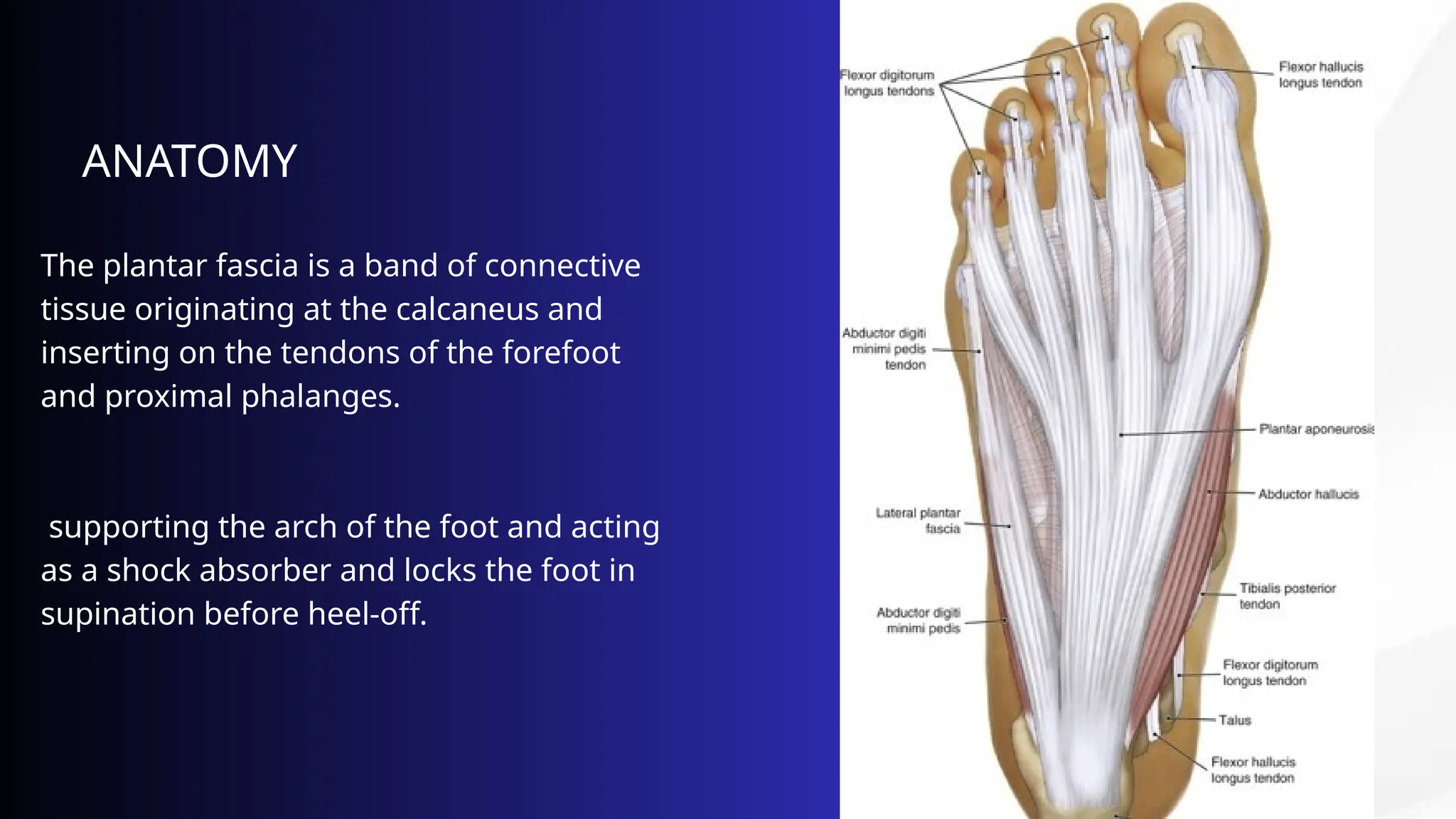
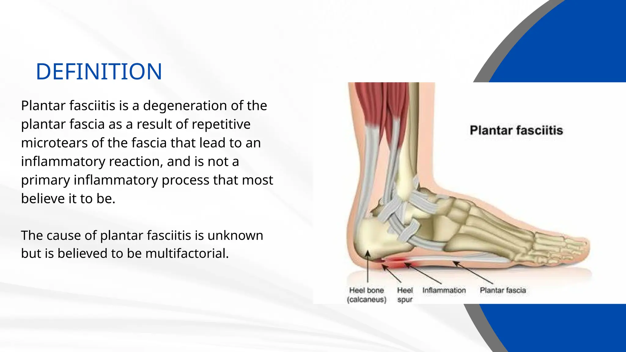
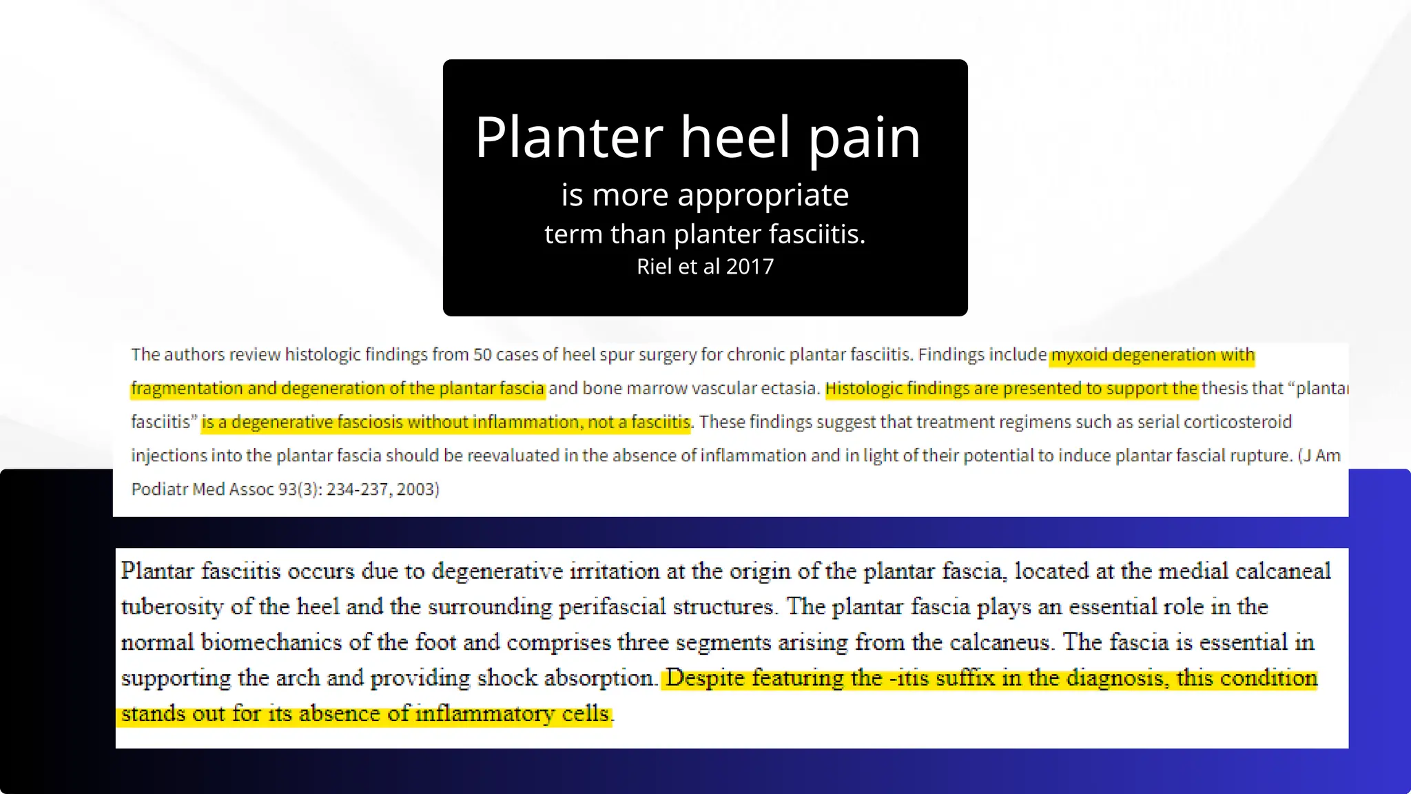
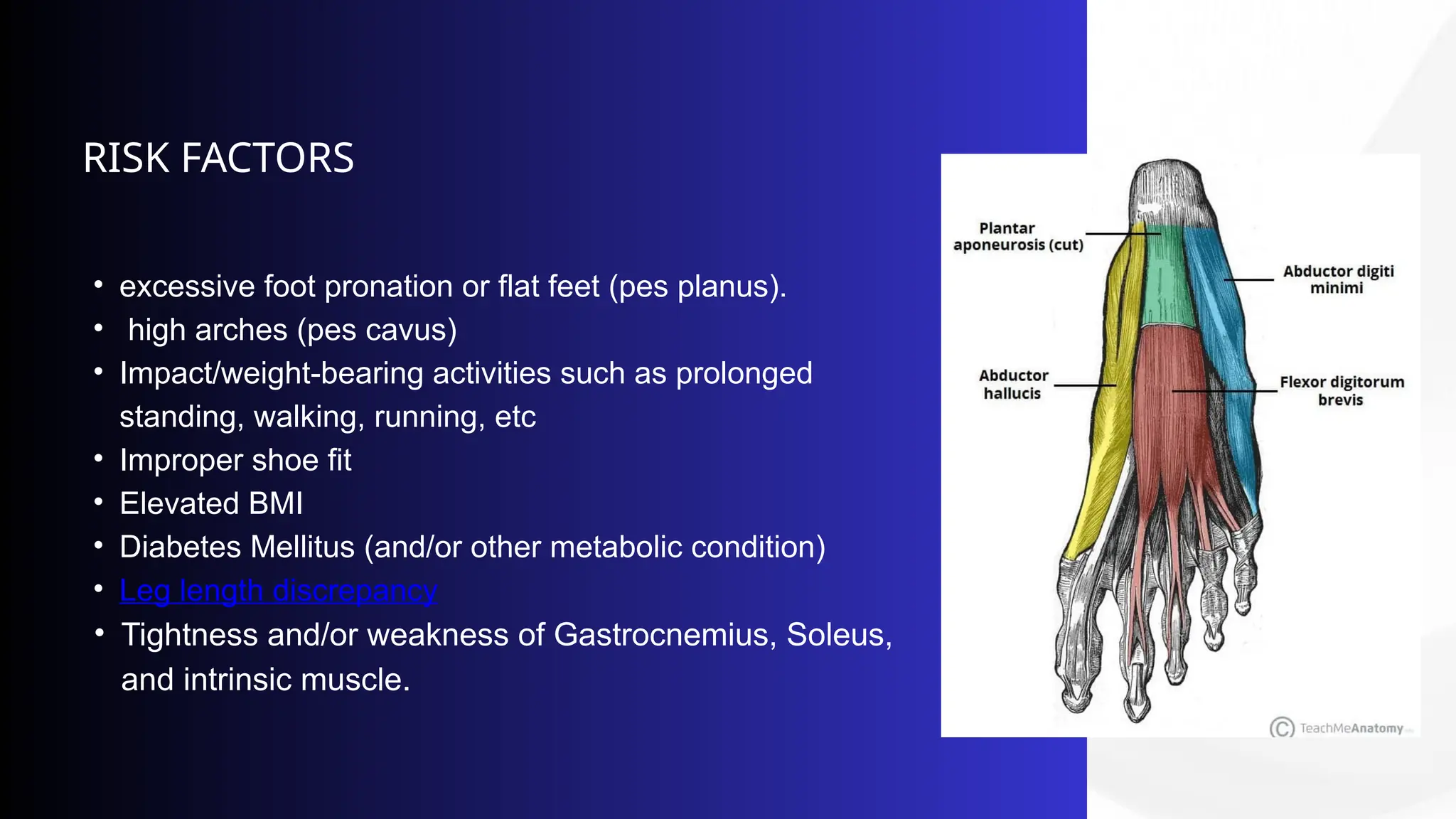
PLANTAR FASCIITIS
OR
“PLANTER HEEL PAIN”
by Inasse Alarousi,PT
ANATOMY
The plantar fascia is a band of connective
tissue originating at the calcaneus and
inserting on the tendons of the forefoot
and proximal phalanges.
supporting the arch of the foot and acting
as a shock absorber and locks the foot in
supination before heel-off.
DEFINITION
Plantar fasciitis is a degeneration of the
plantar fascia as a result of repetitive
microtears of the fascia that lead to an
inflammatory reaction, and is not a
primary inflammatory process that most
believe it to be.
The cause of plantar fasciitis is unknown
but is believed to be multifactorial.
Planter heel pain
is more appropriate
term than planter fasciitis.
Riel et al 2017
• excessive foot pronation or flat feet (pes planus).
• high arches (pes cavus)
• Impact/weight-bearing activities such as prolonged
standing, walking, running, etc
• Improper shoe fit
• Elevated BMI
• Diabetes Mellitus (and/or other metabolic condition)
• Leg length discrepancy
• Tightness and/or weakness of Gastrocnemius, Soleus,
and intrinsic muscle.
RISK FACTORS
PRESENTATION
Many patients complain of pain in the
morning or after rising from prolonged
sitting, with relief with movement.
A physical examination will reveal pain on
medial plantar calcaneal region, pain with
dorsiflexion”windlass test”, and tightness of
the Achilles tendon or the gastrocnemius
muscle.
as we consider a patient's medical history,
physical activity, foot pain symptoms, and
more.
MAKING THE DIAGNOSIS
Plantar fasciitis most often is a clinical
diagnosis
Plain radiographs can rule out bony
lesions or stress fractures
diagnostic Ultrasound can rule out
certain causes of heel pain (such as
plantar fibromatosis, foreign body) and
can aid fascial thickness and the
presence of fascial tears
DIAGNOSTIC ULTRASOUND
Several authors have reported the
thickening of the plantar fascia and
hypoechoic changes as characteristic
features of plantar fasciitis. Multiple
studies have verified that in patients
with symptomatic plantar fasciitis, the
plantar fascia thickness tends to be
greater than 4 mm on diagnostic
ultrasound.
TREATMENT OPTIONS
Between 70% and 80% of patients have reduced pain with
conservative treatment alone as the normal course is within one
year.
.
• activity modification and relative rest of offending activity
• Ice or topical NSAIDs can be used to help alleviate pain.
• Deep friction massage of the arch and insertion.
• Educate patients on proper stretching and rehab.
• orthotics silicone heel cup or arch supports.
Corticosteroid injections were found to be more effective versus
placebo in pain relief at one month, but these effects were not
sustained over longer period.
SURGRY OPTIONS
Consider surgery if the patient has
continued pain after 12 months of
nonsurgical management. Options
include partial or complete plantar fascia
release and gastrocnemius release. These
surgeries may be performed as open or
endoscopically.
THANK YOU
ANY QUESTION?

PLANTAR FASCIITIS or PLANTER HEEL PAIN.pptx

  • 1.
    PLANTAR FASCIITIS OR “PLANTER HEELPAIN” by Inasse Alarousi,PT
  • 2.
    ANATOMY The plantar fasciais a band of connective tissue originating at the calcaneus and inserting on the tendons of the forefoot and proximal phalanges. supporting the arch of the foot and acting as a shock absorber and locks the foot in supination before heel-off.
  • 3.
    DEFINITION Plantar fasciitis isa degeneration of the plantar fascia as a result of repetitive microtears of the fascia that lead to an inflammatory reaction, and is not a primary inflammatory process that most believe it to be. The cause of plantar fasciitis is unknown but is believed to be multifactorial.
  • 4.
    Planter heel pain ismore appropriate term than planter fasciitis. Riel et al 2017
  • 5.
    • excessive footpronation or flat feet (pes planus). • high arches (pes cavus) • Impact/weight-bearing activities such as prolonged standing, walking, running, etc • Improper shoe fit • Elevated BMI • Diabetes Mellitus (and/or other metabolic condition) • Leg length discrepancy • Tightness and/or weakness of Gastrocnemius, Soleus, and intrinsic muscle. RISK FACTORS
  • 6.
    PRESENTATION Many patients complainof pain in the morning or after rising from prolonged sitting, with relief with movement. A physical examination will reveal pain on medial plantar calcaneal region, pain with dorsiflexion”windlass test”, and tightness of the Achilles tendon or the gastrocnemius muscle. as we consider a patient's medical history, physical activity, foot pain symptoms, and more.
  • 7.
    MAKING THE DIAGNOSIS Plantarfasciitis most often is a clinical diagnosis Plain radiographs can rule out bony lesions or stress fractures diagnostic Ultrasound can rule out certain causes of heel pain (such as plantar fibromatosis, foreign body) and can aid fascial thickness and the presence of fascial tears
  • 8.
    DIAGNOSTIC ULTRASOUND Several authorshave reported the thickening of the plantar fascia and hypoechoic changes as characteristic features of plantar fasciitis. Multiple studies have verified that in patients with symptomatic plantar fasciitis, the plantar fascia thickness tends to be greater than 4 mm on diagnostic ultrasound.
  • 9.
    TREATMENT OPTIONS Between 70%and 80% of patients have reduced pain with conservative treatment alone as the normal course is within one year. . • activity modification and relative rest of offending activity • Ice or topical NSAIDs can be used to help alleviate pain. • Deep friction massage of the arch and insertion. • Educate patients on proper stretching and rehab. • orthotics silicone heel cup or arch supports. Corticosteroid injections were found to be more effective versus placebo in pain relief at one month, but these effects were not sustained over longer period.
  • 13.
    SURGRY OPTIONS Consider surgeryif the patient has continued pain after 12 months of nonsurgical management. Options include partial or complete plantar fascia release and gastrocnemius release. These surgeries may be performed as open or endoscopically.
  • 14.