
Recommended
More Related Content
Similar to Histology and Clinical Correlations of the Placenta
Similar to Histology and Clinical Correlations of the Placenta (20)
Recently uploaded
Recently uploaded (20)
Histology and Clinical Correlations of the Placenta
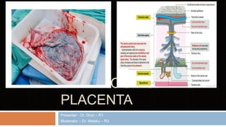
- 1. HISTOLOGY OF PLACENTA Presenter - Dr. Dirar – R1 Moderator – Dr. Melaku – R3
- 2. 1. Gross description 2. Development of placenta 3. Histology of placenta 4. Clinical correlation Outlines
- 3. Gross description Discoid organ 15 to 20 cm in diameter 3 cm thick at the center weighing on average 450 g.
- 4. two surfaces or plates: the chorionic plate - to which the umbilical cord is attached, and the basal plate - that abuts the maternal endometrium.
- 5. Cotyledons 15 to 20 lobes, aka cotyledons formed by invaginations of septa from the basal plate
- 6. Umbilical cord The 50- to 60-cm- long and 12-mm- thick and twisted umbilical cord is attached to the chorionic plate and contains two umbilical arteries and one umbilical vein
- 7. Development of the placenta
- 8. Implantation – on decidua functionalis
- 12. The wall of maternal blood vessels is infiltrated and ruptured by trophoblast cells. And then maternal blood is released into the intervillous space, The uterine spiral arteries are converted to uteroplacental arteries Preeclampsia occurs when there is a reduced development of the branches of the chorionic villus tree and limited fetal growth.
- 13. The decidual reaction provides an immune protective environment for the development of the embryo 1. production of immunosuppressiv e substances 2. Syncytiotrophoblas t cells do not express major histocompatibility complex class II.
- 14. Histology
- 15. Placental lobe - cotyledons
- 16. cytotrophoblast a layer of mitotically active cells immediately around the amnion and yolk sac nuclei are more round and open
- 17. syncytiotrophoblast more superficial, no mitotis mass of multinucleated cytoplasm which invades the surrounding stroma possesses a brush border with microvilli
- 18. Intermediate trophoblast – pleomorphic and hyperchromatic nuclei with amphophilic to eosinophilic cytoplasm Other cell types mesenchymal cells fibroblasts, myofibroblasts, smooth muscle cells Hofbauer cells
- 19. Early placenta
- 20. Compared with early placenta, in late placenta Average villous diameter is much smaller Reflecting the extensive branching growth of the villi
- 21. Late placenta
- 22. placental barrier consists of Syncytiotrophoblast inner cytotrophoblast layer Basal lamina of the trophoblast Connective (mesenchymal) tissue of the villus Basal lamina of the endothelium Endothelium of the fetal placental capillary in the tertiary villus
- 23. Umbilical arteries Vs. umbilical vein
- 24. Umbilical arteries Vs. umbilical vein
- 25. Three regions of the decidua 1. The decidua basalis 2. The decidua capsularis 3. The decidua parietalis chorion laeve Vs
- 27. 1. Chorioamnionitis 2. Complete mole 3. Partial mole 4. Invasive mole 5. Choriocarcinoma Clinical correlation
- 28. Chorioamnionitis A collection of neutrophils (pus) has formed between the amnion and chorion Inflammation in the decidua (oval) may be physiologic and is not sufficient to diagnose chorioamnionitis
- 29. Complete mole Diploid gestation with two paternal genomes no identifiable embryo, cord, or amniotic membranes a ‘bunch of grapes’
- 30. Marked trophoblastic hyperplasia and vesicular swelling circumferential but haphazard arrangement around the individual villi Vessels seem absent or very scanty
- 31. Partial mole Triploid Embryo is present
- 32. grossly vesicular villi are mixed with normal-appearing ones Trophoblastic proliferation is present but lesser than complete mole
- 33. Invasive mole villi penetrate deeply to the myometrium and/or its blood vessels nearly always of the complete type but occasionally of the partial type
- 34. Choriocarcinoma Most cases occur following a complete hydatidiform mole characteristically forms soft, dark red, hemorrhagic, round nodular tumor masses
- 35. clusters of cytotrophoblast separated by streaming masses of syncytiotrophoblast characteristic dimorphic plexiform pattern Villi are characteristically absent
- 36. Reference Wheater`s functional histology 6th ed Histology - A text and atlas 7th ed. Difiore`s atlas of histology 12th ed. Rosai and Akerman surgical pathology Gabbe text book of obstetrics